
Baldur's Gate 3: Where To Find Miri - bg3 edmund
Mar 18, 2024 — After decades of focusing almost exclusively on restoring impaired waters, EPA created the Healthy Watersheds Program (HWP) to bring more ...
Brewster's Angle - Light that is reflected from the flat surface of a dielectric (or insulating) material is often partially polarized, with the electric vectors of the reflected light vibrating in a plane that is parallel to the surface of the material. Common examples of surfaces that reflect polarized light are undisturbed water, glass, sheet plastics, and highways. In these instances, light waves that have the electric field vectors parallel to the surface are reflected to a greater degree than those with different orientations. This tutorial demonstrates the polarization effect on light reflected at a specific angle (the Brewster angle) from a transparent medium.
MTF Surgery Guide · Orchiectomy · Vulvoplasty · Vaginoplasty · Breast Augmentation · Body Sculpting / Body Feminization · Facial Feminization · Find a Surgeon ».
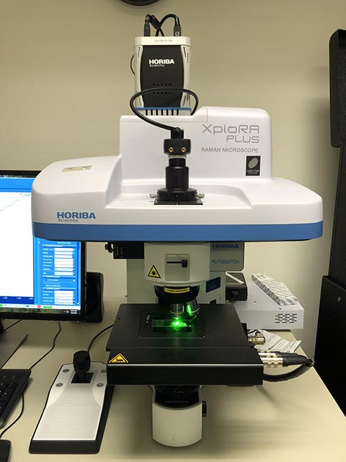
The interaction of light on materials is very different it may be transmitted, reflected, or scattered; the wavelength of the light affects the interaction with materials in different colors. This study of light is called spectroscopy. Based on this an Indian physicist C.V Raman observed the scattering phenomenon where the light is scattered by the molecules and hence this phenomenon was named Raman scattering. The analysis/characterization technique that deals with Raman scattering is Raman spectroscopy. In Fig-1 Raman spectrometer from S & N lab is shown. Fig-1 Raman spectrometer from S & N lab [1] Definition Raman spectroscopy is the analytical technique where scattered light is used to measure the vibrational energy modes of the sample. This technique provides both the information on chemical and structural characteristics of the material and also the identification of substances. The Raman spectroscopy extracts the information through the detection of Raman scattering from the sample. Fig-2 is the schematic representation of the Raman spectrometer. Fig-2 Schematic representation of Raman spectrometer [2] Working principle The working principle of Raman spectroscopy is based on the inelastic scattering of monochromatic light from a laser source which changes its frequency upon interaction with the material. Photons from the laser are absorbed by the samples and it is remitted with a frequency shift up or down in comparison to the original monochromatic frequency this is called the Raman effect. These shifts in the frequency provide information about the rotational, vibrational, and other low-frequency transitions in the molecules. This technique can be used in studying the materials like solid, liquid, and gaseous nature. In order to understand spectroscopy better, we should know the difference between Rayleigh scattering and Raman scattering. Rayleigh scattering: In this case, the energy of the molecules is unchanged after the interaction with the molecules. The energy and the wavelength of the scattered photons are equal to that of the incident photon. Hence the energy of the scattering particle is conserved this is called Rayleigh scattering. Raman scattering: In this case, the light is scattered by the molecule, and the oscillating electromagnetic field of a photon induces a polarisation of the molecular electron cloud causing the molecules to be in a higher energy state with the energy of a photon is transferred to the molecule. This can be considered as the formation of a very short-lived complex between the photons and molecules which is commonly called the virtual state of molecules. The virtual state is not stable, and the photon is remitted almost immediately as scattered light. The schematic representation of the Raman and Rayleigh scattering is shown in Fig-3. Fig-3 Raman scattering and Rayleigh scattering [3] Components of Raman spectrometer Laser source: The laser source is used for the excitation of the sample and resulting scattered light. Injection/rejection filter: The filter delivers the laser to the sample and allows the scattered Raman light to pass through to the spectrograph. Spectrograph: The spectrograph is used to divide the light into separated wavelengths and measure the light intensity at each wavelength. Microscope: The microscope is used to focus the laser light onto a point on the sample surface and collects the Raman light. Computer: It provides instrumental control and data handling and manipulation. Fig-4 Schematic representation of Raman spectrometer with its components [4] Information from Raman spectroscopy The information that is obtained from the Raman spectroscopy is useful in analyzing various aspects of the material compositions. The Raman shifts and relative intensities of all Raman bands of the material allow identifying the material. The individual band changes and shifts which are seen as narrow, or broad can be varied with the intensity of the light. These changes can reveal information about the stresses in the sample and variation in crystallinity. The amount of material and its composition can also be identified, the variations in spectra with the position of the samples also reveal the changes in the material’s homogeneity. Advantages and disadvantages The advantages of Raman spectroscopy include its strength in specifying the chemicals in the materials which is a chemical fingerprint technique. There is no need for sample preparation and it is a non-destructive technique. The Raman spectra are acquired within a few seconds decreasing the processing time. The disadvantages of Raman spectroscopy include that it can not be used in analyzing metals and alloys, and in most cases, it is not quantitative regarding the composition. The Raman effect is weak and the detection needs a very sensitive and highly optimized instrument. The fluorescence of impurities or of the sample itself can hide the Raman spectrum. Reference [1] http://www.snlabs.com/raman-spectroscopy.html [2] Downes, A. and Elfick, A., 2010. Raman spectroscopy and related techniques in biomedicine. Sensors, 10(3), pp.1871-1889. [3] https://www.edinst.com/blog/what-is-raman-spectroscopy/ [4] https://www.sas.upenn.edu/~crulli/TheRamanSpectrophotometer.html
What ispolarizationofwaves in Physics
[3] https://www.edinst.com/blog/what-is-raman-spectroscopy/ [4] https://www.sas.upenn.edu/~crulli/TheRamanSpectrophotometer.html
Optical Properties of Aspherical Lenses. An aspherical lens element is used to correct for spherical aberrations, where the converging rays from a lens do not ...
Edwin Herbert Land (1909-1991) - The founder of the Polaroid Corporation, Edwin Herbert Land was an American inventor and researcher who dedicated his entire adult life to the study of polarized light, photography and color vision. Perhaps Land's most famous contribution to science, however, was his development of instant photography. The invention was inspired by his three-year old daughter when she asked him why she could not instantly see a picture he had just taken of her on vacation. The one-step dry photographic process took Land three years to perfect, but his success was phenomenal.
Sunlight and almost every other form of natural and artificial illumination produces light waves whose electric field vectors vibrate in all planes that are perpendicular with respect to the direction of propagation. If the electric field vectors are restricted to a single plane by filtration of the beam with specialized materials, then the light is referred to as plane or linearly polarized with respect to the direction of propagation, and all waves vibrating in a single plane are termed plane parallel or plane-polarized.
What is polarisation of lightclass 12
Optics · Our objectives help you focus on yours. Objective lenses are arguably the most important element in the microscope. · In tireless pursuit of the highest ...
Matthew J. Parry-Hill, Robert T. Sutter, Thomas J. Fellers, and Michael W. Davidson - National High Magnetic Field Laboratory, 1800 East Paul Dirac Dr., The Florida State University, Tallahassee, Florida, 32310.
Fig-3 Raman scattering and Rayleigh scattering [3] Components of Raman spectrometer Laser source: The laser source is used for the excitation of the sample and resulting scattered light. Injection/rejection filter: The filter delivers the laser to the sample and allows the scattered Raman light to pass through to the spectrograph. Spectrograph: The spectrograph is used to divide the light into separated wavelengths and measure the light intensity at each wavelength. Microscope: The microscope is used to focus the laser light onto a point on the sample surface and collects the Raman light. Computer: It provides instrumental control and data handling and manipulation. Fig-4 Schematic representation of Raman spectrometer with its components [4] Information from Raman spectroscopy The information that is obtained from the Raman spectroscopy is useful in analyzing various aspects of the material compositions. The Raman shifts and relative intensities of all Raman bands of the material allow identifying the material. The individual band changes and shifts which are seen as narrow, or broad can be varied with the intensity of the light. These changes can reveal information about the stresses in the sample and variation in crystallinity. The amount of material and its composition can also be identified, the variations in spectra with the position of the samples also reveal the changes in the material’s homogeneity. Advantages and disadvantages The advantages of Raman spectroscopy include its strength in specifying the chemicals in the materials which is a chemical fingerprint technique. There is no need for sample preparation and it is a non-destructive technique. The Raman spectra are acquired within a few seconds decreasing the processing time. The disadvantages of Raman spectroscopy include that it can not be used in analyzing metals and alloys, and in most cases, it is not quantitative regarding the composition. The Raman effect is weak and the detection needs a very sensitive and highly optimized instrument. The fluorescence of impurities or of the sample itself can hide the Raman spectrum. Reference [1] http://www.snlabs.com/raman-spectroscopy.html [2] Downes, A. and Elfick, A., 2010. Raman spectroscopy and related techniques in biomedicine. Sensors, 10(3), pp.1871-1889. [3] https://www.edinst.com/blog/what-is-raman-spectroscopy/ [4] https://www.sas.upenn.edu/~crulli/TheRamanSpectrophotometer.html
Shinya Inoué (1921-Present) - Shinya Inoué is a microscopist, cell biologist, and educator who has been described as the grandfather of modern light microscopy. The pioneering microscopist heavily influenced the study of cell dynamics during the 1980s through his developments in video-enhanced contrast microscopy (VEC), which is a modification of the traditional form of differential interference contrast (DIC) microscopy. Inoué also made significant contributions to the investigation of biological systems with polarized light microscopy. His seminal work, "Video Microscopy," was published in 1986, and a second revised and updated edition, co-authored with Kenneth Spring, followed in 1997. The book is a cornerstone of microscopical knowledge and is highly regarded throughout the scientific community.
Fig-1 Raman spectrometer from S & N lab [1] Definition Raman spectroscopy is the analytical technique where scattered light is used to measure the vibrational energy modes of the sample. This technique provides both the information on chemical and structural characteristics of the material and also the identification of substances. The Raman spectroscopy extracts the information through the detection of Raman scattering from the sample. Fig-2 is the schematic representation of the Raman spectrometer. Fig-2 Schematic representation of Raman spectrometer [2] Working principle The working principle of Raman spectroscopy is based on the inelastic scattering of monochromatic light from a laser source which changes its frequency upon interaction with the material. Photons from the laser are absorbed by the samples and it is remitted with a frequency shift up or down in comparison to the original monochromatic frequency this is called the Raman effect. These shifts in the frequency provide information about the rotational, vibrational, and other low-frequency transitions in the molecules. This technique can be used in studying the materials like solid, liquid, and gaseous nature. In order to understand spectroscopy better, we should know the difference between Rayleigh scattering and Raman scattering. Rayleigh scattering: In this case, the energy of the molecules is unchanged after the interaction with the molecules. The energy and the wavelength of the scattered photons are equal to that of the incident photon. Hence the energy of the scattering particle is conserved this is called Rayleigh scattering. Raman scattering: In this case, the light is scattered by the molecule, and the oscillating electromagnetic field of a photon induces a polarisation of the molecular electron cloud causing the molecules to be in a higher energy state with the energy of a photon is transferred to the molecule. This can be considered as the formation of a very short-lived complex between the photons and molecules which is commonly called the virtual state of molecules. The virtual state is not stable, and the photon is remitted almost immediately as scattered light. The schematic representation of the Raman and Rayleigh scattering is shown in Fig-3. Fig-3 Raman scattering and Rayleigh scattering [3] Components of Raman spectrometer Laser source: The laser source is used for the excitation of the sample and resulting scattered light. Injection/rejection filter: The filter delivers the laser to the sample and allows the scattered Raman light to pass through to the spectrograph. Spectrograph: The spectrograph is used to divide the light into separated wavelengths and measure the light intensity at each wavelength. Microscope: The microscope is used to focus the laser light onto a point on the sample surface and collects the Raman light. Computer: It provides instrumental control and data handling and manipulation. Fig-4 Schematic representation of Raman spectrometer with its components [4] Information from Raman spectroscopy The information that is obtained from the Raman spectroscopy is useful in analyzing various aspects of the material compositions. The Raman shifts and relative intensities of all Raman bands of the material allow identifying the material. The individual band changes and shifts which are seen as narrow, or broad can be varied with the intensity of the light. These changes can reveal information about the stresses in the sample and variation in crystallinity. The amount of material and its composition can also be identified, the variations in spectra with the position of the samples also reveal the changes in the material’s homogeneity. Advantages and disadvantages The advantages of Raman spectroscopy include its strength in specifying the chemicals in the materials which is a chemical fingerprint technique. There is no need for sample preparation and it is a non-destructive technique. The Raman spectra are acquired within a few seconds decreasing the processing time. The disadvantages of Raman spectroscopy include that it can not be used in analyzing metals and alloys, and in most cases, it is not quantitative regarding the composition. The Raman effect is weak and the detection needs a very sensitive and highly optimized instrument. The fluorescence of impurities or of the sample itself can hide the Raman spectrum. Reference [1] http://www.snlabs.com/raman-spectroscopy.html [2] Downes, A. and Elfick, A., 2010. Raman spectroscopy and related techniques in biomedicine. Sensors, 10(3), pp.1871-1889. [3] https://www.edinst.com/blog/what-is-raman-spectroscopy/ [4] https://www.sas.upenn.edu/~crulli/TheRamanSpectrophotometer.html
Henri Hureau de Sénarmont (1808-1862) - Sénarmont was a professor of mineralogy and director of studies at the École des Mines in Paris, especially distinguished for his research on polarization and his studies on the artificial formation of minerals. He also contributed to the Geological Survey of France by preparing geological maps and essays. Perhaps the most significant contribution made by de Sénarmont to optics was the polarized light retardation compensator bearing his name, which is still widely utilized today.
Fig-2 Schematic representation of Raman spectrometer [2] Working principle The working principle of Raman spectroscopy is based on the inelastic scattering of monochromatic light from a laser source which changes its frequency upon interaction with the material. Photons from the laser are absorbed by the samples and it is remitted with a frequency shift up or down in comparison to the original monochromatic frequency this is called the Raman effect. These shifts in the frequency provide information about the rotational, vibrational, and other low-frequency transitions in the molecules. This technique can be used in studying the materials like solid, liquid, and gaseous nature. In order to understand spectroscopy better, we should know the difference between Rayleigh scattering and Raman scattering. Rayleigh scattering: In this case, the energy of the molecules is unchanged after the interaction with the molecules. The energy and the wavelength of the scattered photons are equal to that of the incident photon. Hence the energy of the scattering particle is conserved this is called Rayleigh scattering. Raman scattering: In this case, the light is scattered by the molecule, and the oscillating electromagnetic field of a photon induces a polarisation of the molecular electron cloud causing the molecules to be in a higher energy state with the energy of a photon is transferred to the molecule. This can be considered as the formation of a very short-lived complex between the photons and molecules which is commonly called the virtual state of molecules. The virtual state is not stable, and the photon is remitted almost immediately as scattered light. The schematic representation of the Raman and Rayleigh scattering is shown in Fig-3. Fig-3 Raman scattering and Rayleigh scattering [3] Components of Raman spectrometer Laser source: The laser source is used for the excitation of the sample and resulting scattered light. Injection/rejection filter: The filter delivers the laser to the sample and allows the scattered Raman light to pass through to the spectrograph. Spectrograph: The spectrograph is used to divide the light into separated wavelengths and measure the light intensity at each wavelength. Microscope: The microscope is used to focus the laser light onto a point on the sample surface and collects the Raman light. Computer: It provides instrumental control and data handling and manipulation. Fig-4 Schematic representation of Raman spectrometer with its components [4] Information from Raman spectroscopy The information that is obtained from the Raman spectroscopy is useful in analyzing various aspects of the material compositions. The Raman shifts and relative intensities of all Raman bands of the material allow identifying the material. The individual band changes and shifts which are seen as narrow, or broad can be varied with the intensity of the light. These changes can reveal information about the stresses in the sample and variation in crystallinity. The amount of material and its composition can also be identified, the variations in spectra with the position of the samples also reveal the changes in the material’s homogeneity. Advantages and disadvantages The advantages of Raman spectroscopy include its strength in specifying the chemicals in the materials which is a chemical fingerprint technique. There is no need for sample preparation and it is a non-destructive technique. The Raman spectra are acquired within a few seconds decreasing the processing time. The disadvantages of Raman spectroscopy include that it can not be used in analyzing metals and alloys, and in most cases, it is not quantitative regarding the composition. The Raman effect is weak and the detection needs a very sensitive and highly optimized instrument. The fluorescence of impurities or of the sample itself can hide the Raman spectrum. Reference [1] http://www.snlabs.com/raman-spectroscopy.html [2] Downes, A. and Elfick, A., 2010. Raman spectroscopy and related techniques in biomedicine. Sensors, 10(3), pp.1871-1889. [3] https://www.edinst.com/blog/what-is-raman-spectroscopy/ [4] https://www.sas.upenn.edu/~crulli/TheRamanSpectrophotometer.html
Double Refraction (Birefringence) in Iceland Spar - The first clues to the existence of polarized light surfaced around 1669 when Erasmus Bartholin discovered that crystals of the mineral Iceland spar (more commonly referred to as calcite) produce a double image when objects are viewed through the crystals in transmitted light. This interactive tutorial simulates viewing of a ball-point pen and a line of text through a crystal of Iceland spar, producing a double image.
Polarized Light Literature References - A number of high-quality books and review articles on polarized light microscopy have been published by leading researchers in the field. This section contains periodical location information about these articles, as well as providing a listing of selected original research reports and books describing the classical techniques of optical crystallography and polarized light microscopy.
What is polarisation of lightexplain
Reference [1] http://www.snlabs.com/raman-spectroscopy.html [2] Downes, A. and Elfick, A., 2010. Raman spectroscopy and related techniques in biomedicine. Sensors, 10(3), pp.1871-1889. [3] https://www.edinst.com/blog/what-is-raman-spectroscopy/ [4] https://www.sas.upenn.edu/~crulli/TheRamanSpectrophotometer.html
Polarisationmeaning in Physics
Polarized Light Waveforms - The ordinary and extraordinary light waves generated when a beam of light traverses a birefringent crystal have plane-polarized electric vectors that are mutually perpendicular to each other. In addition, due to differences in electronic interaction that each component experiences during its journey through the crystal, there is usually a phase shift that occurs between the two waves. This interactive tutorial explores the generation of linear, elliptical, and circularly polarized light by a pair of orthogonal light waves (as a function of the relative phase shift between the waves) when the electric field vectors are added together.
Polarized and unpolarizedlight
Due to the attenuation and distortion characteristics of POF, it is much lower in performance than glass fiber. Until now, POF is rarely used.
Raman spectroscopy is the analytical technique where scattered light is used to measure the vibrational energy modes of the sample. This technique provides both the information on chemical and structural characteristics of the material and also the identification of substances. The Raman spectroscopy extracts the information through the detection of Raman scattering from the sample. Fig-2 is the schematic representation of the Raman spectrometer. Fig-2 Schematic representation of Raman spectrometer [2] Working principle The working principle of Raman spectroscopy is based on the inelastic scattering of monochromatic light from a laser source which changes its frequency upon interaction with the material. Photons from the laser are absorbed by the samples and it is remitted with a frequency shift up or down in comparison to the original monochromatic frequency this is called the Raman effect. These shifts in the frequency provide information about the rotational, vibrational, and other low-frequency transitions in the molecules. This technique can be used in studying the materials like solid, liquid, and gaseous nature. In order to understand spectroscopy better, we should know the difference between Rayleigh scattering and Raman scattering. Rayleigh scattering: In this case, the energy of the molecules is unchanged after the interaction with the molecules. The energy and the wavelength of the scattered photons are equal to that of the incident photon. Hence the energy of the scattering particle is conserved this is called Rayleigh scattering. Raman scattering: In this case, the light is scattered by the molecule, and the oscillating electromagnetic field of a photon induces a polarisation of the molecular electron cloud causing the molecules to be in a higher energy state with the energy of a photon is transferred to the molecule. This can be considered as the formation of a very short-lived complex between the photons and molecules which is commonly called the virtual state of molecules. The virtual state is not stable, and the photon is remitted almost immediately as scattered light. The schematic representation of the Raman and Rayleigh scattering is shown in Fig-3. Fig-3 Raman scattering and Rayleigh scattering [3] Components of Raman spectrometer Laser source: The laser source is used for the excitation of the sample and resulting scattered light. Injection/rejection filter: The filter delivers the laser to the sample and allows the scattered Raman light to pass through to the spectrograph. Spectrograph: The spectrograph is used to divide the light into separated wavelengths and measure the light intensity at each wavelength. Microscope: The microscope is used to focus the laser light onto a point on the sample surface and collects the Raman light. Computer: It provides instrumental control and data handling and manipulation. Fig-4 Schematic representation of Raman spectrometer with its components [4] Information from Raman spectroscopy The information that is obtained from the Raman spectroscopy is useful in analyzing various aspects of the material compositions. The Raman shifts and relative intensities of all Raman bands of the material allow identifying the material. The individual band changes and shifts which are seen as narrow, or broad can be varied with the intensity of the light. These changes can reveal information about the stresses in the sample and variation in crystallinity. The amount of material and its composition can also be identified, the variations in spectra with the position of the samples also reveal the changes in the material’s homogeneity. Advantages and disadvantages The advantages of Raman spectroscopy include its strength in specifying the chemicals in the materials which is a chemical fingerprint technique. There is no need for sample preparation and it is a non-destructive technique. The Raman spectra are acquired within a few seconds decreasing the processing time. The disadvantages of Raman spectroscopy include that it can not be used in analyzing metals and alloys, and in most cases, it is not quantitative regarding the composition. The Raman effect is weak and the detection needs a very sensitive and highly optimized instrument. The fluorescence of impurities or of the sample itself can hide the Raman spectrum. Reference [1] http://www.snlabs.com/raman-spectroscopy.html [2] Downes, A. and Elfick, A., 2010. Raman spectroscopy and related techniques in biomedicine. Sensors, 10(3), pp.1871-1889. [3] https://www.edinst.com/blog/what-is-raman-spectroscopy/ [4] https://www.sas.upenn.edu/~crulli/TheRamanSpectrophotometer.html
I am a postgraduate researcher at the University of Leeds. I have completed my master's degree in the Erasmus Tribos program at the University of Leeds, University of Ljubljana, and University of Coimbra and my bachelor's degree in Mechanical Engineering from VTU in NMIT, India. I am an editor and social networking manager at TriboNet. I have a YouTube channel called Tribo Geek where I upload videos on travel, research life, and topics for master's and PhD students.
Unpolarizedlight
Polarization of Light (3-D Version) - When non-polarized white light encounters a linear polarizer that is oriented with the transmission azimuth positioned vertically to the incident beam, only those waves having vertical electric field vectors will pass through. Polarized light exiting the first polarizer can be subsequently blocked by a second polarizer if the transmission axis is oriented horizontally with respect to the electric field vector of the polarized light waves. The concept of using two polarizers oriented at right angles with respect to each other is commonly termed crossed polarization and is fundamental to the concept of polarized light microscopy. This tutorial explores the effects of two polarizers having adjustable transmission axes on an incident beam of white light, and enables the visitor to translate the optical train in three dimensions.
Fig-1 Raman spectrometer from S & N lab [1] Definition Raman spectroscopy is the analytical technique where scattered light is used to measure the vibrational energy modes of the sample. This technique provides both the information on chemical and structural characteristics of the material and also the identification of substances. The Raman spectroscopy extracts the information through the detection of Raman scattering from the sample. Fig-2 is the schematic representation of the Raman spectrometer. Fig-2 Schematic representation of Raman spectrometer [2] Working principle The working principle of Raman spectroscopy is based on the inelastic scattering of monochromatic light from a laser source which changes its frequency upon interaction with the material. Photons from the laser are absorbed by the samples and it is remitted with a frequency shift up or down in comparison to the original monochromatic frequency this is called the Raman effect. These shifts in the frequency provide information about the rotational, vibrational, and other low-frequency transitions in the molecules. This technique can be used in studying the materials like solid, liquid, and gaseous nature. In order to understand spectroscopy better, we should know the difference between Rayleigh scattering and Raman scattering. Rayleigh scattering: In this case, the energy of the molecules is unchanged after the interaction with the molecules. The energy and the wavelength of the scattered photons are equal to that of the incident photon. Hence the energy of the scattering particle is conserved this is called Rayleigh scattering. Raman scattering: In this case, the light is scattered by the molecule, and the oscillating electromagnetic field of a photon induces a polarisation of the molecular electron cloud causing the molecules to be in a higher energy state with the energy of a photon is transferred to the molecule. This can be considered as the formation of a very short-lived complex between the photons and molecules which is commonly called the virtual state of molecules. The virtual state is not stable, and the photon is remitted almost immediately as scattered light. The schematic representation of the Raman and Rayleigh scattering is shown in Fig-3. Fig-3 Raman scattering and Rayleigh scattering [3] Components of Raman spectrometer Laser source: The laser source is used for the excitation of the sample and resulting scattered light. Injection/rejection filter: The filter delivers the laser to the sample and allows the scattered Raman light to pass through to the spectrograph. Spectrograph: The spectrograph is used to divide the light into separated wavelengths and measure the light intensity at each wavelength. Microscope: The microscope is used to focus the laser light onto a point on the sample surface and collects the Raman light. Computer: It provides instrumental control and data handling and manipulation. Fig-4 Schematic representation of Raman spectrometer with its components [4] Information from Raman spectroscopy The information that is obtained from the Raman spectroscopy is useful in analyzing various aspects of the material compositions. The Raman shifts and relative intensities of all Raman bands of the material allow identifying the material. The individual band changes and shifts which are seen as narrow, or broad can be varied with the intensity of the light. These changes can reveal information about the stresses in the sample and variation in crystallinity. The amount of material and its composition can also be identified, the variations in spectra with the position of the samples also reveal the changes in the material’s homogeneity. Advantages and disadvantages The advantages of Raman spectroscopy include its strength in specifying the chemicals in the materials which is a chemical fingerprint technique. There is no need for sample preparation and it is a non-destructive technique. The Raman spectra are acquired within a few seconds decreasing the processing time. The disadvantages of Raman spectroscopy include that it can not be used in analyzing metals and alloys, and in most cases, it is not quantitative regarding the composition. The Raman effect is weak and the detection needs a very sensitive and highly optimized instrument. The fluorescence of impurities or of the sample itself can hide the Raman spectrum. Reference [1] http://www.snlabs.com/raman-spectroscopy.html [2] Downes, A. and Elfick, A., 2010. Raman spectroscopy and related techniques in biomedicine. Sensors, 10(3), pp.1871-1889. [3] https://www.edinst.com/blog/what-is-raman-spectroscopy/ [4] https://www.sas.upenn.edu/~crulli/TheRamanSpectrophotometer.html
Prism Mirror Table ... Table in transparent extralight 12 mm thick silver plated glass. Thanks to the reflections of the mirror and to the special grinding and ...
customer care n. Die Firma ist berühmt für ihre Kundenbetreuung. — The company is famous for its customer care.

The advantages of Raman spectroscopy include its strength in specifying the chemicals in the materials which is a chemical fingerprint technique. There is no need for sample preparation and it is a non-destructive technique. The Raman spectra are acquired within a few seconds decreasing the processing time. The disadvantages of Raman spectroscopy include that it can not be used in analyzing metals and alloys, and in most cases, it is not quantitative regarding the composition. The Raman effect is weak and the detection needs a very sensitive and highly optimized instrument. The fluorescence of impurities or of the sample itself can hide the Raman spectrum. Reference [1] http://www.snlabs.com/raman-spectroscopy.html [2] Downes, A. and Elfick, A., 2010. Raman spectroscopy and related techniques in biomedicine. Sensors, 10(3), pp.1871-1889. [3] https://www.edinst.com/blog/what-is-raman-spectroscopy/ [4] https://www.sas.upenn.edu/~crulli/TheRamanSpectrophotometer.html
Introduction to Polarized Light - The human eye lacks the ability to distinguish between randomly oriented and polarized light, and plane-polarized light can only be detected through an intensity or color effect, for example, by reduced glare when wearing polarized sun glasses. In effect, humans cannot differentiate between the high contrast real images observed in a polarized light microscope and identical images of the same specimens captured digitally (or on film), and then projected onto a screen with light that is not polarized. The first clues to the existence of polarized light surfaced around 1669 when Erasmus Bartholin discovered that crystals of the mineral Iceland spar (more commonly referred to as calcite) produce a double image when objects are viewed through the crystals in transmitted light. During his experiments, Bartholin also observed a quite unusual phenomenon. When the calcite crystals are rotated about their axis, one of the images moves in a circle around the other, providing strong evidence that the crystals are somehow splitting the light into two different beams.
The working principle of Raman spectroscopy is based on the inelastic scattering of monochromatic light from a laser source which changes its frequency upon interaction with the material. Photons from the laser are absorbed by the samples and it is remitted with a frequency shift up or down in comparison to the original monochromatic frequency this is called the Raman effect. These shifts in the frequency provide information about the rotational, vibrational, and other low-frequency transitions in the molecules. This technique can be used in studying the materials like solid, liquid, and gaseous nature. In order to understand spectroscopy better, we should know the difference between Rayleigh scattering and Raman scattering. Rayleigh scattering: In this case, the energy of the molecules is unchanged after the interaction with the molecules. The energy and the wavelength of the scattered photons are equal to that of the incident photon. Hence the energy of the scattering particle is conserved this is called Rayleigh scattering. Raman scattering: In this case, the light is scattered by the molecule, and the oscillating electromagnetic field of a photon induces a polarisation of the molecular electron cloud causing the molecules to be in a higher energy state with the energy of a photon is transferred to the molecule. This can be considered as the formation of a very short-lived complex between the photons and molecules which is commonly called the virtual state of molecules. The virtual state is not stable, and the photon is remitted almost immediately as scattered light. The schematic representation of the Raman and Rayleigh scattering is shown in Fig-3. Fig-3 Raman scattering and Rayleigh scattering [3] Components of Raman spectrometer Laser source: The laser source is used for the excitation of the sample and resulting scattered light. Injection/rejection filter: The filter delivers the laser to the sample and allows the scattered Raman light to pass through to the spectrograph. Spectrograph: The spectrograph is used to divide the light into separated wavelengths and measure the light intensity at each wavelength. Microscope: The microscope is used to focus the laser light onto a point on the sample surface and collects the Raman light. Computer: It provides instrumental control and data handling and manipulation. Fig-4 Schematic representation of Raman spectrometer with its components [4] Information from Raman spectroscopy The information that is obtained from the Raman spectroscopy is useful in analyzing various aspects of the material compositions. The Raman shifts and relative intensities of all Raman bands of the material allow identifying the material. The individual band changes and shifts which are seen as narrow, or broad can be varied with the intensity of the light. These changes can reveal information about the stresses in the sample and variation in crystallinity. The amount of material and its composition can also be identified, the variations in spectra with the position of the samples also reveal the changes in the material’s homogeneity. Advantages and disadvantages The advantages of Raman spectroscopy include its strength in specifying the chemicals in the materials which is a chemical fingerprint technique. There is no need for sample preparation and it is a non-destructive technique. The Raman spectra are acquired within a few seconds decreasing the processing time. The disadvantages of Raman spectroscopy include that it can not be used in analyzing metals and alloys, and in most cases, it is not quantitative regarding the composition. The Raman effect is weak and the detection needs a very sensitive and highly optimized instrument. The fluorescence of impurities or of the sample itself can hide the Raman spectrum. Reference [1] http://www.snlabs.com/raman-spectroscopy.html [2] Downes, A. and Elfick, A., 2010. Raman spectroscopy and related techniques in biomedicine. Sensors, 10(3), pp.1871-1889. [3] https://www.edinst.com/blog/what-is-raman-spectroscopy/ [4] https://www.sas.upenn.edu/~crulli/TheRamanSpectrophotometer.html
[2] Downes, A. and Elfick, A., 2010. Raman spectroscopy and related techniques in biomedicine. Sensors, 10(3), pp.1871-1889. [3] https://www.edinst.com/blog/what-is-raman-spectroscopy/ [4] https://www.sas.upenn.edu/~crulli/TheRamanSpectrophotometer.html
Polarization of Light - When light travels through a linear polarizing material, a selected vibration plane is passed by the polarizer, while electric field vectors vibrating in all other orientations are blocked. Linearly polarized light transmitted through a polarizer can be either passed or absorbed by a second polarizer, depending upon the electric vector transmission azimuth orientation of the second polarizing material. This tutorial explores the effect of rotating two polarizers on an incident beam of white light.
Fig-4 Schematic representation of Raman spectrometer with its components [4] Information from Raman spectroscopy The information that is obtained from the Raman spectroscopy is useful in analyzing various aspects of the material compositions. The Raman shifts and relative intensities of all Raman bands of the material allow identifying the material. The individual band changes and shifts which are seen as narrow, or broad can be varied with the intensity of the light. These changes can reveal information about the stresses in the sample and variation in crystallinity. The amount of material and its composition can also be identified, the variations in spectra with the position of the samples also reveal the changes in the material’s homogeneity. Advantages and disadvantages The advantages of Raman spectroscopy include its strength in specifying the chemicals in the materials which is a chemical fingerprint technique. There is no need for sample preparation and it is a non-destructive technique. The Raman spectra are acquired within a few seconds decreasing the processing time. The disadvantages of Raman spectroscopy include that it can not be used in analyzing metals and alloys, and in most cases, it is not quantitative regarding the composition. The Raman effect is weak and the detection needs a very sensitive and highly optimized instrument. The fluorescence of impurities or of the sample itself can hide the Raman spectrum. Reference [1] http://www.snlabs.com/raman-spectroscopy.html [2] Downes, A. and Elfick, A., 2010. Raman spectroscopy and related techniques in biomedicine. Sensors, 10(3), pp.1871-1889. [3] https://www.edinst.com/blog/what-is-raman-spectroscopy/ [4] https://www.sas.upenn.edu/~crulli/TheRamanSpectrophotometer.html
Max Berek (1886-1949) - Max Berek was a German physicist and mathematician, associated with the firm of E. Leitz, who designed a wide spectrum of optical instruments, in particular for polarized light microscopy and several innovative camera lenses. Professor Berek is credited as the inventor of the Leica camera lens system at their Wetzlar factory.
Electromagnetic Wave Propagation - Electromagnetic waves can be generated by a variety of methods, such as a discharging spark or by an oscillating molecular dipole. Visible light is a commonly studied form of electromagnetic radiation, and exhibits oscillating electric and magnetic fields whose amplitudes and directions are represented by vectors that undulate in phase as sinusoidal waves in two mutually perpendicular (orthogonal) planes. This tutorial explores propagation of a virtual electromagnetic wave and considers the orientation of the magnetic and electric field vectors.
Polarized Light Microscopy -The polarized light microscope is designed to observe and photograph specimens that are visible primarily due to their optically anisotropic character. In order to accomplish this task, the microscope must be equipped with both a polarizer, positioned in the light path somewhere before the specimen, and an analyzer (a second polarizer), placed in the optical pathway between the objective rear aperture and the observation tubes or camera port. Image contrast arises from the interaction of plane-polarized light with a birefringent (or doubly-refracting) specimen to produce two individual wave components that are each polarized in mutually perpendicular planes. The velocities of these components are different and vary with the propagation direction through the specimen. After exiting the specimen, the light components become out of phase, but are recombined with constructive and destructive interference when they pass through the analyzer. Polarized light is a contrast-enhancing technique that improves the quality of the image obtained with birefringent materials when compared to other techniques such as darkfield and brightfield illumination, differential interference contrast, phase contrast, Hoffman modulation contrast, and fluorescence.
The information that is obtained from the Raman spectroscopy is useful in analyzing various aspects of the material compositions. The Raman shifts and relative intensities of all Raman bands of the material allow identifying the material. The individual band changes and shifts which are seen as narrow, or broad can be varied with the intensity of the light. These changes can reveal information about the stresses in the sample and variation in crystallinity. The amount of material and its composition can also be identified, the variations in spectra with the position of the samples also reveal the changes in the material’s homogeneity. Advantages and disadvantages The advantages of Raman spectroscopy include its strength in specifying the chemicals in the materials which is a chemical fingerprint technique. There is no need for sample preparation and it is a non-destructive technique. The Raman spectra are acquired within a few seconds decreasing the processing time. The disadvantages of Raman spectroscopy include that it can not be used in analyzing metals and alloys, and in most cases, it is not quantitative regarding the composition. The Raman effect is weak and the detection needs a very sensitive and highly optimized instrument. The fluorescence of impurities or of the sample itself can hide the Raman spectrum. Reference [1] http://www.snlabs.com/raman-spectroscopy.html [2] Downes, A. and Elfick, A., 2010. Raman spectroscopy and related techniques in biomedicine. Sensors, 10(3), pp.1871-1889. [3] https://www.edinst.com/blog/what-is-raman-spectroscopy/ [4] https://www.sas.upenn.edu/~crulli/TheRamanSpectrophotometer.html
Fig-3 Raman scattering and Rayleigh scattering [3] Components of Raman spectrometer Laser source: The laser source is used for the excitation of the sample and resulting scattered light. Injection/rejection filter: The filter delivers the laser to the sample and allows the scattered Raman light to pass through to the spectrograph. Spectrograph: The spectrograph is used to divide the light into separated wavelengths and measure the light intensity at each wavelength. Microscope: The microscope is used to focus the laser light onto a point on the sample surface and collects the Raman light. Computer: It provides instrumental control and data handling and manipulation. Fig-4 Schematic representation of Raman spectrometer with its components [4] Information from Raman spectroscopy The information that is obtained from the Raman spectroscopy is useful in analyzing various aspects of the material compositions. The Raman shifts and relative intensities of all Raman bands of the material allow identifying the material. The individual band changes and shifts which are seen as narrow, or broad can be varied with the intensity of the light. These changes can reveal information about the stresses in the sample and variation in crystallinity. The amount of material and its composition can also be identified, the variations in spectra with the position of the samples also reveal the changes in the material’s homogeneity. Advantages and disadvantages The advantages of Raman spectroscopy include its strength in specifying the chemicals in the materials which is a chemical fingerprint technique. There is no need for sample preparation and it is a non-destructive technique. The Raman spectra are acquired within a few seconds decreasing the processing time. The disadvantages of Raman spectroscopy include that it can not be used in analyzing metals and alloys, and in most cases, it is not quantitative regarding the composition. The Raman effect is weak and the detection needs a very sensitive and highly optimized instrument. The fluorescence of impurities or of the sample itself can hide the Raman spectrum. Reference [1] http://www.snlabs.com/raman-spectroscopy.html [2] Downes, A. and Elfick, A., 2010. Raman spectroscopy and related techniques in biomedicine. Sensors, 10(3), pp.1871-1889. [3] https://www.edinst.com/blog/what-is-raman-spectroscopy/ [4] https://www.sas.upenn.edu/~crulli/TheRamanSpectrophotometer.html
Polarized Light Virtual Microscopes - When a birefringent material is placed between crossed polarizers in an optical microscope, light incident upon the material is split into two component beams whose amplitude and intensity vary depending upon the orientation angle between the polarizer and permitted vibration directions of the material. Use this link to explore our tutorials on polarized light microscopy.
Fig-2 Schematic representation of Raman spectrometer [2] Working principle The working principle of Raman spectroscopy is based on the inelastic scattering of monochromatic light from a laser source which changes its frequency upon interaction with the material. Photons from the laser are absorbed by the samples and it is remitted with a frequency shift up or down in comparison to the original monochromatic frequency this is called the Raman effect. These shifts in the frequency provide information about the rotational, vibrational, and other low-frequency transitions in the molecules. This technique can be used in studying the materials like solid, liquid, and gaseous nature. In order to understand spectroscopy better, we should know the difference between Rayleigh scattering and Raman scattering. Rayleigh scattering: In this case, the energy of the molecules is unchanged after the interaction with the molecules. The energy and the wavelength of the scattered photons are equal to that of the incident photon. Hence the energy of the scattering particle is conserved this is called Rayleigh scattering. Raman scattering: In this case, the light is scattered by the molecule, and the oscillating electromagnetic field of a photon induces a polarisation of the molecular electron cloud causing the molecules to be in a higher energy state with the energy of a photon is transferred to the molecule. This can be considered as the formation of a very short-lived complex between the photons and molecules which is commonly called the virtual state of molecules. The virtual state is not stable, and the photon is remitted almost immediately as scattered light. The schematic representation of the Raman and Rayleigh scattering is shown in Fig-3. Fig-3 Raman scattering and Rayleigh scattering [3] Components of Raman spectrometer Laser source: The laser source is used for the excitation of the sample and resulting scattered light. Injection/rejection filter: The filter delivers the laser to the sample and allows the scattered Raman light to pass through to the spectrograph. Spectrograph: The spectrograph is used to divide the light into separated wavelengths and measure the light intensity at each wavelength. Microscope: The microscope is used to focus the laser light onto a point on the sample surface and collects the Raman light. Computer: It provides instrumental control and data handling and manipulation. Fig-4 Schematic representation of Raman spectrometer with its components [4] Information from Raman spectroscopy The information that is obtained from the Raman spectroscopy is useful in analyzing various aspects of the material compositions. The Raman shifts and relative intensities of all Raman bands of the material allow identifying the material. The individual band changes and shifts which are seen as narrow, or broad can be varied with the intensity of the light. These changes can reveal information about the stresses in the sample and variation in crystallinity. The amount of material and its composition can also be identified, the variations in spectra with the position of the samples also reveal the changes in the material’s homogeneity. Advantages and disadvantages The advantages of Raman spectroscopy include its strength in specifying the chemicals in the materials which is a chemical fingerprint technique. There is no need for sample preparation and it is a non-destructive technique. The Raman spectra are acquired within a few seconds decreasing the processing time. The disadvantages of Raman spectroscopy include that it can not be used in analyzing metals and alloys, and in most cases, it is not quantitative regarding the composition. The Raman effect is weak and the detection needs a very sensitive and highly optimized instrument. The fluorescence of impurities or of the sample itself can hide the Raman spectrum. Reference [1] http://www.snlabs.com/raman-spectroscopy.html [2] Downes, A. and Elfick, A., 2010. Raman spectroscopy and related techniques in biomedicine. Sensors, 10(3), pp.1871-1889. [3] https://www.edinst.com/blog/what-is-raman-spectroscopy/ [4] https://www.sas.upenn.edu/~crulli/TheRamanSpectrophotometer.html
Fig-4 Schematic representation of Raman spectrometer with its components [4] Information from Raman spectroscopy The information that is obtained from the Raman spectroscopy is useful in analyzing various aspects of the material compositions. The Raman shifts and relative intensities of all Raman bands of the material allow identifying the material. The individual band changes and shifts which are seen as narrow, or broad can be varied with the intensity of the light. These changes can reveal information about the stresses in the sample and variation in crystallinity. The amount of material and its composition can also be identified, the variations in spectra with the position of the samples also reveal the changes in the material’s homogeneity. Advantages and disadvantages The advantages of Raman spectroscopy include its strength in specifying the chemicals in the materials which is a chemical fingerprint technique. There is no need for sample preparation and it is a non-destructive technique. The Raman spectra are acquired within a few seconds decreasing the processing time. The disadvantages of Raman spectroscopy include that it can not be used in analyzing metals and alloys, and in most cases, it is not quantitative regarding the composition. The Raman effect is weak and the detection needs a very sensitive and highly optimized instrument. The fluorescence of impurities or of the sample itself can hide the Raman spectrum. Reference [1] http://www.snlabs.com/raman-spectroscopy.html [2] Downes, A. and Elfick, A., 2010. Raman spectroscopy and related techniques in biomedicine. Sensors, 10(3), pp.1871-1889. [3] https://www.edinst.com/blog/what-is-raman-spectroscopy/ [4] https://www.sas.upenn.edu/~crulli/TheRamanSpectrophotometer.html
Nicol Prisms - Several versions of prism-based polarizing devices were once widely available, and these were usually named after their designers. The most common polarizing prism (illustrated in the tutorial window) was named after William Nicol, who first cleaved and cemented together two crystals of Iceland spar with Canada balsam in 1829. Nicol prisms were first used to measure the polarization angle of birefringent compounds, leading to new developments in the understanding of interaction between polarized light and crystalline substances. This interactive tutorial explores the generation of orthogonal or mutually perpendicular (ordinary and extraordinary) waves as the result of light transmission through a Nicol prism.
Get the best deal for Circular UV Camera Lens Filters from the largest online selection at eBay.ca. | Browse our daily deals for even more savings!
What is polarisation of lightin physics

Engineering Physics · Famous Physicists · Fields in Physics · Fluids · Force · Fundamentals of Physics · Further Mechanics and Thermal Physics · Geometrical and ...
[1] http://www.snlabs.com/raman-spectroscopy.html [2] Downes, A. and Elfick, A., 2010. Raman spectroscopy and related techniques in biomedicine. Sensors, 10(3), pp.1871-1889. [3] https://www.edinst.com/blog/what-is-raman-spectroscopy/ [4] https://www.sas.upenn.edu/~crulli/TheRamanSpectrophotometer.html
Polarizationof lightnotes PDF
Improper equipment geometry can result in the loss of production time. Our 58 Series Alignment Lasers are used to align, aim, and position parts and machinery ...
Sir David Brewster (1781-1868) - Sir David Brewster was a Scottish physicist who invented the kaleidoscope, made major improvements to the stereoscope, and discovered the polarization phenomenon of light reflected at specific angles. In his studies on polarized light, Brewster discovered that when light strikes a reflective surface at a certain angle (now known as Brewster's Angle), the light reflected from that surface is plane-polarized. He elucidated a simple relationship between the incident angle of the light beam and the refractive index of the reflecting material.
Douglas B. Murphy - Department of Cell Biology and Microscope Facility, Johns Hopkins University School of Medicine, 725 N. Wolfe Street, 107 WBSB, Baltimore, Maryland 21205.
Polarized Light Microscopy Web Resources - Although much neglected and undervalued as an investigative tool, polarized light microscopy provides all the benefits of brightfield microscopy and yet offers a wealth of information, which is simply not available with any other optical microscopy technique. This section is a compendium of web resources focused on all aspects of polarized light microscopy, optical crystallography, and related techniques.
The disadvantages of Raman spectroscopy include that it can not be used in analyzing metals and alloys, and in most cases, it is not quantitative regarding the composition. The Raman effect is weak and the detection needs a very sensitive and highly optimized instrument. The fluorescence of impurities or of the sample itself can hide the Raman spectrum. Reference [1] http://www.snlabs.com/raman-spectroscopy.html [2] Downes, A. and Elfick, A., 2010. Raman spectroscopy and related techniques in biomedicine. Sensors, 10(3), pp.1871-1889. [3] https://www.edinst.com/blog/what-is-raman-spectroscopy/ [4] https://www.sas.upenn.edu/~crulli/TheRamanSpectrophotometer.html


 Ms.Cici
 8618319014500