
Applications of infrared radiation - infrared light
2. In the medical field, the fluorescence microscope can use fluorescent reagents to detect the presence and distribution of bacteria and viruses, or to assist in labeling surgical targets to facilitate surgery.
We use cookies to enhance your browsing experience, serve personalized ads or content, and analyze our traffic. By clicking "Accept", you consent to our use of cookies.
What is a biomarker
3. In the field of mineralogy, the fluorescence microscope is often used to study substances with spontaneous fluorescence properties, such as asphalt, petroleum, coal, graphene oxide and other minerals.
4. In materials science, the fluorescence microscope can be used in the textile industry or the paper industry to analyze fiber-based materials.
Research output: Chapter in Book/Report/Conference proceeding › Conference article in proceedings › Scientific › peer-review
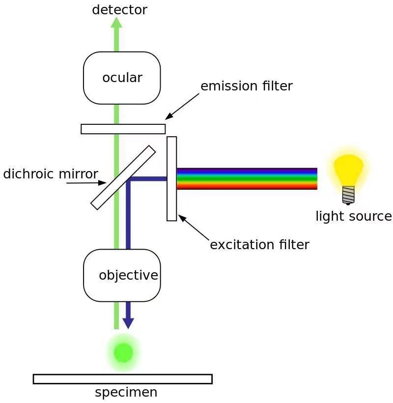
It is the most common type of fluorescence microscope. The excitation of the fluorophore and detection of the fluorescence are done through the same light path (i.e. through the objective). The majority of fluorescence microscopes, especially those used in the life sciences, are of the epifluorescence design.
We use cookies to help provide and enhance our service and tailor content. By continuing you agree to the use of cookies
Stress biomarkersin biological fluids and their point-of-use detection
Confocal Fluorescence Microscope: This type of fluorescence microscope combines laser scanning with fluorescent illumination to produce an image. It can be used in wide range of applications, such as studying cells and tissues, detecting proteins and other substances within cells, and measuring the thickness of materials.
The emission filter passes only the wavelengths emitted by the fluorophore and blocks all undesired light outside this band – especially the excitation wavelengths.
This kind of microscope’s light source and condenser are located on the top, facing downwards. The angle of illumination must be at 90 degrees with respect to the surface of the specimen being examined.
All content on this site: Copyright © 2024 Elsevier B.V. or its licensors and contributors. All rights are reserved, including those for text and data mining, AI training, and similar technologies. For all open access content, the Creative Commons licensing terms apply
A fluorescence microscope uses a mercury or xenon lamp to produce ultraviolet light. The light comes into the microscope and hits a dichroic mirror — a mirror that reflects one range of wavelengths and allows another range to pass through. The dichroic mirror reflects the ultraviolet light up to the specimen. Some specimens fluoresce naturally under ultraviolet light because they contain fluorescent substances such as chlorophyll. If the specimen to be viewed does not naturally fluoresce, it can be stained with fluorescent dyes called fluorochromes.
The most common light sources are mercury, xenon, and LEDs. Mercury provides the best quality of light for fluorescence microscope. LEDs are becoming more popular because they are less expensive than other sources and they consume less power.
Anni Ranta-Lassila* (Corresponding author), Duc Le, Teemu Sipola, Mikko Karppinen, Jarno Petäjä, Minna Kehusmaa, Sanna Aikio, Tian Long Guo, Matthieu Roussey, Jussi Hiltunen, Alexey Popov*Corresponding author for this work
A fluorescence microscope is a type of optical microscope that uses a high-intensity light source to illuminate the specimen and excite fluorochromes in the sample. The illumination of the specimen is usually done with a light source that emits ultraviolet light. They are widely used in biological, medical and industrial fields.
-The cells are susceptible to the phototoxic effect after staining with fluorescent dyes, as the fluorophore molecules absorb the high energy photons from the short-wavelength light.
The excitation filter is essential for the operation of a fluorescence microscope. It passes the light of a shorter wavelength, which the fluorescent dye could absorb. Also, it blocks the other sources of exciting light.
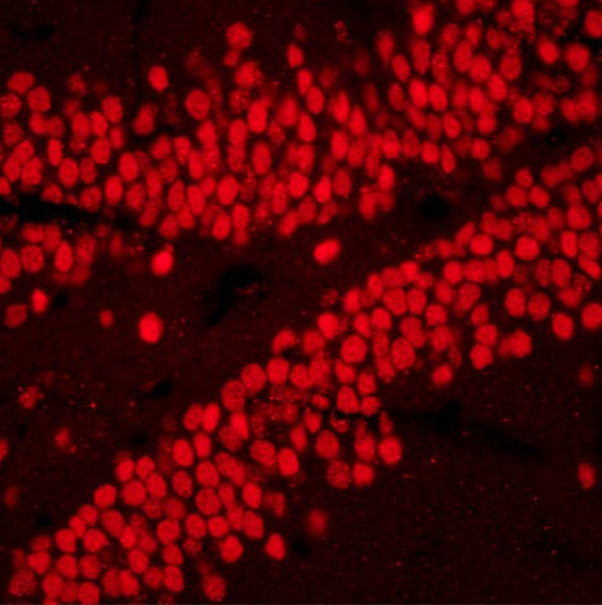
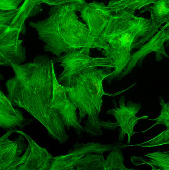
1. In the field of biology, the fluorescence microscope enables accurate and detailed identification of cellular and submicroscopic cellular components and activities with the help of fluorescent dye labeling.
Stressmarkers in blood
Fluorescent dyes are organic compounds that possess a property of fluorescence, by which they can form a fluorescent image by emitting highly contrast visible green light after getting excited by the highly illuminating ultraviolet light. Commonly used fluorescent dyes are; DAPI (49,6-diamidino-2-phenylindole), acridine orange, auramine-rhodamine, Alexa Fluors, or DyLight 488.
The resources are collected and organized on the Internet, and are only used for learning and communication. If there is any infringement, please contact us to delete.
Research output: Chapter in Book/Report/Conference proceeding › Conference article in proceedings › Scientific › peer-review
AB - Stress is a widely spread phenomenon in the modern society. Only work-related stress was estimated to cost US companies more than $300 billion a year in healthcare costs, absences and decreased performance. Early diagnosis of stress conditions and therefore improved recovery and reduced costs could potentially be achieved with continuous monitoring of stress biomarkers using wearable devices. Compared to the conventional electrochemical and optical sensing methods used in current wearable devices, plasmonic sensing could offer higher sensitivity, better stability and faster data collection. Our developed plasmonic sensor chip represents a nanograting structured polymer on a silicon substrate, covered with gold. The sensing method is based on detecting a surface plasmon resonance wavelength shift due to refractive index change caused by presence of analytes in the vicinity of the plasmonic grating. The sensitivity of the chip was tested with two different stress-related biomarkers: cortisol and creatinine. With the tested range from 0 to 265 mM, in the current version of the system, without a receptor layer, the detection limits for cortisol and creatinine were 10.65 and 7.09 mM, respectively, which are close to the physiological ranges of these analytes in body fluids. When integrated into a wearable device, this approach has a potential in future healthcare applications paving the way to continuous stress monitoring.
Fluorescence microscopes are widely used in various fields of research and application including biochemistry, cell biology, microbiology, immunology, and medicine.
The dichroic mirror is a type of optical filter that reflects light at certain wavelengths while transmitting others. It is used in fluorescence microscopes to separate the excitation and emission wavelengths.
Stress is a widely spread phenomenon in the modern society. Only work-related stress was estimated to cost US companies more than $300 billion a year in healthcare costs, absences and decreased performance. Early diagnosis of stress conditions and therefore improved recovery and reduced costs could potentially be achieved with continuous monitoring of stress biomarkers using wearable devices. Compared to the conventional electrochemical and optical sensing methods used in current wearable devices, plasmonic sensing could offer higher sensitivity, better stability and faster data collection. Our developed plasmonic sensor chip represents a nanograting structured polymer on a silicon substrate, covered with gold. The sensing method is based on detecting a surface plasmon resonance wavelength shift due to refractive index change caused by presence of analytes in the vicinity of the plasmonic grating. The sensitivity of the chip was tested with two different stress-related biomarkers: cortisol and creatinine. With the tested range from 0 to 265 mM, in the current version of the system, without a receptor layer, the detection limits for cortisol and creatinine were 10.65 and 7.09 mM, respectively, which are close to the physiological ranges of these analytes in body fluids. When integrated into a wearable device, this approach has a potential in future healthcare applications paving the way to continuous stress monitoring.
N2 - Stress is a widely spread phenomenon in the modern society. Only work-related stress was estimated to cost US companies more than $300 billion a year in healthcare costs, absences and decreased performance. Early diagnosis of stress conditions and therefore improved recovery and reduced costs could potentially be achieved with continuous monitoring of stress biomarkers using wearable devices. Compared to the conventional electrochemical and optical sensing methods used in current wearable devices, plasmonic sensing could offer higher sensitivity, better stability and faster data collection. Our developed plasmonic sensor chip represents a nanograting structured polymer on a silicon substrate, covered with gold. The sensing method is based on detecting a surface plasmon resonance wavelength shift due to refractive index change caused by presence of analytes in the vicinity of the plasmonic grating. The sensitivity of the chip was tested with two different stress-related biomarkers: cortisol and creatinine. With the tested range from 0 to 265 mM, in the current version of the system, without a receptor layer, the detection limits for cortisol and creatinine were 10.65 and 7.09 mM, respectively, which are close to the physiological ranges of these analytes in body fluids. When integrated into a wearable device, this approach has a potential in future healthcare applications paving the way to continuous stress monitoring.
-The photobleaching due to the electron excitation during the process of fluorescence may affect reactive molecules of the fluorescent dyes. As a result, the reactive dyes might lose their chemical property of fluorescence emission intensity.


 Ms.Cici
 8618319014500