
Anti-Reflection VS Anti-Glare - What's the difference? - anti glare and anti reflection
Phasecontrastmicroscopeprinciple
Shown at the left is the phase contrast kit used with National Optical model 160 series of microscopes. The kit consists of 4 objective lenses, a centering telescope and a Zernike phase condenser lens. The long silver adjustment screws on either side of the condenser lens push in to engage set screws inside the condenser for proper alignment.
Phasecontrastmicroscopeimages
In order to set up your microscope to view objects with phase contrast you must have phase contrast objective lenses and a phase contrast substage condenser.
Not all phase contrast microscopes are the same but generally they rely on similar techniques to set up the system for optimum results. In the system shown at the right, the phase condenser has five settings that you can rotate through (10x, 20x, 40x, 100x and BF) BF is "brightfield" with no phase. To set up your microscope for phase optics, you first set it at BF and focus on the specimen. Adjust the height of the condenser for optimum image quality. Next, set the condenser turret to the phase setting for that particular lens and remove the specimen. The controls that stick out from both sides on the back are for centering the condenser.
Phasecontrastmicroscopeppt
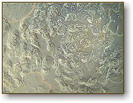
Image above with regular brightfield objectives. Notice the air bubbles at three locations, some cells are visible on the left.
Pairing the analysis with TGA, DSC, and/or DTA can complement the TMA results by providing more insight into the thermal events over the temperature range. There is a glass transition temperature at about 82.4°C, followed shortly by a dilatometric softening temperature (1 mm contraction), and the cold crystallization is evident at 161°C. The glass transition was also observed on the DTA and DSC curves, in good agreement with the TMA result.
Phasecontrastmicroscopediagram
A common activity in high school biology is to observe cheek cells. To do this, one takes a flat toothpick and gently scrapes the inside of their cheek. They smear the specimen on a flat microscope slide and cover it with a cover slip. The cheek cells are epithelial cells and will be seen in large numbers. If you add a drop of iodine to the sample, the nuclei of the cells will become more visible. They will appear as small round dots inside the cell. In the picture on the left, we see the cells as they would appear without iodine with a regular microscope. At the right, you see the same specimen using a phase contrast microscope (actually, we used the same microscope with different lenses). This clearly shows that, for some specimens, phase contrast will greatly improve your image quality.
TMA measures a material’s expansion or contraction when subject to a temperature change. It involves applying a controlled force to a sample while it is heated or cooled and measuring the resulting deformation or movement. TMA is used to study the properties of metals, polymers, ceramics, and composites. It can provide insights into their behavior under different conditions, such as in high-temperature environments or under mechanical load.
PhasecontrastmicroscopePDF
α represents the instantaneous coefficient of linear expansion at temperature T°C and sometimes called the coefficient of derivative linear expansion:In the case of polymers, CTE should be considered as it can predict the material’s behavior under varying conditions of stress and/or temperature fluctuations over areas such as:
Phasecontrastmicroscopeapplication
Next, you will need to remove one of the eyepiece lenses and insert the centering telescope in its place. The set screw is used to focus the centering telescope. When looking through the telescope, you will see two rings. They may or may not be concentric. By turning the centering adjustment screws on the condenser, you align the rings so that they are concentric (see the images below). Finally, remove the centering telescope and replace the eyepiece lens. Put your specimen back on the stage and you are ready for phase observations. When you change objectives, you should go through this centering procedure again (although you may discover that the alignment remains consistent with all objectives).
Phase contrast is a method developed in the early 20th century by Frits Zernike. Zernike discovered that if you can speed up the direct light path, you can cause destructive interference patterns in the viewed image. These patterns create details in the image to appear darker against a light background. To cause these interference patterns, Zernike developed a system of rings located both in the objective lens and in the condenser system. When aligned properly, light waves emitted from the illuminator arrive at your eye 1/2 wavelength out of phase. The image of the specimen then becomes greatly enhanced. Phase is only useful on specimens that do not absorb light (they are called "phase objects") and it is very useful in showing details in certain specimens such as cell parts in protozoans, bacteria, sperm tails and other types of unstained cells. This technique proved to be such an advancement in microscopy that Zernike was awarded the Nobel prize in physics in 1953. You can see the Frits Zernike biography here.
Phasecontrastmicroscopeparts
Thermomechanical analysis (TMA) is a critical tool for measuring a material’s expansion over a temperature range, providing key insights into its integrity and reliability.
A critical parameter TMA characterizes is the coefficient of thermal expansion (CTE). The CTE or average linear expansion % represents the amount of expansion that will occur per unit of length or volume within a specific temperature range. For example, if a material has a CTE of 5 ppm/°C, it will expand by 5 parts per million (ppm) of its original size for every degree Celsius increase in temperature. The CTE can vary depending on the material’s composition and structure and thus is an important material property to characterize. If the sample length is at room temperature and at T°C, the average CTE from room temperature to T°C is
These properties help inform design and processing decisions. For example, glass transition temperature will determine the flexible or rigid application of a material. If a polymer is heated above its glass transition temperature, it will become more flexible, whereas keeping it below the glass transition temperature will enable a rigid format.

As seen in Figure 3, the PET sample is stable until about 77°C where it begins shrinking rapidly until it reaches 136°C. Over the temperature range, it shrinks by almost 1 millimeter. Since PET is used for various applications, from clothing to food packaging, this would be an essential consideration for manufacturing optimization and product quality.
We offer several different microscopes that include phase contrast. They are listed below, with the highest quality found in the Meiji microscope.
C-Therm has developed niche expertise in a number of application areas where thermal conductivity is of critical importance.
Thermal Analysis Labs has a Rigaku TMA that can run the measurements to identify sample CTE and glass transition. Furthermore, our Rigaku DSC Vesta can run DSC tests to provide vital heat flow measurements that can be coupled with TMA results to understand material properties better. For a full overview of our services, consult our 2023 contract testing catalog. Have a question? Reach out to one of our thermal analysis experts to start the discussion.


 Ms.Cici
 8618319014500