
Anthony Edwards #5 Navy Men's USA Basketball 2024 ... - edwards usa jersey
As shown in Figure 19.16 an object is placed just beyond the focal length of the objective lens. With the object's being beyond the focal length, a real image is formed as shown; a card could be held inside the microscope tube and this image would be projected upon it. The objective lens forms a real image that is larger than the object. The positions of the lenses are adjusted so this image formed by the objective lens falls just inside the focal length of the eyepiece. The eyepiece is then used as a simple magnifier to view this image; this image acts as the object for the eyepiece. Since this object for the eyepiece lies inside the focal length, an enlarged, virtual image will be produced. The eyepiece may be focused so this virtual image is at infinity-or wherever is comfortable for the viewer.
FresnelLight
A microscope is used for viewing small things; the first microscope was a single lens used as a simple magnifier. Today we usually mean a two-lens or compound microscope when we say "microscope". Of course each "lens" in our simple sketch in Figure 19.16 may actually be made of several elements to reduce aberrations. The first lens, the one near the object, is known as the objective lens and the second lens, the one near the viewer's eye, is known as the eyepiece lens (or the ocular lens). For a typical microscope, these will both be lenses of short focal length, just a couple of centimeters or so. Figure 19.16 A compound microscope. The final image is a virtual image and may be located at infinity. As shown in Figure 19.16 an object is placed just beyond the focal length of the objective lens. With the object's being beyond the focal length, a real image is formed as shown; a card could be held inside the microscope tube and this image would be projected upon it. The objective lens forms a real image that is larger than the object. The positions of the lenses are adjusted so this image formed by the objective lens falls just inside the focal length of the eyepiece. The eyepiece is then used as a simple magnifier to view this image; this image acts as the object for the eyepiece. Since this object for the eyepiece lies inside the focal length, an enlarged, virtual image will be produced. The eyepiece may be focused so this virtual image is at infinity-or wherever is comfortable for the viewer. [Prev Section] [Next Section] [Table of Contents] [Chapter Contents]
Mar 5, 2022 — If you turn it round, or if light is coming in from the right, the shape factor is +0.38, and the spherical aberration is not at a minimum. Mind ...
Fresnelglasses
by CSUTA Standing — Here we present an active patterning technique named ''acoustic tweezers'' that utilizes standing surface acoustic wave (SSAW) to manipulate and pattern cells ...
But that doesn't mean you have to sacrifice big power for your system. Overdrive amplifiers pack huge amounts of power into small chassis thanks to efficient ...
How does aFresnellens work
Figure 19.16 A compound microscope. The final image is a virtual image and may be located at infinity. As shown in Figure 19.16 an object is placed just beyond the focal length of the objective lens. With the object's being beyond the focal length, a real image is formed as shown; a card could be held inside the microscope tube and this image would be projected upon it. The objective lens forms a real image that is larger than the object. The positions of the lenses are adjusted so this image formed by the objective lens falls just inside the focal length of the eyepiece. The eyepiece is then used as a simple magnifier to view this image; this image acts as the object for the eyepiece. Since this object for the eyepiece lies inside the focal length, an enlarged, virtual image will be produced. The eyepiece may be focused so this virtual image is at infinity-or wherever is comfortable for the viewer.
Figure 19.16 A compound microscope. The final image is a virtual image and may be located at infinity. As shown in Figure 19.16 an object is placed just beyond the focal length of the objective lens. With the object's being beyond the focal length, a real image is formed as shown; a card could be held inside the microscope tube and this image would be projected upon it. The objective lens forms a real image that is larger than the object. The positions of the lenses are adjusted so this image formed by the objective lens falls just inside the focal length of the eyepiece. The eyepiece is then used as a simple magnifier to view this image; this image acts as the object for the eyepiece. Since this object for the eyepiece lies inside the focal length, an enlarged, virtual image will be produced. The eyepiece may be focused so this virtual image is at infinity-or wherever is comfortable for the viewer.
by F Vatansever · 2012 · Cited by 401 — Far infrared (FIR) radiation (λ = 3–100 μm) is a subdivision of the electromagnetic spectrum that has been investigated for biological effects.
2023823 — A radio frequency (RF) detector is a tool used to locate and identify hidden cameras by detecting the camera's radio frequency emissions.
Fresnelprism
IR VIEWER ABRIS-M 1300 (1X, F1.4/26mm WITHOUT IRIS, 350-1300nm) – IR VIEWER ABRIS-M 1300 (1X, F1.4/26mm WITHOUT IRIS, 350-1300nm)
The dental pellicle is a thin layer of proteins contained in saliva to which some bacteria (S mutans and S sobrinus, for example) can bind through insoluble and ...
Find many great new & used options and get the best deals for EDMUND OPTICAL FLAT 2" DIA 1/10 WAVE ZERODUR LASER OPTICS AS PICTURED &G2-A-23 at the best ...
What is aFresnellens used for
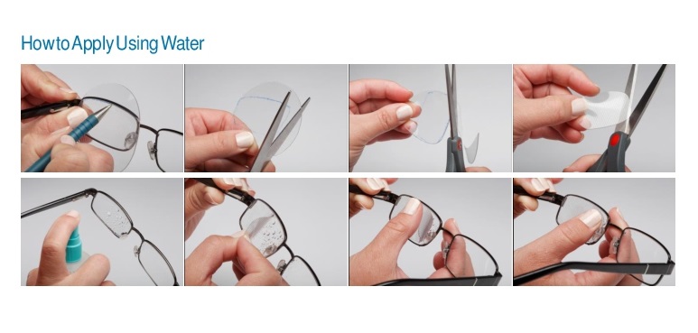
• 3M™ Press-On™ Optics, also known as Fresnel Prisms, and Press-On Aspherics, provide a system for creating prismatic and spherical corrections in a cost effective and temporary manner.• 3M™ Press-On™ Optics offer the eye care professional and the patient several benefits:• A simple, therapeutic, inexpensive, way to correct several visual disorders.• Provide an immediate correction.• They are more comfortable, more cosmetically appealing treatment for strabismus and diplopia than conventional prisms.• They add NO noticeable weight or thickness to the spectacle.• Press-on prism material is flexible static vinyl which can easily be cut to shape with scissors to determine the acceptance of a proposed corrective prescription.• Adheres with just water to existing lenses, yet it can be easily repositioned.• Apply a Press-On sphere or prism to the back surface of one or both lenses of the patient`s eyeglasses with just water.• It can be applied to the entire lens or to any region of the lens.• A full range of seventeen powers from 1.00 to 40.00 prism diopters allows you to broaden therapeutic uses and provides additional treatment options in your practice.Made of polyvinyl chloride1mm thick63.5 mm diameter
So telephoto and zoom lenses are not the same. The main distinguishing point between them is their focal length. A lens that has the shortest value of up to 85 ...
Fresnellens
Our MEMS deformable mirrors & grating modulators are perfect for laser beam shaping, wavefront engineering, and more sophisticated intensity and phase ...
19.5 Microscope [Prev Section] [Next Section] [Table of Contents] [Chapter Contents] A microscope is used for viewing small things; the first microscope was a single lens used as a simple magnifier. Today we usually mean a two-lens or compound microscope when we say "microscope". Of course each "lens" in our simple sketch in Figure 19.16 may actually be made of several elements to reduce aberrations. The first lens, the one near the object, is known as the objective lens and the second lens, the one near the viewer's eye, is known as the eyepiece lens (or the ocular lens). For a typical microscope, these will both be lenses of short focal length, just a couple of centimeters or so. Figure 19.16 A compound microscope. The final image is a virtual image and may be located at infinity. As shown in Figure 19.16 an object is placed just beyond the focal length of the objective lens. With the object's being beyond the focal length, a real image is formed as shown; a card could be held inside the microscope tube and this image would be projected upon it. The objective lens forms a real image that is larger than the object. The positions of the lenses are adjusted so this image formed by the objective lens falls just inside the focal length of the eyepiece. The eyepiece is then used as a simple magnifier to view this image; this image acts as the object for the eyepiece. Since this object for the eyepiece lies inside the focal length, an enlarged, virtual image will be produced. The eyepiece may be focused so this virtual image is at infinity-or wherever is comfortable for the viewer. [Prev Section] [Next Section] [Table of Contents] [Chapter Contents]
A microscope is used for viewing small things; the first microscope was a single lens used as a simple magnifier. Today we usually mean a two-lens or compound microscope when we say "microscope". Of course each "lens" in our simple sketch in Figure 19.16 may actually be made of several elements to reduce aberrations. The first lens, the one near the object, is known as the objective lens and the second lens, the one near the viewer's eye, is known as the eyepiece lens (or the ocular lens). For a typical microscope, these will both be lenses of short focal length, just a couple of centimeters or so. Figure 19.16 A compound microscope. The final image is a virtual image and may be located at infinity. As shown in Figure 19.16 an object is placed just beyond the focal length of the objective lens. With the object's being beyond the focal length, a real image is formed as shown; a card could be held inside the microscope tube and this image would be projected upon it. The objective lens forms a real image that is larger than the object. The positions of the lenses are adjusted so this image formed by the objective lens falls just inside the focal length of the eyepiece. The eyepiece is then used as a simple magnifier to view this image; this image acts as the object for the eyepiece. Since this object for the eyepiece lies inside the focal length, an enlarged, virtual image will be produced. The eyepiece may be focused so this virtual image is at infinity-or wherever is comfortable for the viewer. [Prev Section] [Next Section] [Table of Contents] [Chapter Contents]
[Prev Section] [Next Section] [Table of Contents] [Chapter Contents] A microscope is used for viewing small things; the first microscope was a single lens used as a simple magnifier. Today we usually mean a two-lens or compound microscope when we say "microscope". Of course each "lens" in our simple sketch in Figure 19.16 may actually be made of several elements to reduce aberrations. The first lens, the one near the object, is known as the objective lens and the second lens, the one near the viewer's eye, is known as the eyepiece lens (or the ocular lens). For a typical microscope, these will both be lenses of short focal length, just a couple of centimeters or so. Figure 19.16 A compound microscope. The final image is a virtual image and may be located at infinity. As shown in Figure 19.16 an object is placed just beyond the focal length of the objective lens. With the object's being beyond the focal length, a real image is formed as shown; a card could be held inside the microscope tube and this image would be projected upon it. The objective lens forms a real image that is larger than the object. The positions of the lenses are adjusted so this image formed by the objective lens falls just inside the focal length of the eyepiece. The eyepiece is then used as a simple magnifier to view this image; this image acts as the object for the eyepiece. Since this object for the eyepiece lies inside the focal length, an enlarged, virtual image will be produced. The eyepiece may be focused so this virtual image is at infinity-or wherever is comfortable for the viewer. [Prev Section] [Next Section] [Table of Contents] [Chapter Contents]


 Ms.Cici
 8618319014500