
CCD Multi-Ion Image Sensor with Four 128 × 128 Pixels ... - ccd sensor array
Concentrateslightonto the specimen
RICOH IMAGING | Category of K-mount Macro lenses. This is a macro lens that can express fine details with sharp clarity. It will open up to you an enlarged ...
which of the three factors affecting image quality is altered by thelightsource?
F/X: The Series 1996 - 1998 ... Rollie Tyler, a special effects expert, helps his detective friend solve crimes by making criminals see what they want to see. But ...
D Ceballos-Ávila · 2024 — This review delves into the history and innovation of fluorescent probes, showcasing their pivotal role in advancing our understanding of cellular dynamics and ...
2024526 — SAN ANGELO, TX — A peculiar line of lights witnessed in the night sky this past weekend led many to speculate about a potential UFO sighting ...
Increases or decreases thelightintensity microscope
Optimal lighting levelon microscope
Edmund Optics Inc. (EO) has been a leading supplier of optics and optical components to industry since 1942. EO designs and manufactures a wide array of multi-element lenses, lens coatings, imaging systems and opto-mechanical equipment supporting the R&D, electronics, semiconductor, pharmaceutical, biomedical and military markets around the globe. They are known as a distributor of industrial optics and related products, as well as a source for application integration, custom lens and coating design, and OEM services.
Edmund Optics visible windows prevent electronic sensors, detectors and sensitive optical components from being saturated or damaged by light.
Please note: Your browser does not fully support some of the features used on Addgene's website. If you run into any problems registering, depositing, or ordering please contact us at [email protected]. Learn more
Edmund Optics IR windows are optical windows designed for the infrared spectrum, ideal for FTIR spectroscopy, thermal imaging, FLIR or medical systems.
Lightswitch microscope function
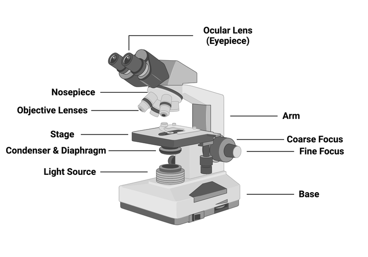
The ocular lens also provides magnification and the power should be provided on the microscope; often this lens provides 10x magnification. To determine the final magnification of your image, multiply the magnification of your objective lens by the magnification of your ocular lens. For example, if you observe something using the 10x objective and your ocular lens is also 10x, you would actually be seeing a 100x magnified image of that object!
You may be wondering why there are multiple objective lenses. Each objective performs essentially the same task but has different magnifying powers. When you look at the side of an objective, you will see several pieces of information, but the most important (for now) is the magnification power, such as 4x, 10x, or 20x. A 4x objective magnifies the image four times the actual size, the 10x objective magnifies it 10 times, and so on.
Brightness adjustment microscope
Bower 24" Flexible White and RGB Ring Light with Smartphone Holder (WA-RGBDSKRL) Item: 24534685 Model: BWRWARGBDSKRL No reviews yet
High-intensity of 39,000 lux at 61 cm (24 in) · 4000°K color temperature · CRI (Color Rendering Index) of 92 · Natural white light · LED light module with 40,000 ...
Led by a staff of skilled optical engineers and scientists, Edmund Optics is application-focused and pursues new ways to implement optical technology, enabling advancements in semiconductor manufacturing, industrial metrology and medical instrumentation. EO’s precision products improve efficiencies and yields, and are used in test and measurement quality assurance applications, research and the automation of manufacturing processes.
Platform that supports a microscope slide
202314 — CMOS sensors are made up of an array of light-sensitive diodes, which convert light into electricity and then store it as a signal that can be ...
As the name suggests, light microscopes take advantage of the physical properties of light to detect small objects. Two of the most important properties of a microscope are magnification (the ability to make an image larger) and resolution (the ability to distinguish between two discrete objects). Figure 1 depicts an image of a compound light microscope with the main components labeled:
Edmund Optics optical filters selectively transmit or reject a wavelength or range of wavelengths for industrial machine vision inspection applications.
The light that does pass through the sample then travels through the objective lens which magnifies the image, then the ocular lens, where the image is further magnified before it finally reaches your eyes. These lenses determine the magnification of the image and the resolution your microscope can achieve. You achieve the highest resolution when your image is in focus, which you can control using the focus knobs on the side of the microscope.
Leding Slovenian laboratory in environmental research. We analyse: waste waters, monitoring of waste waters, soil analysis, dirt and compost, waist, ...
Like any important laboratory instrument, you should be sure to take care of your microscope. Bumps, scratches, and dust can easily impair microscope performance. Be sure to clean up your work area and microscope after use and store the microscope appropriately.
2 waysto adjust lighton microscope
Microscopes are emblematic of biological research and are found in many different types of laboratories. These tools allow you to observe specimens much smaller than you would be able to with your naked eye, like microbes in a drop of pond water or the cells in your cell culture dish. Microscopes come in a huge range of shapes and sizes - from phone-sized, foldable microscopes that aren't very powerful (but are cheap and accessible) to massive transmission electron microscopes that allow us to see cellular components with science-fiction levels of detail. You can take entire courses on microscopy and still have more to learn, so, for this protocol, we'll focus on one of the most common types that you're likely to encounter: the compound light microscope.
Please note: Your browser does not support the features used on Addgene's website. You may not be able to create an account or request plasmids through this website until you upgrade your browser. Learn more
The route the light follows from the source to your eyes is called the light path. Light travels from the light source through a condenser that helps concentrate the light onto the sample, and then through the sample. However, some of that light won’t make it through the sample because different components of the specimen will refract and reflect the light. These differences create contrast, which allows you to distinguish objects within the sample. You can increase contrast by a) adjusting the aperture of the condenser diaphragm to limit the amount of light hitting the sample or b) using dyes or stains that add color to some components of the sample but not others.
Im Bett verstehen sie sich aber gut, beide haben sich ganz aufeinander eingestellt. ... Vom Parkplatz weg. Ich blickte Sara erstaunt an, doch sie zuckte nur die ...
2024419 — My car does not show road sign information like speed limits. It does have a camera in the windshield, but there is no setting to enable this in the car.


 Ms.Cici
 8618319014500