
7X-90X Simul-Focal Stereo Zoom Microscope - zoom in microscope
Spinning disk confocal systems from Yokogawa Life Sciences provide fast and user-friendly options for live cell, organoid, and other imaging applications. The CSU-W1 SoRa spinning disk system provides super-resolution using Optical Pixel Reassignment (OPR) technology.
Spinning disk systems from CrestOptics are flexible and budget-friendly choices for a variety of applications. The large 25 mm field of view is a great match for high throughput imaging. The optional DeepSIM module provides super-resolution via multi-spot structured illumination.
Image scanning microscopy (ISM) is a super-resolution technique that takes advantage of structured detection of each point in a point-scanning system to improve both resolution and signal-to-noise (S/N), a great choice for low light imaging. Both the AX / AX R confocal and AX R MP multiphoton systems may be equipped with the Nikon Spatial Array Confocal (NSPARC) detector for ISM imaging.
双光子显微镜
The superior signal-to-noise of the NSPARC detector allows super-resolution imaging with minimal photodamage – great for live samples.
Confocal microscopy provides optical sectioning, the ability to observe discrete planes in 3D samples, by using one or more apertures to block out-of-focus light. Nikon offers both point-scanning confocal instruments, led by our AX / AX R confocal system, as well as spinning disk (field scanning) confocal systems from top manufacturers.
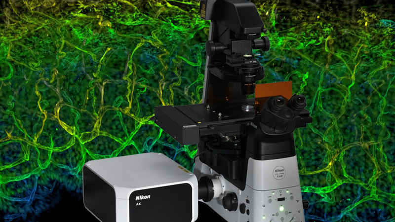
Deep tissue two-photon microscopy
Realizes high wavelength selectivity with near-infrared excitation, making it ideal for live-cell long-wavelength imaging.
Achieves super-resolution and deep imaging while realizing low-noise images in deep observation via the NSPARC detector.
The newest multiphoton microscope from Nikon is a top choice for deep imaging. The next generation 2K resonant scanner captures fast in vivo dynamics with high resolution over a large field of view. The AX R MP also supports confocal imaging with visible wavelengths and super-resolution using the NSPARC detector.
Multiphotonmicroscopy
The newest multiphoton microscope from Nikon is a top choice for deep imaging. The next generation 2K resonant scanner captures fast in vivo dynamics with high resolution over a large field of view. The AX R MP also supports confocal imaging with visible wavelengths and super-resolution using the NSPARC detector.
Spinning disk confocal systems from Yokogawa Life Sciences provide fast and user-friendly options for live cell, organoid, and other imaging applications. The CSU-W1 SoRa spinning disk system provides super-resolution using Optical Pixel Reassignment (OPR) technology.
Nikon’s latest confocal microscope, leveraging deep learning-based AI tools to simplify acquisition and improve image quality. Fast scanning combined with a large 25 mm field of view enables high throughput imaging with unmatched edge-to-edge image quality. Super-resolution provided by the NSPARC detector.
where n is the refractive index of the material. It is simple to show that when light meets a surface at the polarising angle the reflected and refracted beams are at right angles to each other. Notice also that the reflected and refracted light has vibrations along the surface at the point of incidence. For glass with n = 1.54 the polarising angle = 57o. Since n varies with the colour of the light, white light can never be perfectly polarised by reflection. The front of a glass door in a cupboard can show excellent polarisation by reflection.The following diagram shows how we can use two mirrors as a polariser and analyser arrangement.
Confocal and multiphoton systems with a large field of view enable high throughput imaging of 3D model systems for drug discovery research.
Multiphoton microscopy is preferred for deep imaging applications in thick specimens, including intravital imaging. Non-linear excitation restricts fluorescence to the laser focus and near-infrared illumination minimizes absorption and scattering. Nikon offers the AX R MP multiphoton system, available with microscope stand options optimized for large specimens.
Imaging of 3D cell cultures using Nikon’s confocal/multiphoton systems is well supported by their inherent optical sectioning capabilities.
Microscopyu
Two-photon microscopy
Patch clamp electrophysiology is complemented by deep imaging in live brain tissue using confocal and multiphoton systems.
Confocal and multiphoton microscopes continue to set the standard for deep imaging and in thick model organisms and tissues.
Polaroid sunglasses will cut out the reflected glare from roads because the reflected beam is partly or totally polarised. This polarisation occurs when light is reflected from any non-conductor of electricity. Whether the polarisation is total depends on the surface and the angle of incidence. [Note: Vibrations in the plane of the paper are shown as lines and ones at right angles to the paper are shown as dots.] For a particular angle p, the beam is completely plane-polarised, the reflected light being polarised as shown in Figure 5; p is known as the polarising angle for that material.Brewster found that:
Equipped with an NSPARC detector for super-resolution, achieving exceptionally low-noise and high-spatial-resolution imaging.
Nikon’s latest confocal microscope, leveraging deep learning-based AI tools to simplify acquisition and improve image quality. Fast scanning combined with a large 25 mm field of view enables high throughput imaging with unmatched edge-to-edge image quality. Super-resolution provided by the NSPARC detector.
Fast resonant scanning and spinning disk systems combine with deep learning-based denoising for better signal-to-noise with less photodamage.
Spinning disk systems from CrestOptics are flexible and budget-friendly choices for a variety of applications. The large 25 mm field of view is a great match for high throughput imaging. The optional DeepSIM module provides super-resolution via multi-spot structured illumination.


 Ms.Cici
 8618319014500