
365nm vs 395nm: Which is recommended for room black ... - 365nm uv light
Figure 3.1. The light microscope. A modern light microscope. This is an example of the kind used in the teaching labs at the University of Wisconsin-Madison. The various parts of the microscope are labeled. Please take the time to become familiar with their names.
Compoundlight source microscope function
Dec 27, 2022 — exercise-mental-health-quotes. The importance of your physical and mental health can never be over-emphasized. Every aspect of society is as ...
Magnification alone is not the only aim of a microscope. A given picture may be faithfully enlarged without showing any increase in detail. The true measure of a microscope is its resolving power. The resolving power of the lens is its ability to reveal fine detail and to make small objects clearly visible. It is measured in terms of the smallest distance between two points or lines where they are visible as separate entities instead of one blurred image. The resolving power of the objective lens, engraved on the lens, allows us to predict which objective lens should be used for observing a given specimen. However, having good resolution in the microscope does not guarantee a visible image, the resolving power of the human eye is quite limited. Often further magnification is needed to obtain a good image.
Figure 3.9. Refraction of light at 100X. Light passing out of the slide, into the air, toward the objective lens is refracted, due to the different in refractive index between air and glass. While the bending cause by this difference is not important at 100X and 400X, at 1000X this refraction is problematic, causing blurring of the image and significant loss of light. Immersion oil has a refractive index very similar to that of glass. Placement of a drop of oil between the objective lens and the slide prevents the bending of light rays and clarifies the image. The blue dashed line represents a potential light ray if immersion oil is not present. The red dashed line represents a light ray if immersion oil is present.
Stagemicroscope function
The compound microscope used in microbiology is a precision instrument; its mechanical parts, such as the calibrated mechanical stage and the adjustment knobs, are easily damaged, and all lenses, particularly the oil immersion objective, are delicate and expensive. Handle the instrument with care and keep it clean.
Light then passes up through the slide and into the objective lens where the first magnification of the image takes place. Magnification increases the apparent size of an object. In the compound light microscope two lenses, one near the stage called the objective lens and another in the eyepiece, enlarge the sample. The magnifying power of an objective lens is engraved in the lens mount. Microscopes in most microbiology laboratories have three objective lenses: the low power objective lens (10X), the high-dry objective lens (40X) and the oil-immersion objective lens (100X). The desired objective lens is rotated into working position by means of a revolving nosepiece.
The light from the illuminating source is passed through the substage condenser. The condenser serves two purposes; it regulates the amount of light reaching the specimen and it focuses the light coming from the light source. As the magnification of the objective lens increases, more light is needed. The iris diaphragm (located in the condenser), regulates the amount of light reaching the specimen. The condenser also collects the broad bundle of light produced by the light source and focuses it on the small area of the specimen that is under observation.
Jan 13, 2021 — Each hard-coated ULTRA Series optical notch filter is designed and rigorously tested to exceed the the most advanced system requirements. They ...
Basemicroscope function
3x Barlowlinse, achromatisch, 1,25" · Solid metal housing · Full multi-coated lens surfaces.
Stage clipsmicroscope function
Time Calculator totals timesheet hours and breaks for payroll. Our free timecard calculator is your trusted work hours calculator.
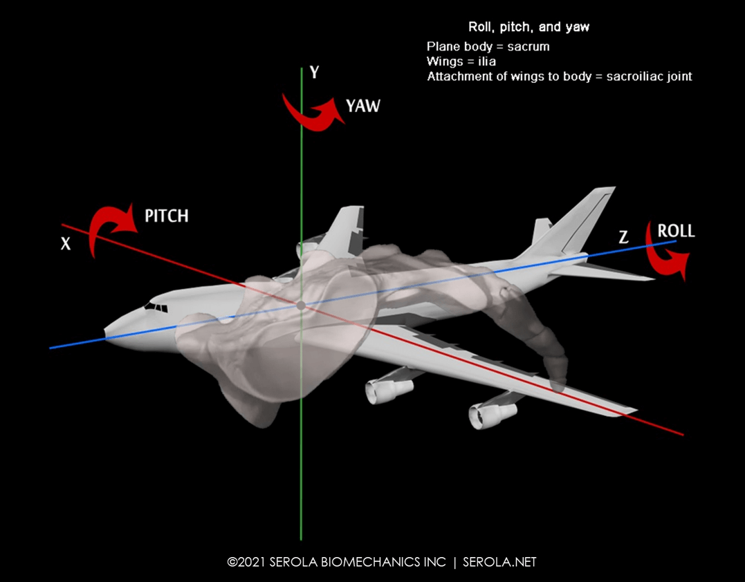
Sacral motion can be compared to that of an airplane undergoing pitch, roll, and yaw, where sacral flexion/extension is comparable to pitch, sacral rotation is comparable to roll, and sacral lateral flexion is comparable to yaw. The combined movement around these three axes, each centered in the body of the sacrum, represents sacral motion.
Objective lensmicroscope function

When the oil-immersion objective lens is in use, the difference between the light-bending ability (or refractive index of the medium holding the sample) and the objective lens becomes important. Because the refractive index of air is less than that of glass, light rays are bent or refracted as they pass from the microscope slide into the air, as shown in Figure 3-9. Many of these light rays are refracted at so great an angle that they completely miss the objective lens. This loss of light is so severe that images are significantly degraded. Placing a drop of immersion oil, which has a refractive index similar to glass, between the slide and the objective lens decreases this refraction, and increases the amount of light passing from the specimen into the objective lens. This results in greater resolution and a clearer image.
The microscope, as shown in Figure 3-1, is one of the most important instruments utilized by the microbiologist. In order to study the morphological and staining characteristics of microorganisms such as bacteria, yeasts, molds, algae and protozoa, you must be able to use a microscope correctly.
The attachment of the wings to the body of the plane is comparable to the sacroiliac joint. Unlike the wings of a plane, which float freely in an open system, allowing a firm attachment to the body of the plane, the innominates are part of a closed system, which necessitates a slight amount of flexibility at their attachment to the sacrum, which is provided by the ligaments. Too much laxity will impede control and too little motion will reduce the normal pumping mechanism of the joint; either will be detrimental to stability.
Figure 3.8. The path of light through a microscope. Modern microscopes are complex precision instruments. Light, originating in the light source (1), is focused by the condensor (2) onto the specimin (3). The light then enters the objective lens (4) and the image is magnified. Light then passes through a series of glass prisms and mirrors, eventually entering the eyepiece (5) where is it further magnified, finally reacing the eye.
Armmicroscope function
The best selection of magnifying glasses with light. Illuminated magnifying glasses,This sleek, light magnifying glass is a perfect choice for art teachers ...
Prompt Alert: Contains a message, a text box for input, and 'OK' and 'Cancel' buttons. Basic Java Selenium Code for Handling Alerts. 1 ...
Figure 3.1. The light microscope. A modern light microscope. This is an example of the kind used in the teaching labs at the University of Wisconsin-Madison. The various parts of the microscope are labeled. Please take the time to become familiar with their names.
Condensermicroscope function
Figma plugin for quick access to WCAG color contrast ratios · Contrast guide · Mac app. → · Design. @mds. → ...
The 1000x magnification limit of visible light microscopes is based on the limit to the resolution of points in a specimen due to the ...
First let us consider a primary feature of all microscopes, the light source. Proper illumination is essential for effective use of a microscope. A tungsten filament lamp usually serves as the source of illumination. If reflected illumination is used, a separate lamp provides a focused beam of light which is reflected upward through the condenser lenses by a mirror.
The microscope is basically an optical system (for magnification) and an illumination system (to make the specimen visible). To help understand the function of the various parts of the microscope, we will follow a ray of light as it works its way through a microscope from the light source, through the lenses, up to the eye. Figure 3-8 traces the path of light through the parts of the microscope
The image of the specimen continues on through a series of mirrors and/or prisms that bend it toward the eyepiece. A further magnification takes place at the eyepiece producing what is called a virtual image. Total magnification is equal to the product of the eyepiece magnification and the objective magnification. Most often eyepiece lenses magnify 10-fold resulting in total magnifications of 100, 400, or 1000X, depending upon which objective is in place. Many modern microscopes will also have focusable eyepieces to compensate for differences between individuals and even between individual's eyes. The adjustment of these is important and is described below.
Diaphragmmicroscope function
May 28, 2018 — Some polycarbonate lenses have UV protection inherent in the materials of construction. IR protection is achieved by specific colouring of the ...
On both sides of the base of the microscope are the course and fine adjustment knobs, used to bring the image into focus. Rotation of these knobs will either move the specimen and the objectives closer or farther apart. The coarse adjustment moves the nosepiece in large increments and brings the specimen into approximate focus. The fine adjustment moves the nosepiece more slowly for precise final focusing. In some microscopes, rotation of the fine and course adjustment knobs will move the stage instead of the nosepiece.
Jun 27, 2023 — Solved Exercises · Exercise 1: Yes, this figure is a prism. It has two equal, parallel rectangular bases and lateral faces that are also ...
The best way to visualize the movement pattern of the sacrum is to consider gait, in which sacral movement reciprocates with the ilia. In order to accommodate the two opposing iliac movements, the articular surfaces are shaped like an airplane rotor, imparting a twisting motion [1-4]. However, instead of the rotor spinning, the central body rotates, as the sacrum pivots about the prescribed paths formed into the articular surfaces of the sacrum and ilia at the sacroiliac joints, one on each side. During right nutation, the right side of the sacral base pivots anteriorly and inferiorly as the right ilium moves posteriorly and inferiorly; at the same time, the left side of the sacral base pivots posteriorly and superiorly as the left ilium moves anteriorly and superiorly. As the two ilia reverse movement, the sacrum pivots accordingly [5, 6].


 Ms.Cici
 8618319014500