
123 Ignition Electronic 4 Cylinder Dist - 123MG4RVPOS - 123 electronic
Human lysed whole blood was stained with the indicated antibodies and unmixed on a 5-laser Cytek™ Aurora Cytometer using cells. All plots are gated on lymphocytes.
Fluorophore structure
(Left) Anti-human CD3 (clone UCHT1) BV421™ and anti-human T-bet (clone 4B10) KIRAVIA Blue 520™ staining on human PBMCs. (Right) Titration curve with FITC or KIRAVIA Blue 520™ conjugated to anti-T-bet antibody.
Anti-human CD4 (clone SK3), conjugated to FITC (red), Alexa Fluor® 488 (blue), BD Horizon™ Brilliant Blue 515 (green), or KIRAVIA Blue 520™ (purple) was used to stain human lysed whole blood.
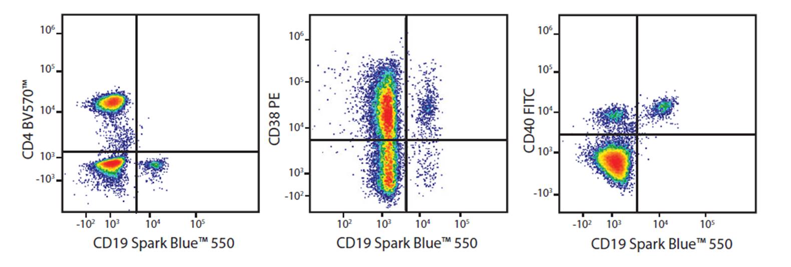
Human whole blood was stained with anti-CD19 conjugated to Spark Blue™ 550, anti-CD4 BV570™, anti-CD38 PE, and anti-CD40 FTIC. Samples were unmixed on a Cytek™ Aurora Cytometer using compensation beads and cells. All plots are gated on lymphocytes.
Simple organic fluorophores encompass a variety of synthetically-derived dyes. Because of their small size, multiple simple organic fluorophores can be conjugated to a single antibody. Due to their conjugation chemistry, these dyes are flexible and can be easily used to generate custom antibodies. Unlike protein-based fluors, simple organic dyes are not sensitive to organic solvents, like those used in phospho-flow, making them advantageous for these applications. Organic dyes are also soluble, meaning they are less susceptible to aggregation and precipitation. Simple organic dyes are often used in microscopy due to their discrete excitation and emission profiles and are chosen for their limited spillover into other imaging channels. Simple organic dyes are great to stain intracellular or intranuclear targets.
Fluorophores in fluorescence microscopy
Image from the RCSB PDB (rcsb.org) of PDB ID 1EYX Contreras-Martel, C. et al. 2001. Crystallization and 2.2 A resolution structure of R-phycoerythrin from Gracilaria chilensis: a case of perfect hemihedral twinning. Acta Cystallogr. Sect.D.57:52-60.
To demonstrate that APC/Fire™ 810 can be clearly unmixed from other fluorophores with heavily overlapping emission spectra, we created parallel staining panels where CD4 APC/Fire™ 810 was replaced with CD4 BV510™. Human lysed whole blood was stained with the indicated antibodies and the two panels were compared in a side-by-side experiment.
Human whole blood was stained for 20 min with CD3 (SK7) conjugates of APC/Fire™ 750, APC/Cyanine7, APC-H7 followed by RBC lysis and wash steps. Cells were then treated with either PBS control, 1%PFA in PBS, 4%PFA followed by.1%saponin, BioLegend's True-Nuclear™ Fix/Perm Buffer Set, BD's Transcription Factor Buffer Set, or eBioscience's FoxP3/Transcription Factor Staining Buffer Set prior to analysis.
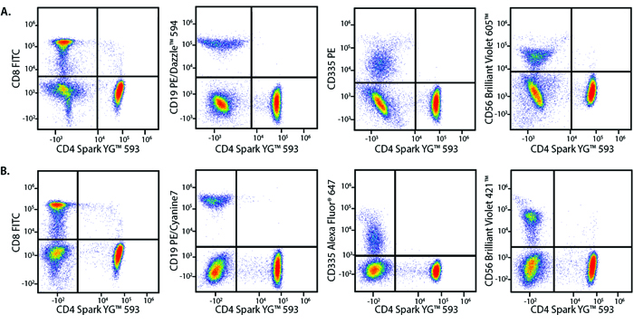
Human lysed whole blood was stained with the indicated antibodies in panels with overlapping (A) or non-overlapping (B) fluorophore conjugates. Samples were unmixed on a 5-laser Cytek™ Aurora Cytometer using cells.
Anti-human CD4 (clone SK3), conjugated to FITC (red), Alexa Fluor® 488 (blue), BD Horizon™ Brilliant Blue 515 (green), or KIRAVIA Blue 520™ (purple) was used to stain human lysed whole blood.
Fluorophores in flow cytometry
Human peripheral blood lymphocytes stained with anti-human CD3 (clone UCHT1) conjugated to PE/Dazzle™ 594 or other equivalent fluorophores.
RBC lysed and washed human blood was stained with optimal test concentrations of the indicated antibodies. Staining was performed in the presence of appropriate blocking buffers, including True-Stain Monocyte Blocker™.
Lysed human whole blood was stained with the indicated antibodies in the presence of Brilliant Stain Buffer. Samples were unmixed on a Cytek™ Aurora Cytometer using compensation beads and cells. All plots are gated on lymphocytes.
A vial of APC/Cyanine7 or APC/Fire™ 750 was stored in the dark at either 4°C or 37°C for 81 days. At certain intervals, an aliquot of reagent was used to stain Veri-Cells™, lyophilized human PBMCs. Cells were stained for 15 min at room temp in cell staining buffer.
A Canadian owned and operated online industrial distributor. We carry over 16,000 items in-stock, ranging from HSS taps and carbide end mills, to indexable inserts and bearings. Buy tooling at low prices directly from our store - no account needed.We ship to all locations in Canada & the United States. All stock orders ship within 24hrs.Shop today & save money on the tools you need!
Fluorophores pronunciation
Two of the most commonly used fluorophores in flow cytometry, R-phycoerythrin (R-PE) and allophycocyanin (APC), are phycobiliproteins originally derived from algae. Both proteins contain fluorescent phycobilin chromophores embedded into a protein scaffolding which accounts for their large size. When phycobiliproteins are exposed to organic solvents like MeOH and EtOH, they denature and are no longer able to fluoresce. Both R-PE and APC are fairly bright fluors and are often used in flow cytometry to detect proteins with low levels of expression.
How do fluorophores work
Our diverse array of fluorophores allows you to expand the limits of flow cytometry and develop larger multicolor panels. High level panels (>22 colors) can be built using cytometers capable of spectral detection or the ability to unmix a fluorescent spectrum across an array of emission detectors such as the Cytek™ Aurora or Northern Lights. Some fluorophores, like the Spark Dyes, are specifically designed to be used on these cytometers.
(Left) Anti-human CD3 (clone UCHT1) BV421™ and anti-human T-bet (clone 4B10) KIRAVIA Blue 520™ staining on human PBMCs. (Right) Titration curve with FITC or KIRAVIA Blue 520™ conjugated to anti-T-bet antibody.
At BioLegend, we've spent nearly two decades crafting new fluorophores for flow cytometry. With each new option, researchers and colleagues have come to trust the performance of our products, as evidenced by this CiteAb report indicating we are the most cited company for flow cytometry antibodies.
Is GFP a fluorophore
Brilliant Violet™ dyes are organic, light-emitting, diode polymers whose brightness is related to the length of the polymer, but whose spectra remain stable no matter the polymer’s length. These dyes have a high extinction coefficient and quantum yield resulting in high intensity brightness. Brilliant Violet™ dyes are effective over a wide pH range and are compatible with a variety of fixatives.
Human PBMCs were incubated with no monocyte blocker (left) or with 5 µL of True-Stain Monocyte Blocker (right) and then stained with 5 µL/test of the indicated antibodies in 100 µL of cells at 1 x 10⁶cells/mL.
Fluorophores list
Image from the RCSB PDB (rcsb.org) of PDB ID 1EYX Contreras-Martel, C. et al. 2001. Crystallization and 2.2 A resolution structure of R-phycoerythrin from Gracilaria chilensis:a case of perfect hemihedral twinning. Acta Cystallogr. Sect.D. 57:52-60.
RBC lysed and washed human blood was stained with a multicolor panel with an intentionally high degree of spectral complexity and overlap. Cells were analyzed on a 5-laser or 3-laser Cytek™ Aurora. Cells were gated on CD19-/CD56- events, and CD154 (CD40L) PE/Fire™ 640 is displayed against CD4 PE/Fire™ 700 or CD8 PerCP. This panel is able to be unmixed on both a 3-laser and a 5-laser Aurora. However, the spreading error of PE/Fire™ 640 is less substantial using a system that has both a 488 nm blue laser and a 561 nm yellow/green laser. Spreading error only poses a problem when the brightness of the two fluors being plotted is not of sufficient amplitude to be resolved from the spreading error of the two populations.
Sign up for our newsletter and receive a curated flyer, once per week, containing specials on industrial supply items & surplus cutting tools.
Another protein-based dye, PerCP, a peridinin-chlorophyll protein complex, is derived from photosynthetic dinoflagellates. Other fluorescent proteins, like eGFP, mCherry, and tdTomato can be fused to a protein of interest to visualize or track cell processes. These are typically derived from sea anemone and jellyfish, and exist as either monomers, dimers, or trimers. All of these protein-based dyes can be used as the donor molecule in FRET pairs. Generally, antibodies conjugated to protein-based flours are not commonly used in standard microscopy as they are more susceptible to photobleaching.
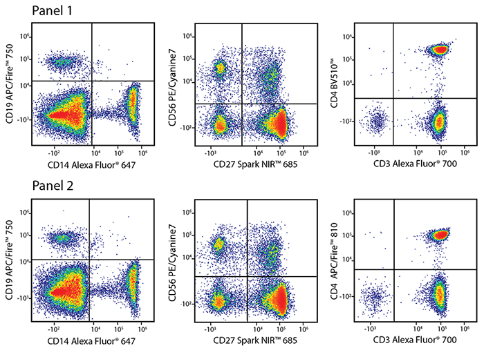
Whole blood from the same donor was stained with either panel A or B. Panel A included the following antibodies: anti-CD4 Spark NIR™ 685, anti-CD19 Alexa Fluor® 700, anti-CD8 APC/Fire™ 750, anti-CD3 PE, and anti-CD56 APC. Panel B is shown as a reference panel using pre-optimized fluorophore selections and includes the following antibodies: anti-CD4 FITC, anti-CD19 Alexa Fluor® 700, anti-CD8 Pacific Blue™, anti-CD3 PE, and anti-CD56 PE/Cyanine7. Cells were washed and fixed with Fluorofix™ prior to analysis.
Fluorochrome vs fluorophore
Introducing the KIRAVIA Dyes™, a brand new fluorescent chemistry to advance applications in multicolor flow cytometry and beyond. This class of dyes employs a unique organic backbone that separates fluorophores to minimize quenching effects, thus allowing optimal and higher fluorophore to protein (F:P) ratios, surpassing what is possible with direct conjugations of single fluorophore like FITC.
Learn more about the unique advantages offered by each member among the various families of fluorophores and how to best use them in multicolor panels:
Human PBMCs were incubated with no monocyte blocker (left) or with 5 µL of True-Stain Monocyte Blocker (right) and then stained with 5 µL/test of the indicated antibodies in 100 µL of cells at 1 x 10⁶cells/mL.


 Ms.Cici
 8618319014500