
Bright 2: sequência com Will Smith é cancelada pela ... - serie bright
If your Mac laptop has a backlit keyboard, you can manually adjust the level of backlighting or turn it off, or set options to do so automatically.
If your Mac has a Touch Bar: In the Touch Bar, expand the Control Strip, then tap or . To turn off backlighting, touch and hold .
An optical microscope can generate a micrograph using standard light-sensitive cameras. Photographic film was traditionally used to capture the images.
Tip: The light sensor is located at the top of your computer near the camera; make sure that area isn’t covered when you use your Mac in low light conditions.
The power of magnification of a compound optical microscope depends on the ocular and the objective lenses. It is equal to the product of the powers of these lenses (e.g. for a 10x ocular lens and 100x objective lens used together, the final magnification is 1000x.)
Yolanda graduated with a Bachelor of Pharmacy at the University of South Australia and has experience working in both Australia and Italy. She is passionate about how medicine, diet and lifestyle affect our health and enjoys helping people understand this. In her spare time she loves to explore the world and learn about new cultures and languages.
Smith, Yolanda. 2023. What is Optical Microscopy?. News-Medical, viewed 09 November 2024, https://www.news-medical.net/life-sciences/What-is-Optical-Microscopy.aspx.
Technological developments have now enabled digital images to be taken with CMOS and charge-couple device (CCD) cameras for optical microscopes. As a result, the image can be projected onto a computer screen in real time to examine a sample with these digital microscopes. This increases the convenience of use as eyepieces are no longer needed.
In this interview, conducted at SfN 2024 in Chicago, News Medical speaks with Joel Svensson and Carolyn Marks of Atlas Antibodies, about their new launch, the MolBoolean™, as well as how conferences like SfN are helping shape the future of neuroscience research.
The objective lens should be brought close to the study sample to allow the light inside the tube of the microscope. This creates an enlarged, inverted image of the sample, which can be viewed through the eyepiece of the microscope.
If your Mac has keyboard brightness keys: On the keyboard, press the increase brightness key or the decrease brightness key . To turn off backlighting, keep pressing .
There are some instances when optical microscopy is not well suited to the task at hand due to limitations of the technique. For example, at very high magnifications airy disks may be visible, which are fuzzy discs surrounded by diffraction rings, which appear in place of point objects.
Your questions, but not your email details will be shared with OpenAI and retained for 30 days in accordance with their privacy principles.
In this interview, News Medical speaks with Dr Martin Biggs from Synoptics about Syngene's G:Box, a high-quality imaging and gel documentation system.
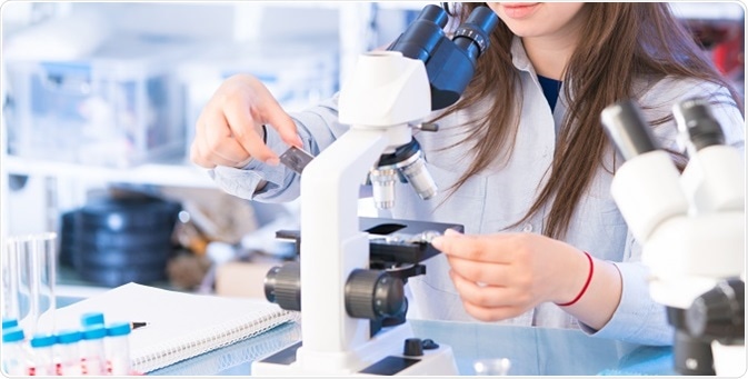
An optical microscope, also sometimes known as a light microscope, uses one or a series of lenses to magnify images of small samples with visible light. The lenses are placed between the sample and the viewer’s eye to magnify the image so that it can be examined in greater detail.
Registered members can chat with Azthena, request quotations, download pdf's, brochures and subscribe to our related newsletter content.
The intersection of arts and neuroscience reveals transformative effects on health and learning, as discussed by Susan Magsamen in her neuroaesthetics research.
Smith, Yolanda. (2023, July 19). What is Optical Microscopy?. News-Medical. Retrieved on November 09, 2024 from https://www.news-medical.net/life-sciences/What-is-Optical-Microscopy.aspx.
If you added keyboard brightness to Control Center: Click in the menu bar, click , then drag the slider to increase or decrease the brightness. If you don’t see , you can add it. See Customize Control Center.
On all Mac computers: Choose Apple menu > System Settings, then click Keyboard in the sidebar (you may need to scroll down). Drag the “Keyboard brightness” slider to adjust the brightness. Drag it all the way to the left to turn off backlighting.
Smith, Yolanda. "What is Optical Microscopy?". News-Medical. https://www.news-medical.net/life-sciences/What-is-Optical-Microscopy.aspx. (accessed November 09, 2024).
Smith, Yolanda. "What is Optical Microscopy?". News-Medical. 09 November 2024. .
Optical microscopy is a technique employed to closely view a sample through the magnification of a lens with visible light. This is the traditional form of microscopy, which was first invented before the 18th century and is still in use today.
If you added keyboard brightness to the menu bar: Click or in the menu bar, then drag the slider to increase or decrease the brightness. If you don’t see or , you can add it. See Customize Control Center.
News-Medical.Net provides this medical information service in accordance with these terms and conditions. Please note that medical information found on this website is designed to support, not to replace the relationship between patient and physician/doctor and the medical advice they may provide.
Optical microscopy is commonly used in many research areas including microbiology, microelectronics, nanophysics, biotechnology and pharmaceutical research. It can also be useful to view biological samples for medical diagnoses, known as histopathology.
While we only use edited and approved content for Azthena answers, it may on occasions provide incorrect responses. Please confirm any data provided with the related suppliers or authors. We do not provide medical advice, if you search for medical information you must always consult a medical professional before acting on any information provided.
There are many types of optical microscopes. They can vary from a very basic design to a high complexity that offers higher resolution and contrast. Some of the types of optical microscopes include the following:
Other types of optical microscopes include petrographic, polarizing, phase contrast, epifluorescence, and confocal microscopes.


 Ms.Cici
 8618319014500