
Online Terminvereinbarung - Schritt 1 von 6 - e mail termin vereinbaren
Specimens imaged under the proper conditions of darkfield illumination are quite spectacular in appearance (try, for instance, a drop of fresh blood). Often specimens containing very low inherent contrast in brightfield microscopy are readily observable in darkfield, and this type of illumination is ideal for revealing outlines, edges, boundaries, and refractive index gradients. Unfortunately, darkfield illumination is less useful for revealing internal details. Other types of specimens, including many that have been stained with dyes, also respond well to illumination under darkfield conditions. These include plant and tree thin sections (stained and unstained), diatoms, radiolarians, fossils, bone sections, embryos, and hair (both human and animal).
Noise is dependent of camera make and model as well as settings. Different types of noise makeup the overall noise profile for a given image.
As the bit depth of the system increases the degree of precision which information is communicated from the real world into the digital world also increases.
Selecting the best overall camera settings (ISO, shutter speed, f-stop) and image quality attributes (dynamic range, noise, bit-depth, sensor size) is impossible without a basic understanding of how a camera sensor works.
No matter the camera, higher ISO values will always produce more overall noise and less overall dynamic range in the final RAW file.
The 14-bit file contains 2^14th power of possible variations for each of the 3 color channels. That’s 16,384 possible choices per color channel.
Image ofdark field microscope
A pixel can only display a single color, including black, white, greyscale, and RGB Color values. The color of each pixel is determined by the amount & kind of light information it collects.
Over a 3-day weekend, I can teach you everything I know, plus provide 1 on 1 feedback that will quickly improve your skills.
The 2-year-old child, seeing the same thing, has a hard time describing the scene accurately, having a limited vocabulary.
Understanding camera sensor size and why it actually matters is one of the most important aspects of learning photography.
Smaller camera sensors such as a standard 22.3mm width, APS-C Sensor ( see graphic above ), would have a crop factor of approximately 1.6.
Smaller pixel pitch (width), combined with larger sensor size, and the latest software & hardware, will produce the best image quality.
We have surpassed the 2-year-old child that can barely speak, we have surpassed the adult with a vivid and detailed vocabulary, we have arrived at a degree of precision that only machines can record and communicate.
For example, Red ( 12 ), Blue ( 6 ), Green ( 15 ) would create unique color and Red ( 1 ), Blue ( 2 ), Green ( 4 ) would create another unique color.
Unlike megapixel counts, having a larger dynamic range is always a positive camera attribute. Dynamic range is provided in stops, which is a measure of light. For each stop increase the amount of light information collected doubles.
Canon continues to produce their own sensors which significantly lack in dynamic range, comparatively, rated at approximately 11.8 stops for their top model cameras. They also produce a much larger amount of noise at high ISO values.
The crop factor is a dimensionless reference number, associated with image sensors. It compares the diagonal distance across each specific camera sensor to the diagonal distance across the full frame camera sensor.
Along with the number of pixels, sensors are also rated in terms of physical sensor size or surface area. The sensor surface area also determines the size of each pixel.
Humans perceive green as the brightest, red as the second brightest, and blue as the darkest, out of the three primary colors. This perception of color brightness, known as lightness or luminosity, is only a function of the eye’s physiology. Remember the Bayer Filter!
NOTE: Not all cameras process color the same way. The following example allows you to conceptualize this concept. It’s not meant to be technically accurate for a specific camera.
Lightfieldmicroscopy
When the correct camera settings, shutter speed, ISO and f-stop, are selected, each pixel on the sensor grid will collect & record the exact color of the corresponding square on the image grid.
Becoming a histogram expert is critical to understanding why camera sensor size matters, in turn producing the best image quality.
There is a much smaller difference between the APS-H vs APS-C. These cameras will produce close to the same image quality, with slight variations.
Total Color Choices Per Pixel = 16*16*16 or 16^3 which equals 4096 total choices. 16 represents the number of color choices per channel. There are 3 primary color channels.
Phase contrast microscopy
If the rear of the objective in a stereomicroscope operating in darkfield illumination is viewed using a Bertrand lens or eyepiece telescope, it will appear filled with light. The faint diffracted light is reconstituted into a visible image at the plane of the eyepiece diaphragm with its contrast reversed to produce a bright image on a dark background. Because darkfield microscopy eliminates the bright, undiffracted zeroth order light, this form of illumination is very wasteful of light and thus demands a high intensity illumination source. Stereomicroscope illumination stands that are equipped for darkfield illumination take this factor into account, and high-intensity tungsten halogen bulbs are provided to produce sufficient light flux for the purpose.
In some shooting scenarios such as star, Milky Way & night sky photography, the light levels are so low that the image noise will be very high. Even the best camera sensor for low light, such as models made by Sony, still produce some noise.
Dark fieldmicroscopy
Think of a sensor like the sail on a boat. The larger the sail, the greater the surface area, the more wind it will catch.
JPEG files are usually 8-bit whereas RAW files are usually 12 to 16 bit. Some cameras have the ability to change their current bit rating through user defined settings.
Darkfield microscopy is a simple and popular method for rendering unstained and transparent specimens clearly visible. Good candidates for darkfield observation often have refractive indices very close in value to that of their surroundings and are difficult to image with conventional brightfield techniques. As an example, small aquatic organisms, oocytes, and cells in tissue culture have a refractive index ranging from 1.2 to 1.4, resulting in a negligible optical difference from the surrounding aqueous medium (refractive index of 1.3). These and similar specimens are ideal candidates for observation with darkfield illumination techniques.
They both see and collect the same real world information, but one can describe it in vivid detail, while the other cannot.
For example, a camera with high dynamic range capability could shoot directly into the bright sunlight & still collect information from dark shadow regions, without producing much noise. This is shown in the video above.
Darkfield microscopy is still an excellent tool for both biological and medical investigations. The technique can be effectively utilized to view a wide spectrum of biomedical and industrial specimens and can often reveal details that are not visible with other illumination methodology.
I am a landscape & outdoor photographer. I don’t shoot weddings, for clients or do product work. Therefore I can’t recommend cameras that I haven’t personally tested.
Ideal candidates for darkfield illumination in stereomicroscopy include minute living aquatic organisms, diatoms, small insects, bone, fibers, hair, unstained bacteria, yeast, and protozoa. Non-biological specimens include minerals, chemical crystals, colloidal particles, inclusions and porosity in glass, ceramics, polymer thin sections, and refractive index gradients. Care should be taken in preparing specimens for darkfield microscopy because features that lie above and below the plane of focus, especially fingerprints, dust, fibers, and cleaning residue, can also scatter light and contribute to image degradation. Specimen thickness and microscope slide thickness are also very important and, in general, a thin specimen is desirable to eliminate the possibility of diffraction artifacts that can interfere with image formation.
ISO determines the amplification the light information receives as it’s conveyed into the digital world, where it’s stored on a memory card as a picture file.
The combination of the following, provide a reasonable estimate of a camera’s image quality. They are discussed in further detail below.
Dynamic Range is defined as the difference or range between the strongest undistorted signal (brightest tonal value) & the weakest undistorted signal (darkest tonal value) captured by an image sensor, in a single photo.
Each electron produced during the Photon sensor collision carries a small electric charge. The more electrons a pixel collects, the more charge the pixel well contains. Electric charge is a physical measured value.
Mega is the mathematical term denoting 10^6 also stated as “10 to the 6th power” which can be written as 1,000,000 or 1 million.
Dark field microscopeuses
The 3 & 4 bit systems provide a larger selection of choices used to communicate varying tonal values within the tonal range.
When photons collide or interact with certain materials, such as silicon CMOS image sensors, free electrons are released from the sensor material, producing a small electric charge. This is known as the Photoelectric effect.
Digital cameras can be broken up into 3 different categories for sensor sizes, largest to smallest respectively, Medium Format, Full Frame, and Crop.
CMOS camera sensors and pixels inherently produce a small amount of noise. This is similar to radio static heard at low volumes in headphones. Even the best cameras with the optimal settings create small amounts of noise.
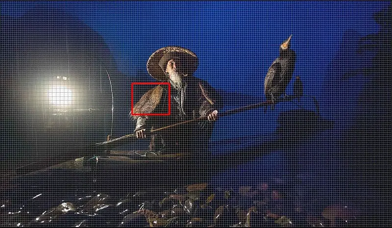
NOTE: This image was captured before twilight, in the pouring rain, on the Li River in China. This shooting scenario is the ultimate test for a camera sensor.
It only means that each generation of camera will get slightly better in the areas noted above, as software, hardware, and engineering improves.
Electrons counts can’t determine specific color information, therefore, a color filter is placed over each pixel helping to determine it’s color. This is discussed in detail below.
The example below shows the tonal values of black to white communicated with varying degrees of precision, by different bit depth systems.
The human eye, the second most (known) complex object on the planet, after the brain, has no problem discerning approximately 12 million different colors.
Each pixel can only collect the primary color information of it’s assigned red, green or blue filter, along with the number of electrons collected in the pixel well, which determine tonal value.
TECHNICAL NOTE: Although each of the color channels have the same amount of steps, the variation of green can still be seen all the way down to 1, where it’s hard to tell any difference in red at 1, and blue drops off at 2.
The digital language takes the form of zeros and ones ( bits ) & communicates the values of color ( Red, Green, Blue ) & tone collected by each pixel.
For the following example, assume the latest pro model full frame camera from Nikon or Sony. The exact model does not matter.
The stereomicroscope illustrated in Figure 1 produces an oblique cone of illumination using a specially-designed seven-sided toroidal mirror (Figure 2) that substantially reduces the stray light entering the large common main objective front lens. The toroidal mirror operates in a manner similar to high numerical aperture reflecting darkfield condensers that are equipped with internal mirror surfaces having a variety of curvature geometries.
Camera sensor size is the most important factor in determining overall camera performance & image quality, given the optimal focus, f-stop, ISO, and shutter speed settings have already been obtained.
Using this information, and a series of algorithms & interpolations, the camera can determine the color of each pixel contained on the sensor grid.
Most, but not all, CMOS sensors use a Bayer Filter which looks like a quilt of Red, Green, and Blue screens, with a single color screen covering each pixel as shown in the graphic.
During the first half of the twentieth century, darkfield microscopy (both compound and stereo) had a very strong following and a great deal of effort was expended in optimizing darkfield condenser systems and illuminators. This intense interest slowly began to fade with the emergence of more advanced contrast-enhancing techniques such as phase contrast, differential interference contrast, and Hoffman modulation contrast. Recently, new stereomicroscope illumination techniques, such as Nikon's oblique coherent contrast, which dramatically increase the contrast of transparent specimens, are being introduced and will ultimately probably displace a significant amount of interest in darkfield stereomicroscopy. However, a renewed interest in transmitted darkfield microscopy has arisen due to its advantage when used in combination with fluorescence microscopy.
The aperture diameter & shutter speed control how much light is captured by each pixel, thus increasing or decreasing the signal strength.
In turn, a digital image is produced, from millions of pixels, which matches the real world composition seen through the viewfinder.
The goal is to fill each pixel well to it’s corresponding tonal value maximum without clipping or losing data off the top end, thus increasing the Signal to Noise Ratio and image quality.
Light is made of photons or small packets for carrying light information. Photons are elementary particles which have no weight but carry information about light.
Larger physical sensor sizes combined with larger megapixel counts provide increased camera performance, with less noise, especially in low-light shooting situations.
Due to this fact, slightly overexposing images, known as Expose to the Right or ETTR , provides higher Signal to Noise Ratios and overall better image quality, provided that the brightest pixels are not “clipped” or “blown out”.
Therefore the number of possible choices, for each pixel, in a small 8-bit file is 256^3 power, or 256*256*256 which equals 16,777,216.
For example, the number of possible choices for a 3-bit system is found by using the binary base 2 and raising it to the power of 3, 2^3 = 8.
There are two popular types of image sensors, CMOS sensors (complementary metal-oxide semiconductor) & CCD sensors (charge-coupled device).
When a pixel well fills to the top with electrons, creating the maximum signal, it’s corresponding tonal value is white, producing a white pixel in the photo.
Digital photography is the process of recording real world color and tones, from a scene or composition, using individual pixels.
To the human eye, the perceived brightness of green is greater than that of red or blue, thus green filtered pixels are represented twice as often in the Bayer Filter.
That being said, I’m happy to recommend a few different camera models for landscape & nature photographers. These may not be specific to you, but they can help in cutting down decision fatigue. They may also work for other niches of photography, but I can’t guarantee it:)
Pretend the following graphics are the real world scenes that you’re seeing through the camera viewfinder or on the back of your camera live view screen.
The bit depth determines how many steps or possible choices within the tonal range can be communicated. Each step or possible choice is known as a bin. The more bins the more choices.
One of the most popular darkfield condenser designs, heavily utilized for high magnification compound microscopy prior to the emergence of phase contrast, is the paraboloid condenser, which has a curved and mirrored cardioidal internal surface. Illuminating light passes through the condenser and reflects from a single surface that is made from a paraboloid truncated by a light stop oriented perpendicular to the condenser and microscope optical axis. This system is free from spherical, chromatic, and coma aberrations and produces a sharply focused cone of illumination for the specimen from all azimuths. Although the stereomicroscope toroidal mirror design illustrated in Figures 2 and 3 does not operate with the sophistication and precision of the paraboloid condenser, it is far more effective for illuminating specimens in darkfield than conventional reflection mirrors that have a cylindrical geometry. The diagrams in Figure 3 compare the toroidal mirror design with a more conventional cylindrical mirror found in a majority of stereomicroscopes. In addition to providing more even illumination from all azimuths, the toroidal condenser design substantially reduces the amount of stray light entering the objective front lens, which leads to a significant enhancement of contrast between the specimen and background.
Type ofmicroscope
Physical sensor sizes are provided in terms of width and height, usually in millimeters. A standard sensor size such as 36mm × 24 mm is known as a full frame 35mm format camera.
Take a look at different objects around you. If you look closely, under a large amount of magnification, everything becomes a single color on a very small scale.
The number of electrons each pixel well collects determines it’s brightness, also known as value, on a scale of black to white. The scale of black to white is known as the tonal range or tonal scale.
Larger sensor widths yield larger sensor surface areas providing more area for the capture of light information over a standard interval, known as exposure time.
By gaining an understanding of how camera sensors actually work, and experimenting on your own, is the best way to figure out which camera and sensor size best fits your needs.
Imagine an image composition, seen through a camera viewfinder, with an imaginary overlaid grid, containing millions of tiny uniformly sized squares, as seen in the graphic below.
Each photographer has different sensor size requirements to produce the images they desire. I’m not going to tell you what camera to buy, but will provide some of my personal favorites.
The camera sensor, also known as an image sensor, is an electronic device that collects light information, consisting of color & intensity after it passes through the lens opening, known as the aperture.
When making the following comparisons of image sensors, assume that each sensor compared is from the same fabrication year.
None of the information the pixel collected prior to filling can be recovered or used in the final image. It’s gone forever!
As a sensor collects more light, producing a larger signal, less overall noise is seen in the final image. The Signal to Noise Ratio (SNR or S/N) is used to describe the phenomena.
In photography, the number of bits determines the possibilities of color or tone a single pixel can display, known as bit depth.
The precision and accuracy which this information is communicated and displayed in the final image is determined by the bit depth.
Bit depth specifies the number of unique color & tonal choices that are available to create an image. These color choices are denoted using a combination of zeros and ones, known as bits, which form binary code.
In darkfield microscopy, contrast is greatly enhanced by the superposition of a brightly shining specimen on a dark background. Blocking of zeroth order light rays by an opaque stop enables only higher order light rays to bathe the specimen with illumination. Highly oblique light rays, diffracted by the specimen and yielding first, second, and higher diffracted orders at the rear focal plane of the objective, proceed onto the image plane where they interfere with one another to produce an image of the specimen.
These color photographs are produced using the three primary colors, red, green, and blue determined by the Bayer filter.
Advantages and disadvantages ofdark field microscope
The red channel can display 255 different variations of red, the green can display 255 variations of green and the blue, 255 variations of blue.
Illumination of specimens by darkfield requires blocking out of the central light rays along the optical axis of the microscope, which ordinarily pass through and around (surrounding) the specimen. Blocking these light rays allows only those oblique rays originating at large angles to strike the specimen positioned on the microscope stage. In a compound microscope equipped with a simple condenser system, the condenser (Abbe-style) top lens is spherically concave, enabling light rays emerging from the surface in all azimuths to form an inverted hollow cone of illumination having an apex centered in the specimen plane. If no specimen is present on the stage, and the numerical aperture of the condenser is greater than that of the objective, the oblique rays cross and miss entering the objective front lens because of their obliquity. The field of view will appear dark.
Sony currently produces some of the highest dynamic range sensors on the market for full frame cameras. These camera sensors are rated at approximately 14.8 stops. Many Nikon cameras use Sony sensors due to this fact.
For example, the diagonal distance or hypotenuse of the 36mm by 24mm full frame can be found as follows: =SQRT((24^2)+(36^2)). The outcome is approximately 43.3mm.
The example below shows the 4-bit color scale for RGB Primary colors red, green and blue. Bin 15 in each of the color channels is pure fully saturated color, also known as hue.
The pixel specific tonal value is determined from the number of electrons ( charge ) collected and the color is determined using the Bayer Filter.
That’s almost 17 million different possible choices for every single pixel. There are millions of pixels on each sensor! This is a small JPEG file that the worst modern digital cameras can capture.
When a transparent specimen is placed on the glass microscope stage and observed under darkfield illumination, the oblique light rays cross the specimen and are diffracted, reflected, and/or refracted by optical discontinuities (such as the cell membrane, nucleus, and internal organelles) allowing these faint rays to enter the objective. The specimen then appears bright on an otherwise black background. In terms of Fourier optics, darkfield illumination removes the zeroth order (unscattered light) from the diffraction pattern formed at the rear focal plane of the objective. This results in an image formed exclusively from higher order diffraction intensities scattered by the specimen, and is also responsible for the main limitation of darkfield observation. Because the image is composed entirely from scattered light from the specimen, it is rich in glare and can even be distorted to varying degrees, so it cannot be considered a faithful geometrical reproduction of the specimen.
For example, although a crop sensor usually provides less quality & detail than a full frame sensor, a crop sensor from 2017 would most likely provide more quality and detail than a full frame sensor from the year 2000.
Red light passes through the red filtered pixels, while green and blue light do not. Blue light passes through the blue filtered pixels, while red and green light do not. You get the gist…
An adult and a 2-year-old child looking at the same landscape see close to the same thing, consisting of color and tonal values (light intensity).
PRO TIP: If you really enjoy photography buy the best camera you can afford. Then you don’t have to upgrade multiple times over the coming years. It saves money in the end. I know from experience…
The “rainbow colored” rectangle on the graphic shows the sensor grid. Pixels are so small that it’s hard to see each individual unit.
The best way to improve quickly is by learning firsthand from someone that’s optimized their skills, over a decade or more through trial and error.
Images that contain larger proportions of lighter tonal values will have higher Signal to Noise Ratios revealing less visible noise.

Consider this like a child that only speaks two words, yes & no, black & white. You wouldn’t depend on this child to communicate a landscape scene with a large degree of accuracy or precision.
As the ISO becomes larger less overall light (signal) is required to produce the same tonal value. As the ISO increases the noise levels are amplified creating more overall noise in the image.
In the graphic below, the pixel wells on the left have lower signal to noise ratios where the pixel wells moving towards the right have higher signal to noise ratios.
The graphic below shows an 8-bit system with 256 or ( 2^8 ) different bins. Due to the vast number of possible tonal choices the transition from one to the next isn’t discernible to the human eye. A JPEG image is 8-bit.
The photographer’s goal is to select the correct camera settings relaying this information with precision & accuracy, producing a digital image that matches what they see through the viewfinder.
Each tonal value, on the scale of black to white, has a corresponding signal required to produce it. Specific signal levels produce specific tonal values. The more electrons a pixel collects the stronger the signal it creates.
Darkfield observation in stereomicroscopy requires a specialized stand containing a reflection mirror and light-shielding plate to direct an inverted hollow cone of illumination towards the specimen at oblique angles. The principal elements of darkfield illumination are the same for both stereomicroscopes and more conventional compound microscopes, which often are equipped with complex multi-lens condenser systems or condensers having specialized internal mirrors containing reflecting surfaces oriented at specific geometries.
The free electrons are collected and counted by individual pixels on the sensor grid. Each pixel well has a maximum capacity of electrons it can collect. This maximum is known as full well capacity.
Thomas J. Fellers and Michael W. Davidson - National High Magnetic Field Laboratory, 1800 East Paul Dirac Dr., The Florida State University, Tallahassee, Florida, 32310.
For diagonal distance think of a straight line from the top right corner to bottom left corner. This is also known as the hypotenuse.
Each SMZ stereo microscope from Nikon features industry-leading optics, large zoom ranges, and wide fields of view for bridging macro- to micro-imaging.
The precision of the communication is rated on the scale of bit depth. Larger bit depth systems allow more precision in describing the information collected by each pixel.
CMOS Sensors are defined by their physical size ( surface area for capturing light information ) and the number of light information collecting pixels which make up this surface area.
The tonal values produced by each signal are combined with collected color information to produce each pixel’s final color within the photo.
The digital images in Figure 4 illustrate the effects of darkfield and brightfield illumination on fibers in whole mount specimens prepared using Canada balsam and a microscope slide and coverslip. Figure 4(a) and 4(b) compare nylon fibers under conditions of brightfield (Figure 4(a)) and darkfield (Figure 4(b)) illumination. The fibers imaged with brightfield are seriously lacking in contrast and minute details are difficult to distinguish against the white background. In contrast, when the fibers are illuminated with darkfield techniques (Nikon SMZ1500 with a toroidal mirror illuminator), internal fiber detail is discernable to a higher degree and depth of field emphasis becomes more pronounced. A situation where fibers have too much contrast in brightfield is presented in Figure 4(c) for pineapple fibers, which are not transparent and almost opaque when visualized under brightfield illumination. Viewing the same pineapple fiber specimen with darkfield illumination reveals far more intricate detail (Figure 4(d)) and exposes longitudinal splits in the fibers that are not apparent in the brightfield image.
In terms of color & tone, machines have bypassed the precision that the human eye, engineered by trial and error, through millions of years of evolution, can discern.
Images that contain larger proportions of dark tonal values will inherently have lower Signal to Noise Ratios revealing more visible noise. This is one reason low light and night sky images contain so much noise.
Due to the higher performance, especially in low light, and lower cost the CMOS Sensor is found in nearly all modern digital cameras.
The number of electrons collected by each pixel well produces a corresponding tonal value for that pixel. This tonal value is displayed in the final photo, along with the color.
The configuration presented in Figure 1 illustrates a Nikon SMZ1500 stereomicroscope equipped with an advanced stand containing provisions for both brightfield and darkfield illumination through a clear glass stage mounted on the top of the stand. Also depicted is a digital Internet camera system (Nikon Dn100) capable of transferring images collected by the microscope to remote observers. Details of the darkfield illumination mechanism are discussed below. Many current Nikon stereomicroscopes are also compatible with Darkfield illumination.
This charge is used to transfer the light information, collected by each pixel, into digital information which cameras & computers can understand.
I think Sony makes great low-end models with great sensors. Their high end models have fantastic sensors but are plastic and cheaply made. I prefer Nikon at the high end, with metal bodies, and the same Sony sensors. This is my personal preference.
Notice the massive difference in light collecting surface area between the APS-C vs full frame camera sensors. These cameras will produce much different overall image quality, with the larger far exceeding the smaller.
When a pixel well contains no electrons it produces no signal. The corresponding tonal value is a black, producing a black pixel in the photo.
After the exposure time, defined by shutter speed, has elapsed, the signal information produced by each pixel is processed & converted into a digital language known as binary code.
Since color information can’t be determined directly by the number of electrons in each pixel well, a color filter is placed over each pixel.
When the incorrect camera settings are selected, the squares on the pixel grid don’t match the squares image grid, producing a digital image that doesn’t match the scene being photographed.
Each individual square pixel represents a small sample of the image composition as a whole, consisting of a single color. No more.
Fluorescencemicroscope

This is a scientific fact, there is no dispute. Sony makes better sensors than Canon for landscape and outdoor photography.
Digital photography is the process of recording real world color information represented by the image grid, and relaying it into the digital world represented by the pixel grid.
Explore how mirror shape affects the amount of light entering the objective in darkfield stereoscopic microscopy. This tutorial demonstrates lightpath differences between conventional and toroidal mirrors.
As shown in the graphic below, a 1-bit system can only communicate black and white. A 2-bit system can communicate black, white and two tones of gray.
A number of aftermarket products are currently available for retrofitting stereomicroscopes with transmitted darkfield illumination. In addition, many of the microscope manufacturers offer illumination accessories that can be conveniently utilized to achieve darkfield conditions for their stereo systems. Typical aftermarket darkfield illuminators are presented in Figures 5 and 6. The design illustrated in Figure 5 utilizes a fiber optic ring light to provide illumination for a specially crafted stage that contains an internal mirror system and an opaque light stop. Light from the ring light illuminator is reflected from the internal cylindrical mirror with the central (zeroth order) rays being blocked by the light stop to form an inverted cone of illumination. Specimens are placed directly onto a glass plate resting above the stage aperture and can then be visualized with darkfield illumination. The ring light is equipped with an external light source that contains a voltage supply and a high-intensity tungsten-halogen lamp. Another darkfield condenser design, which also contains provisions for brightfield illumination, is presented in Figure 6. This condenser system utilizes a slider to rotate between brightfield and darkfield illumination and also contains a light source coupled to the condenser by a fiber optic bundle.
Specimens that have smooth reflective surfaces produce darkfield images that are primarily due to reflection of light into the objective. In situations where the specimen refractive index is different from the surrounding medium or where refractive index gradients occur (as in the edge of a membrane), light is refracted by the specimen. Both instances of reflection and refraction produce relatively small angular changes in the direction of light, enabling some rays to enter the objective. In contrast, some light striking the specimen is also diffracted, producing a 180-degree arc of light that passes through the entire numerical aperture range of the objective. The resolving power of the objective is the same in darkfield illumination as that achieved under brightfield conditions, but the optical character of the image (as discussed above) is not as accurately reproduced.
The ISO determines the amplification of the signal & inherent noise. The ISO also determines how much light is required for optimal exposure.
Each pixel is covered with a color filter, either red, green or blue. The color of each pixel is determined by the color of light (frequency of light wave) which passes through this filter.


 Ms.Cici
 8618319014500