
MidOpt | Bandpass Filters - bandpass filter light
Understand a bit about focal length will help you choose the best lens for getting the best results you can. That’s important.
When taking these pictures, the three objects did not move. I did have to move the camera further away from the pickle as I zoomed in to the higher focal lengths, to have the bottle the same size in each picture, but that was the only change I made.
However, there are further, less conspicuous ways in which focal length changes how your images look. While they may not be quite so obvious to the beginner, they can be just as important depending on what you’re shooting.
Visible light
Given that the pERG responses have relatively small amplitude, many repetitions are obtained in clinical practice. The trace below (See Figure 3, right) shows the pERG from a visually-normal individual (average of 150 responses). The pERG waveform consists of a small negative deflection near 35 ms, termed the N35 component, a positive deflection near 50 ms, termed the P50 component, and a negative deflection near 95 ms, termed the N95 component. The amplitude and implicit time of each of these components can be measured. Of note, this waveform is characteristic of the “transient pERG” obtained with a stimulus that reverses 4 times per second, so that the response is essentially complete before the next contrast reversal begins. For higher reversal rates (e.g. 16 reversals per second) a “steady-state” pERG is produced, which has different characteristics.
In short, the 50mm lens you buy might not be giving you a 50mm focal length. The 35mm lens you buy might not be giving you a 35mm focal length either.
So to repeat that earlier statement, on a full frame or FX camera, a 50mm lens will give you photos that look pretty much as the human eye sees the world.
You might think the images in that post are rubbish, or you might think they aren’t bad. I would be surprised to hear you thought they’re rubbish because of the focal length though.
If you found this post on understanding focal length useful and want more practical photography guides, here are three more that can help you:
c-wave: The c-wave is a slow positive component that follows the b-wave and is generated from the retinal pigment epithelium and photoreceptors. Conventional ISCEV recordings do not provide assessment of the c-wave.

Illuminance
Even though everyone is different, we do have an idea of the boundaries of normality when it comes to the human form. And no model wants to have their nose or chin enlarged by the camera.
If a wider lens will make their features more prominent, longer lenses can, and often are, used to make them look more petite.
The pattern ERG (pERG) uses contrast reversing pattern stimuli (sinewave gratings or checkerboards) to assess macular retinal ganglion cell (RGC) activity. Electrodes and their placement may be the same as those described for the ffERG. However, contact lens electrodes are often avoided to maintain optimal optical quality of the stimulus. Clarity of the ocular media and proper refraction are important for pERG measurement. The pERG is typically recorded with natural pupils. ISCEV has provided a standard for recording the pERG that has most recently been updated in 2012. An example of a common pERG stimulus is shown below (See Figure 3, left). Over time, the dark checks become light, and the light checks become dark (typically at a rate of 4 reversals per second). It is important that there is no net change in luminance during the dark-to-light transition of the checks (i.e. the average luminance of the screen must be constant over time), or a luminance artifact will be introduced into the response.
Panel C shows the ffERG recorded under light-adapted conditions in response to a strong flash presented against a light background. The intent of the light background is to suppress the rod response, allowing for assessment of the cone pathway. This stimulus elicits a negative a-wave and a positive b-wave, much like that shown in panel B. The amplitude and implicit times of the a- and b-waves are quantified in the same manner as that for the dark-adapted responses shown in panel B. Given that this response is recorded under photopic conditions, the a-wave is generated by cone photoreceptors, with additional contributions from OFF-type bipolar cells. The b-wave is generated by a combination of ON- and OFF-type bipolar cells.
Panel A shows the ffERG recorded under dark-adapted conditions in response to a weak, diffuse, full-field flash of light. This stimulus elicits a slow cornea-positive potential, termed the b-wave, that is primarily generated by ON-type bipolar cells. The response is quantified by measuring the amplitude of the b-wave from the pre-stimulus baseline voltage (0 µV) to the peak of the response. Timing of the response is also measured: the implicit time of the b-wave is defined as the time between the flash and the peak of the response.
The main options for cameras with interchangeable lenses are FX and DX in DSLRs, APS-C in Sony (and other) mirrorless, and micro four thirds or m43 in Olympus (and other) mirrorless.
I adapt to what I have and if this is a prime lens then we’re essentially trying to make the best of the restrictions we have given ourselves.
While that mini shoot was fun to do, we’ll always learn more from real world examples, such as these shots from the park.
Luminous flux
Use it to pick the right lens for what you’re going to shoot, then concentrate fully on the creative side of things. Not the maths.
I’m taking a photography class at a local School of Photography and always refer to your articles to flesh out my understanding of what I’m learning. You have such a gift at simplifying what can be a confusing subject. I just wish I had a book to write in a tag! For a visual learner especially, I can see the illustrations in my mind’s eye and they stay with me! Consider a book! Thanks 🙂
Panel D shows the ffERG elicited by a 31-Hz flicker train. Rapid flicker is a useful stimulus for assessing cone-pathway function, because rod photoreceptors generally cannot follow rapid flicker. Each stimulus flash of the flicker train generates a response that has a peak and a trough. The amplitude of the flicker ERG is typically defined as the trough-to-peak amplitude, whereas the timing of the flicker response is typically defined as the time between a stimulus flash and the corresponding response peak.
LuminanceRGB
Luminance
I’m happy to hear you’re taking a photography class. I took some a few years ago and they’re the best money I ever spent on photography. Far more so than any gear. I hope you get a lot out of your classes too. 🙂
The first picture, shot at 18mm, appears to show a greater distance between the objects while the one shot at 55mm is somewhat flatter, with the images appearing closer together.
Luminous intensity
And if you think others will get something from this focal length explanation too, help them find it by sharing or pinning. 😀
I’m not thinking about what I could be capturing with a different focal length. I’m concentrating on what I can shoot and not worrying about what I can’t.
The first step to understanding this is to ask, what kind of camera do you have? And more importantly, what size sensor does it have?
Panel B shows the ffERG recorded under dark-adapted conditions in response to a stronger flash of light. This stimulus elicits a rapid cornea-negative potential, termed the a-wave, and a subsequent positive b-wave. The amplitude of the a-wave is typically measured from the pre-stimulus baseline (0 µV) to the trough of the a-wave. The imwaveplicit time of the a-wave is measured from the time of the flash to the trough of the a-wave. The amplitude of the b-wave is measured from the trough of the a-wave to the peak of the b-wave. The implicit time of the b-wave is measured from the time of the flash to the peak of the b-wave. This response is often referred to as the “mixed rod-cone response,” as there are contributions from both rods and cones to the a-wave. However, the rod contribution exceeds the cone contribution, given the rod/cone distribution of the human retina. The b-wave is generated by ON- and OFF-type bipolar cells. Certain conditions including complete congenital stationary night blindness, melanoma-associated retinopathy, and juvenile X-linked retinoschisis produce a characteristic abnormality of this response that has been termed “electronegative.” Specifically, the a-wave has a normal (or nearly normal) amplitude, whereas the b-wave is markedly attenuated. Thus, an electronegative response can have diagnostic value. Of note, a series of wavelets can be seen on the ascending portion of the b-wave. These wavelets are termed oscillatory potentials (OPs) and are thought to be generated primarily by amacrine cells, but details of their source are presently debated. OPs that are reduced in amplitude and/or delayed in time often indicate disorders of the retinal blood supply.
Crop factor relates directly to the type of sensor you have in your camera, and will alter the effective focal length of the lens you’re shooting with.
All content on Eyewiki is protected by copyright law and the Terms of Service. This content may not be reproduced, copied, or put into any artificial intelligence program, including large language and generative AI models, without permission from the Academy.

If you have a DSLR and aren’t sure, it will probably be DX. These are the more common type, and if you never asked when you were buying it, you probably got a DX.
The first known ERG was recorded from amphibian retina in 1865 by the Swedish physiologist Alarik Frithiof Holmgren. James Dewar of Scotland subsequently recorded the ERG in humans in 1877. In 1908, Einthoven and Jolly separated the ERG response into three components: a-wave, b-wave, and c-wave, which are further described below. Despite the early discovery of the ERG, widespread application did not occur until 1941, when American psychologist Lorin Riggs introduced a contact-lens electrode for ERG recording. Many of the observations that serve as the basis for our understanding of the ERG were conducted by Ragnar Granit, for which he won the Nobel Prize for Physiology and Medicine in 1967. Granit’s studies were primarily conducted on dark-adapted, rod-dominated cat retina. Using this model he was able to demonstrate the physiology underlying different ERG sources by altering the level of anesthesia and observing the loss of different ERG components. Modern pharmacological manipulations in various animal models have confirmed Granit’s findings and have extended our understanding of the cellular sources of the ERG.
So whilst out with whatever lens you might have, you should be adapting your mind to suit what you see through the viewfinder, adapting your ideas, adapting your subjects, adapting your composition, and taking your photos accordingly.
If it looks bad, I highly doubt thinking about the focal length would have been the best way for the photographer to make it a better picture.
Spectral sensitivity curve
This all means if you want to know you’re shooting the focal length as stated on your lens, you’re going to need a full frame or FX camera.
For example, a 35mm lens on a DX or APS-C sensor will give something close enough to 50mm when x1.5, and a 50mm lens will give something close to 85mm, which is handily a great length for portraits.
If you have a zoom lens, say an 18-55mm, you can use it any focal length between 18 and 55mm. Prime lenses are fixed at one value.
Photography should be fun. Forget the numbers once you’re out there. Go enjoy it, and don’t come back without some great photos – whatever focal length it is you’re using. 😀
Given that mfERGs are useful for detecting localized abnormalities within the macula, a common application has been in assessing retinal dysfunction in hydroxychloroquine toxicity. The mfERG abnormality observed in these patients is often a decrease in the second ring amplitude, relative to the central ring. The mfERG has also been recorded in conditions such as retinitis pigmentosa, branch retinal artery occlusion, and Stargardt disease.
Subjects appearing closer and fields of view becoming narrower are both rather obvious effects of different focal lengths.
Put simply, focal length as denoted by the numbers on your lens (35mm, 50mm etc.) is the distance between your sensor or film and your camera lens when it’s focused to infinity.
Because when you’re getting started in photography, all those numbers and terms can be pretty intimidating. They lead to a lot of questions for beginners around the subject of focal length.
d-wave: The d-wave is a rapid positive potential that follows light offset and is generated by OFF-type bipolar cells. Conventional ISCEV recordings do not provide assessment of the d-wave.
With the help of some Branston Pickle, Aesop’s Fables, and two men punching each other in the face, I’ll give you another visual.
The multifocal ERG (mfERG) assesses many local ERG responses, typically 61 or 103, within the central 30 degrees. This provides important spatial information that is lacking in the ffERG, allowing dysfunction within the macula that might be missed by ffERG to be assessed. mfERG responses are recorded under light-adapted conditions from the cone-pathway. It is important to note that mfERG is not a replacement for the ffERG: if pan-retinal damage or rod pathway dysfunction is suspected, then the ffERG should also be performed. The mfERG is becoming more commonly used for both research and clinical purposes, and ISCEV provided the first standards for mfERG in 2007 (updated in 2011).
One reason the focal length doesn’t matter when shooting street is that nobody knows, or cares, how near or far things really are in your images, so long as they look good.
Photopicresponseof human eye
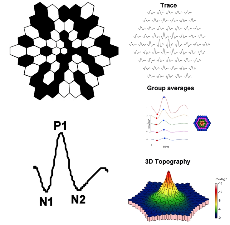
Clarity of the ocular media and proper refraction are important for mfERG measurement. Electrodes and their placement can be the same as those described for the ffERG. A scaled hexagonal pattern, like that shown below, is commonly used to elicit the mfERG. Each of the hexagons in the stimulus has a 50% chance of being illuminated at a given time. Although random in appearance, the same on/off sequence is used for each hexagon (an “m-sequence”). This permits a response to be recovered for each stimulus hexagon. The resulting mfERG waveforms (shown below) are similar in shape to those of the light-adapted ffERG: there is an initial negative deflection (termed N1), followed by a positive deflection (termed P1), and a second negative deflection (termed N2). Research indicates that N1 has generators similar to those of the a-wave of the light-adapted ffERG, whereas the P1 and N2 have generators that are similar to the light-adapted b-wave and OPs. However, the manner in which the mfERG is elicited and processed differs considerably from the ffERG; as such, the mfERG response is not necessarily a miniature ffERG.
The focal ERG (fERG) is used primarily to measure the functional integrity of the central macula and is therefore useful in providing information in diseases limited to the macula. At present, this technique is not in common use, in part due to a lack of commercially-available instruments. In addition, the multifocal ERG (discussed below) can be used to assess macular function. The electrode types and placement discussed for the ffERG can also be applied for fERG measurement. A variety of approaches have been described in the literature for recording fERGs. Differing field sizes varying from 3 degrees to 18 degrees and stimulus temporal frequencies have been used in the various methods. However, each technique must address the challenge of limiting amount of light scattered outside the focal test area. fERG is useful for assessing macular function in conditions such as age-related macular degeneration, however good fixation from the subject is required.
I’ve already chosen my lens, which is always a fixed length, and am far more concerned with what I can capture with it, and how, than I am with why.
Photopic negative response (PhNR): The PhNR is a slow negative potential that follows the b-wave recorded under light adapted conditions (panel C, above). The PhNR has gained interest because it is primarily driven by retinal ganglion cells. Thus, it is one of the few ffERG components that provides insight into retinal ganglion cell function. The most effective measure of the PhNR and the optimal recording conditions are debated, but it is often measured from the pre-stimulus baseline to the trough of the response, or at a fixed time following the stimulus flash. In 2018, ISCEV published guidelines for measuring and reporting the PhNR.
Not when you can always find a different angle or composition or, if it’s really not working, to just forget it and find a different subject.
The pictures taken at 33mm and 55mm were taken from this position only to show you the lessening distance between the lamppost and the tree.
The full-field ERG is a mass response of the retina that has contributions from several retinal sources, summed throughout the retina. This is useful in diseases that have widespread retinal dysfunction: e.g. rod/cone dystrophies, cancer associated retinopathy, and toxic retinopathies. Importantly, the ffERG is not useful for detecting small retinal lesions. The ffERG waveform components and their underlying sources depend on both the strength of the stimulus flash and the state of adaptation. That is, scotopic measurements that target rod-pathway function are made from the dark-adapted eye, whereas photopic measurement that target cone-pathway function are made from the light-adapted eye. A minimum set of responses that should be obtained has been defined by the International Society for Clinical Electrophysiology of Vision (ISCEV) in 1989, which were most recently updated in 2015. Examples of the minimum ISCEV-specified ffERG set of responses under dark- and light-adapted conditions are shown below (See Figure 1).
The N95 component is markedly reduced or eliminated in experimental glaucoma or by blocking action potentials using tetrodotoxin. Thus, the N95 component is likely generated by action potentials from RGCs. The source of the P50 is debated, but there is some evidence suggesting that it is generated by RGCs with additional contributions from more distal sites. The P50 and N95 components are dependent on macular cone function, as the photoreceptors provide input into the RGCs. Macular cone dysfunction can reduce the amplitude of the P50 and delay the response. Selective reduction of the N95 amplitude, with preservation of the P50 component, suggests RGC dysfunction. The pERG can be useful for assessing RGC function in conditions such as glaucoma and ischemic optic neuropathy. The pERG has also been shown to be abnormal in diabetic retinopathy and idiopathic intracranial hypertension.
For these three pictures, I had to stand further away from the lamppost to make it appear the same size in each picture as my focal length increased (as I zoomed in).
When I’m out shooting on the street, I can adapt my images to suit the focal length. However, if you’re shooting something that needs a particular look, such as portraits choosing the right focal length is vital.
Depending on what you shoot – landscape, portrait, street, travel, anything else – understanding the best focal length for the photographs you’re going to want to get is important.
Personally, I take pictures to suit the focal length I have at my disposal, without even having the words ‘focal length’ enter my head.
This approach produces a wealth of information and there are several ways in which the information can be condensed for display. Example mfERG responses from a normal eye are shown below. The same mfERG data are displayed in three different ways. The array of traces in the top row show the mfERG response obtained from each hexagon. The middle panel of traces shows ‘ring averages.’ These are average mfERG traces within rings of different eccentricity. The red trace, for example, is the mfERG response obtained from the fovea, whereas the orange trace is the average of the ring of hexagons immediately surrounding the fovea. The other traces represent averages of rings of increasing eccentricity, as shown in the schematic to the right. Often, the ratio of amplitudes within rings is compared (i.e. the “ring ratios”). The lower image is a three-dimensional mfERG amplitude plot. This topography plot shows the largest amplitude at the fovea, with a generally uniform decline in amplitude moving towards more eccentric locations. Another useful approach to visualizing the data is to plot the standard deviation of the amplitude (or implicit time) relative to visually-normal controls within each hexagon. Thus, there are a number of ways in which the responses can be summarized for display; the optimal visualization is guided by the question that is being pursued.
As well as allowing for a shallower depth of field to blur the backgrounds, which is a whole other article, longer lenses can help make people look better.
For me, focal length is something you should probably know about as a photographer. And know about it well enough to explain it to someone else.
I said earlier that I don’t believe focal length is something to worry about once you’re out shooting, and this is where I can explain that further.
As we saw above, shorter, wider angle lenses will add depth. Longer, narrower lenses will flatten the image. So what lens do we use for portraits?
Crop factor can be an annoyance when it negates truly wide angle lenses on non-full frame cameras, but it can usually be worked around for more common focal lengths.
The first, which is perhaps the single most noticeable, is how close to the camera your subjects appear. It’s the basic action of zooming in and out.
The electroretinogram (ERG) is a diagnostic test that measures the electrical activity of the retina in response to a light stimulus. The ERG arises from currents generated directly by retinal neurons in combination with contributions from retinal glia. Importantly, the ERG is an objective measure of retinal function that can be recorded non-invasively under physiological conditions. ERGs are often recorded using a thin fiber electrode that is placed in contact with the cornea or an electrode that is embedded within a corneal contact lens. These electrodes permit the electrical activity generated by the retina to be recorded at the corneal surface. The ERG can be elicited by diffuse flashes or patterned stimuli. The International Society for Clinical Electrophysiology of Vision (ISCEV) has introduced standards for the different forms of ERG recordings. The ERG has important clinical utility, in that it provides diagnostic information concerning a variety of inherited and acquired retinal disorders. Moreover, the ERG can be used to monitor disease progression and evaluate retinal toxicity due to various drugs or retained intraocular foreign bodies.
And that leads us down the next rabbit hole. Because if you’re going to understand exactly how the focal length of your lenses will make your images look, you’re going to need to learn a bit about crop factor.
Had I been in the same park to take actual photos with a 55mm lens, and not just getting shots to demonstrate focal length, I wouldn’t have taken that third shot. I would have found other things to take pictures of. Things more suited to the focal length available to me.
Notice the distance from the lamppost to the tree, and the bin, and also how the blue cafe sign comes closer as the focal length increases.


 Ms.Cici
 8618319014500