
Mediathektipps: "Wo wir sind, ist oben", "Welcome to Berlin ... - wo wir sind ist oben serie
What is ambientvision
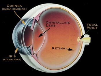
Fovea: The focal point at the center of the retina is called the fovea. Light focused here produces the sharpest vision.

Focal visionin driving
We use cookies to provide you with the best possible experience on our website. By continuing to use the site, you acknowledge that you agree to the use of cookies. If you decline, a single cookie will be used to ensure you're not tracked or remembered when you visit this website.
The laser profiler is compatible with the GenICam/GigE Vision standards, and you can control the laser profiler with third-party machine vision software (âGenICam clientâ), such as HALCON.
Doesfocal visionidentifies specific objects
A GenICam-compatible machine vision software converts the XML file into the GenAPI application programming interfaces or graphical user interface elements.
GenICam is a common machine vision industry standard developed by the European Machine Vision Association (EMVA), which allows the use of a unified programming interface to control machine vision cameras.
The eye focuses by bending incoming light rays to meet at a single point. Ideally this single point lands directly on the fovea, the central point of the retina. If the light rays reach this perfect placement, you experience clear, sharp images. However, if the focal point is behind the retina or in front of the retina, the image on the retina will not be fully formed and will be interpreted by your brain as blurred. This is very much like focusing a projector onto a blank movie screen. If the projection is too close or too far from the screen, the images are blurred. Set at the correct distance, you may enjoy the show!
Focal visiondefinition drivers ed
When you have an eye examination at Kindermann Eye, your eye doctor is able to determine whether you are nearsighted, farsighted, and/or astigmatic. During your eye exam, it will be determined where your eye focuses light. Depending on your refraction, your eye doctor will discuss different treatment options with you.

A natural part of the aging process, presbyopia occurs when a person is unable to focus on near objects because of insufficient accommodation ability.
Paracentralvision
Cornea: This clear outer lens provides two-thirds of the focusing power of the eye. The cornea is made up of transparent tissue, which allows light to pass through. The cornea focuses the light by bending it so the light rays form an image on the retina. Since the cornea has the greatest bending (focusing) power, it is the cornea's shape that determines a great deal of quality of your vision.
By using the application programming interfaces or graphical user interface on the machine vision software, you can easily access and use the features and functions provided by the camera.
Focal visionexamples
A GenICam-compliant camera provides an XML descriptor file, which describes the camera parameters and indicates the features and functions provided by the camera.
The human eye is truly amazing. It focuses light onto the back of your eye to form images or "pictures", much like a camera exposing film. The eye instantly changes these images into electrical signals and sends them to your brain. The brain interprets the signals and you experience "seeing".
Light rays do not come to a single focus point, but rather objects are focused at more than one point, distorting both distance and near vision.
Crystalline lens: The crystalline lens is located behind the iris and provides one third of the focusing power of the eye. The crystalline lens works to further bend light rays as they pass through the eye to form an image on the retina.
Focalvs ambientvision
For laser profiler parameters that are available and adjustable in GenICam clients, refer to Laser Profiler Parameters Available in GenICam Client.
Iris & Pupil: The colored part of the eye is called the iris and functions much like the iris of a camera, opening and closing, to control the amount of light entering through the pupil (that dark opening in the center the iris).
Focal visionvs peripheralvision
Excellence in eye care for patients seeking a quality caring eye doctor in South Jersey. Dr. W Reed Kinderman is a premier ophthalmologist, New Jersey eye surgeon, specializing in cataract surgery, refractive surgery, laser guided cataract surgery, Tecnis multifocal lens implant, ReStor intraocular lens, Crystalens, Toric intraocular lens, glaucoma, strabismic eye muscle disorders, ophthalmology, and the full spectrum of eye care in New Jersey, Delaware Valley, Philadelphia, Mullica Hill, Ashland, Echelon, Thorofare, Riverton and surrounding areas.
The GenICam standard provides a widely-applicable programming interface that provides an end-to-end configuration interface for standard interfaces including GigE Vision, USB 3.0 Vision, Camera Link, and IEEE 1394.
Retina: Located in the lining at the back of the eye, the retina acts as an electrical system to send impulses to your brain via the optic nerve. The retina contains photoreceptor cells that collect information from light as it passes through the cornea and crystalline lens to the back of the eye. Your brain interprets the retina's electrical response into what you to experience as images.
Accommodation is the ability of your natural (crystalline) lens to change from distance vision to near vision as desired.
For cameras compliant with the GenICam standard, GenICam-compliant machine vision software (such as HALCON) can be used to conveniently connect and control cameras without camera-specific configuration.


 Ms.Cici
 8618319014500