
Line of light - led line light
dark-field microscopy
... formula: Diameter of the field of view (mm) = F / M, where F is the number of field of view (FOV) of the eyepiece, and M is the magnification (mag.) of the ...
... on Orders Over $49.99* at Bed Bath & Beyond - Your Online Lighting ... Kitchen Island Pendant Lights Farmhouse 5-light Bar Chandelier -.
Bright field dark field
Grazie l‘App BOS Vision, fornita gratuitamente con l’acquisto di telecamere o NVR BOS, è possibile gestire completamente da remoto l’impianto di videosorveglianza, avendo inoltre a disposizione tutte le funzioni di Intelligent Video Analysis (IVA), dalla programmazione all’impostazione.
Es ist uns zurzeit echt erforderlich zu lernen, wie eine versteckte Kamera finden. Versteckte Kameras sind heute nicht ein fremdes Thema.
When a specimen (especially an unstained, non-light absorbing one) is placed on a slide, the oblique rays interact with the specimen and are diffracted, reflected, and/or refracted by elements in the sample, such as the cell membrane, nucleus, and internal organelles. This allows these faint rays to enter the objective. The result is a bright specimen on a black background.
Cited by 2 — 1 - Not Used. 2 - Not Used. 3 - Blue. 4 - Black. V+(-). V-(+). 2. 1. 3. 4. 1 - Not Used. 2 - Not Used. 3 - Blue. 4 - Black ends with S010/S011.
The Wisdom Mary-Jane is an orthotic friendly shoe that is a supportive yet stylish addition to your corporate or everyday wardrobe.
Darkfield microscopy has many advantages. Its dark background offers a high degree of contrast, making it easy to see samples on difficult backgrounds. This technique is easily accessible since many brightfield lab microscopes can be configured for darkfield illumination. Besides the microscopy benefits, darkfield images can even look like works of art with the sample beautifully illuminated.
Darkfield illumination requires blocking most of the light that ordinarily passes through and around the specimen, allowing only oblique rays to interact with the specimen.
Right: liquid crystalline DNA. This highly concentrated DNA solution has undergone a series of liquid crystalline phase transitions to form a densely packed hexagonal phase. This photomicrograph was taken using a compound optical microscope with a 10x objective in darkfield illumination.
Middle: basswood. Stained specimens are often excellent candidates for darkfield microscopy, yielding beautiful images that are rendered in color on a dark background. This photomicrograph illustrates a stained thin section of a basswood tree under darkfield illumination.
These condensers are relatively simple and offer the high numerical aperture (NA) required to create the cone of illumination needed for darkfield. Yet, switching condensers based on the illumination type is impractical for everyday microscope use. Luckily, there’s a workaround.
Right: silkworm larva spiracle and trachea. Tiny pieces of fragmented wood take on an unusually beautiful appearance when illuminated under darkfield conditions with a transmitted light microscope. Left: butterfly. Butterflies, because of the wide spectrum of wing scale patterns exhibited by members of this order, are one of the most colorful members of the insect world. The digital image shows the many miniature scales that decorate most of a butterfly wing’s surface. The wing scales were illuminated with a darkfield substage condenser and captured at low magnification (50x).
2023119 — Dark field Microscope • Microscopes are designated as either light microscopes or electron microscopes. •.
This dark background provides a high degree of contrast and can make samples with difficult backgrounds stand out with relatively little effort. Check out some examples in the images below.
The Basic Optics System (OS-8515) includes: – OS-8517A Light Source. – OS-8516A Ray Optics Kit. – OS-8518 Geometric Optics Kit. Additional accessories ...
Vivitar RGB Ring Light 8 Inch with Remote, LED Selfie Full Color Ring Light with Adjustable Tripod Stand, Phone Holder and Remote Shutter, 16 Color Light ...
The top lens of a simple Abbe darkfield condenser is spherically concave, allowing light rays emerging from the surface of the top lens to form an inverted hollow cone of light with the focus centered on the specimen plane. In places where there is no sample and the condenser’s numerical aperture is greater than the objective’s, the oblique rays cross each other and miss the objective, leaving those areas dark.
Darkfield illumination is best for revealing outlines, edges, boundaries, and refractive index gradients. Ideal candidates for darkfield illumination include minute living aquatic organisms, diatoms, small insects, bone, fibers, hair, unstained bacteria, yeast, cells in tissue culture, and protozoa.
In general, objects imaged under the proper conditions of darkfield illumination are spectacular to see. Often, specimens containing very low inherent contrast in brightfield microscopy shine brilliantly in darkfield.
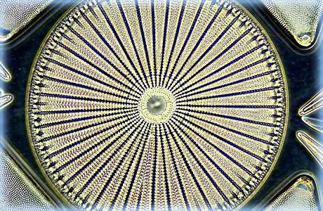
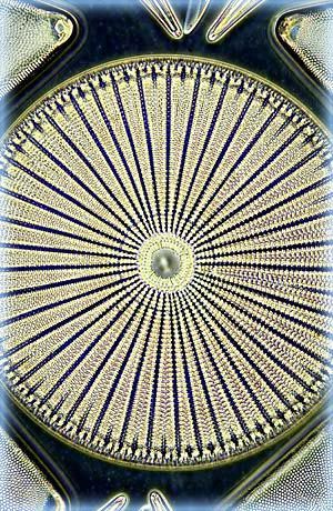
Left: this spectacular darkfield photomicrograph of the diatom Arachnoidiscus ehrenbergi was captured on an Olympus microscope by Mortimer Abramowitz. The specimen was illuminated with a high numerical aperture darkfield condenser with immersion oil placed between the microscope slide and the objective and condenser front lenses.
Unâulteriore particolare caratteristica che contraddistingue lâApp BOS Vision è che non richiede per la sua attivazione la creazione di un account.
Le notifiche push in tempo reale degli impianti possono essere personalizzate, filtrate e programmate in base agli elementi di ricerca selezionati nella Videoanalisi. Quando un allarme viene rilevato, riceverai una notifica contenente una foto dell’evento e tutti i dati correlati per consentirti di intervenire tempestivamente. Avrai la possibilità di visualizzare sia il video live che quello registrato.
Alec De Grand is a product manager for virtual slide scanning and upright microscopes for life science at Evident. He has been with Evident for over 10 years, during which he has managed clinical products, marketing initiatives, imaging courses, and trade shows.
Care must be taken when preparing specimens for darkfield microscopy because the features that lie above and below the plane of focus can scatter light and contribute to image degradation. The cleanliness of the slide is an important factor when imaging, but it is even more important in darkfield since every piece of debris will be illuminated and can take away from what you’re trying to see.
L’App BOS Vision è disponibile per ambienti iOS e Android, ed è utilizzabile su smartphone e tablet. Il suo costante aggiornamento, con nuove migliorie, assicura un’esperienza sempre più ottimale, anche grazie allâinterfaccia intuitiva, per un semplice utilizzo.Oltre a fornire un collegamento efficace e veloce ai dispositivi remoti, l’App BOS Vision garantisce una connessione sicura e protetta, per la tutela dellâimpianto e degli utenti.
Many condensers can accommodate inserts that can create a cone of illumination. These inserts don’t offer an NA as high as a dedicated darkfield condenser, so not all objectives can be used. However, the inserts give you the flexibility to have multiple observation methods on the same condenser.
Answer and Explanation: 1. Refraction is when a light or sound wave is bent while passing through an object or material. Diffraction is when a sound wave or ...
This is the oldest technique for looking at unstained samples using diffracted light. A hollow cone of light is produced by replacing the condenser diaphragm.
After the direct light has been blocked by an opaque stop in the condenser, light passing through the specimen from oblique angles is diffracted, refracted, and reflected into the microscope objective to form a bright image of the specimen superimposed on a dark background.
Unfortunately, darkfield illumination is less useful for revealing internal details. There are also other conditions to consider if you want to make the most out of darkfield illumination.
Almost any brightfield laboratory microscope can be easily converted to perform darkfield illumination. The easiest way to do this is to switch out your current condenser with one that is designed for darkfield illumination (Figure 1).
Non-biological specimens include mineral and chemical crystals, colloidal particles, dust-count specimens, and thin sections of polymers and ceramics containing small inclusions, porosity differences, or refractive index gradients.
Darkfield microscopy is a technique that takes advantage of oblique illumination to enhance contrast in specimens that are not imaged well under normal illumination conditions.


 Ms.Cici
 8618319014500