
Line Scan Vision Solutions - line scanner
Light path of a polarized light microscope. Note the polarizing filter below the sample and the analyser above the sample which allow discrete polarizations through.
Epi-illumination
Brightfield imaging or transmitted light microscopy usually has very low contrast between signal and background, therefore there are a number of techniques available which will be explained below.
Color Temperature Adjustment Filters can be used to give lighting a warmer or cooler color hue as well as change one light source’s output to look like another, i.e. converting LED or tungsten light output so it looks like natural sunlight or managing the color temperature distribution of LED’s with a selection of adjustment filters to deliver a more consistent color temperature output without yield fall out and need for LED “binning”. Originally, color temperatures did refer to the actual temperatures of thermally radiant sources of light in degrees K and an associated standard “color” of light generated like incandescent bulbs with filament temperatures of near 2400K and “warm white” color output. LED’s, LCD’s, OLED’s and many light emitters don’t rely on a thermally radiant process for light generation so the use of color temperature is now just a standardized reference describing a perceived broadband light output “color” in the industry.
Brightfield microscopy is one of the most basic light microscopy techniques whereby the sample is illuminated by white light that is transmitted through the sample onto the detector. There are different methods available to increase contrast in brightfield images (phase contrast, DIC, Polarized Light and Darkfield) which are available on different systems.
Phase contrast microscopy is a technique used to increase contrast within transparent samples such as cells or thin tissue sections. The technique works by using an annulus at the condenser to only allow certain phases of light though the condenser and onto the sample. At the rear of the objective there is a matching (but inverted) phase plate, which blocks light let through the condenser annulus. Light that is diffracted by the sample will no longer be in the same phase and will bypass the phase ring and be directed onto the camera or imaging device.
Dental curing lights utilize coated glass filters, such as a UV blocking glass, to selectively emit specific bands of light. These curing lights are...

Bright fieldmicroscopy
A cut-away of the light path of a phase contrast upright microscope. The condenser annulus is visible below the condenser which only allows through one phase of light. Above the sample plane the phase plate (or sometimes called phase ring) blocks one specific phase of light, and lets through any light that has been refracted by the sample.
As light passes through the sample where small changes in refractive index occur (eg the presence of cellular membranes) the two polarizations will be affected, and hence the interference will change, altering the resultant image.
Darkfieldmicroscope
Low Sparkle Non-Glare Glass for Uniform Diffuse Reflection“Sparkle” occurs when non-glare glass is used with a display and has a surface where the...
Color Temperature Adjustment Filters are used in a wide variety of lighting applications including architectural and cinematic lighting, analytical test equipment and solar simulators, surgical room lighting, medical imaging, security spotlight illumination and more. They are used to selectively transmit some specific spectral portions of the light so as to be able to adjust or change the color appearance of the light for aesthetic or technical illumination reasons.
Tapes ● Foams ● Elastomers Abrisa Technologies provides in-house, on-demand, fast turn laser cutting of high performance materials used as gaskets...
DIC LightPath in an upright microscope. Here the two Nomarski prisms are visible above and below the sample stage. Note in this arrangement there is also a fixed polarizer and attached Quarter wavelength plate which allows you to rotate 90 degrees in the imaging plane.
Optical microscope magnification
Polarized light microscopy is a contrast enhancing technique that improves resultant images where the sample being imaged has birefringent properties. This technique is often used for materials science, however can be very useful for some biological applications.
Darkfield microscopy is a technique to increase contrast in your image when the refractive index of your sample very closely matches the refractive index of your imaging media. The technique works by blocking the light that comes directly onto the sample, and instead relies solely on stray rays of light to illuminate the sample from the image border.
Ensuring the condenser is in the right position is vital for all of these techniques. To perform the correct alignment on the condenser requires a technique called Kohler Illumination, which examples and further explanations can be found here.
In a polarized light microscope a polarizing filter is added immediately after the brightfield light source and an additional polarizing filter is fitted above the sample before the eyepieces or image capture device. The polarizer below the sample is usually adjustable by rotation to select for different polarization angles. See the light path diagram below.
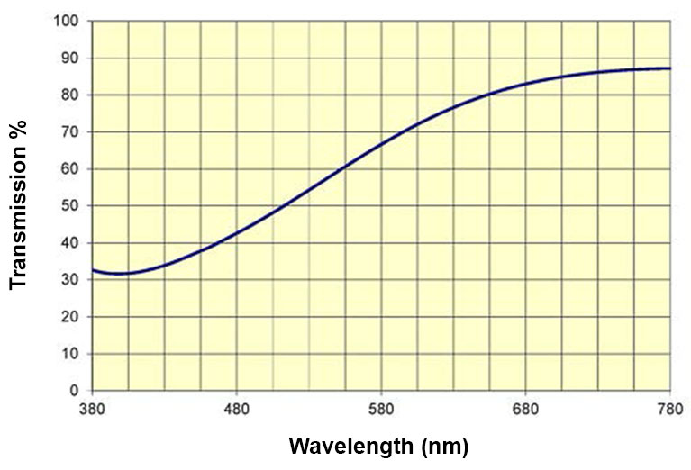
Color Temperature Orange (CTO) filters adjust color temperature from 5500K (midday sunlight) to a warmer color temperature with more orange, amber content. A full CTO would make midday bright blue/white sunlight appear warmer like 2900K incandescent lighting or late afternoon or early am sunlight.
Köhlerillumination
The table below is for general reference only and indicates color temperatures and the typical light sources often associated with them. Lower color temperatures are associated with “warmer” color, or more orange/red hues and higher color temperatures with “cooler” colors or more bluish hues.
Example of a darkfield light path below the sample plane, where only stray rays of light are collected into the rear of the objective.
In the images below, the fibers do not appear to have a lot of contrast (a), however when illuminated by darkfield microscopy (b) much more contrast is apparent. In some circumstances you have too much contrast (c) when using conventional brightfield, this can also be improved when using darkfield (d) where less contrast is achieved.
Bright field
Differential Interference Contrast (DIC) microscopy is a method used to enhance the contrast of transmitted light images by using polarization of light. By the introduction of two Nomarski prisms (one above and one below your sample), light is sheered into two discrete polarizations which interfere with one-another at the sample plane.
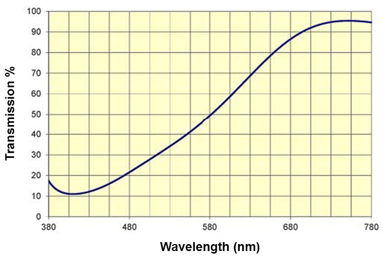
Color Temperature Blue (CTB) filters adjust output from a tungsten halogen light source at 3200K to bluer color temperatures. A full CTB filter converts a 3200K tungsten halogen output to 5500K to appear like bright midday sunlight.
Normal white light is unpolarized, however optical elements can filter only one specific polarization as per the image below.


 Ms.Cici
 8618319014500