
UV Lamp Stockist - ultraviolet light replacement bulb
This technique is used to visualize living unstained cells. This is affected by the way illumination is done on the specimen in that, when a hollow cone beam of light is transmitted to the specimen, deviated light (unreflected/unrefracted) rays do not pass through the objectives but the undeviated (reflected/refracted) light passes through the objectives to the specimen forming an image.

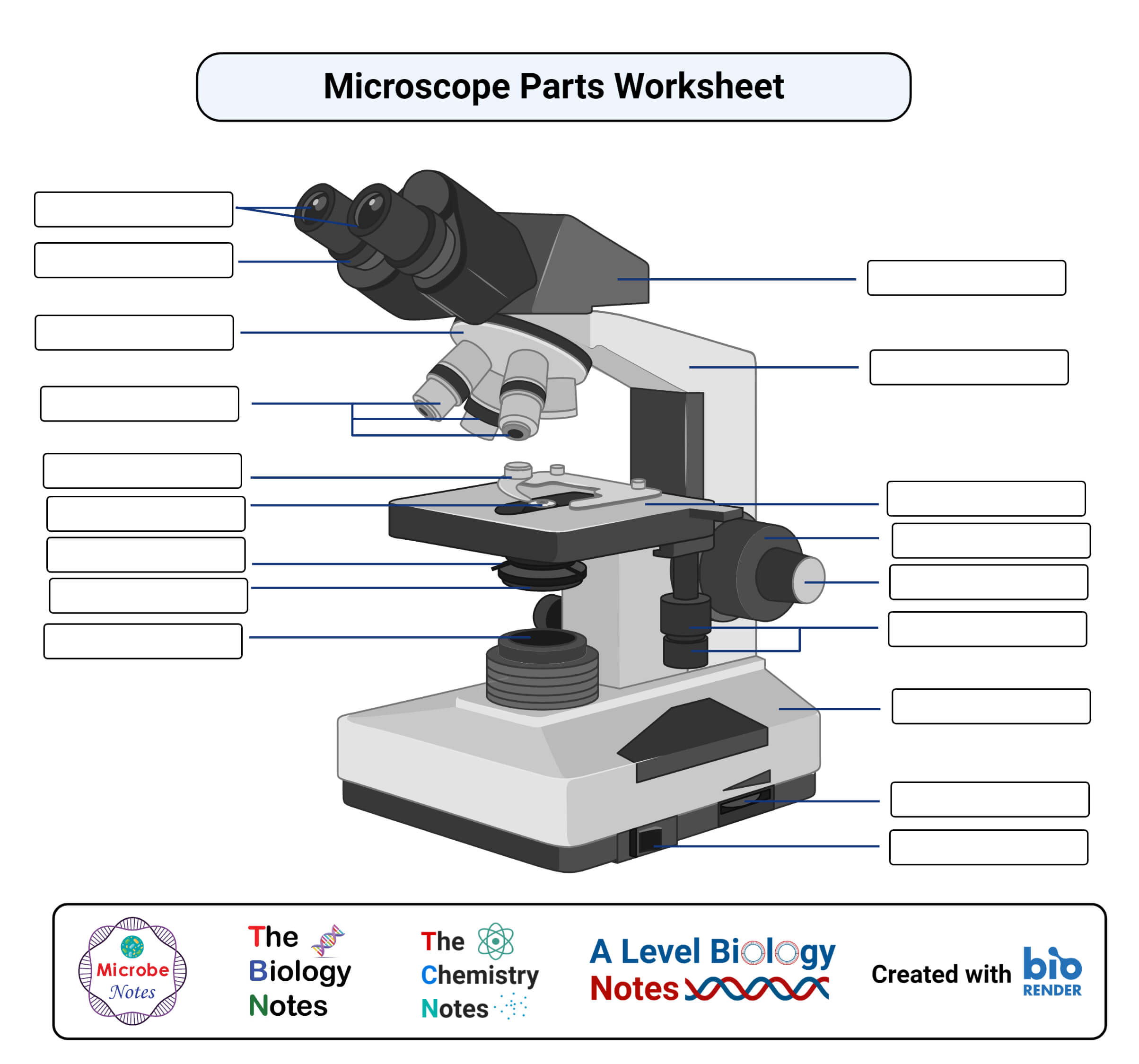
Stagemicroscope function
The evolution of the Microbiology field put to perspective the need to identify, view, observe and understand microorganisms, including their structural morphologies and mechanisms. Microbiology’s scope is to study organisms and minute agents that can only be examined and observed with a microscope.
In the case of the fluorescent Microscope, the specimen emits light. How? By adding a dye molecule to the specimen. This dye molecule will normally become excited when it absorbs light energy, hence it releases any trapped energy as light. The light energy that is released by the excited molecule has a long wavelength compared to its radiating light. The dye molecule is normally a fluorochrome, that fluoresces when exposed to the light of a certain specific wavelength. The image formed is a fluorochrome-labeled image from the emitted light
If an object is put between these two mediums i.e between water and air, in this case, a prism, the prism will bend the light at an angle. This is how the microscopic lenses work, they bend the light at an angle. The lens (convex) on receiving the light rays, focuses the rays at a specific point known as the focal point (F-point). The measure of distance from the center of the lens and the focal point is known as the focal length.
The objective lens plays a vital role in not only enlarging the image but also making it clear for viewing, a feature known as resolution. Resolution according to Prescott, is the ability of a lens to separate or distinguish between small objects closely linked together.
used to view specimens are both simple and compound light microscopes, all using lenses. The difference is simple light microscopes use a single lens for magnification while compound lenses use two or more lenses for magnifications. This means, that a series of lenses are placed in an order such that, one lens magnifies the image further than the initial lens.
Condensermicroscope function
The principle behind this working mechanism is that the fluorescent microscope will expose the specimen to ultra or violet or blue light, which forms an image of the specimen that is emanated by the fluorescent light. They have a mercury vapor arc lamp that produces an intense beam of light that passes through an exciter filter. The exciter filter functions to transmit a specific wavelength to the fluorochrome stained specimen, producing the fluorochrome-labeled image, at the objective.
Armmicroscope function
Besides the above-discussed microscopes, there is one not commonly used microscope known as the Differential Interference Contrast Microscopy. It is very similar to the phase-contrast microscope whereby the images are formed from the variations in the light either deviated and or undeviated. The difference is, here two beams of light are emitted to the specimen and focused by a prism. One beam passes through the prism to the specimen while another passes through the glass slide clear area without the specimen. The two beams then combine and interfere with each other to form an image. It can be used to view cell structures such as endospores, bacterial cell walls, nuclei, and granules for unstained specimens.
Microscopeparts and functions
Vastly used in Microbiology, this microscope is used to view fixed and live specimens, that have been stained with basic stains. This gives contrast for easy visibility under the microscope. Therefore it can be used to identify basic bacteria cells and parasitic protozoans such as Paramecium.
Whereas the eyepiece magnifies the image at the end of the viewing, its magnification range is lower than that of the objective lens at 8X-12X (10X standard) and that of the objective lens at 40X-100X, magnification, and resolution of the microscope is highly dependant on the objective lens.
From the History of Microbiology, Antony Van Lewnehoueek an amateur Microbiologist made the first simple microscope, that enabled him to observe the presence of tiny living organisms in pond water that appeared like dots. His simple microscope was made up of a double convex glass lens that was held between two silver plates.
A light microscope is a biology laboratory instrument or tool, that uses visible light to detect and magnify very small objects and enlarge them.
A microscope uses lenses whose strength is predetermined, in that, the strength of a lens is directly related to the focal length i.e short focal length magnifies objects more than lenses with a long focal length.
A minimum distance (d) between two objects that distinguishes them to be two separate entities, determined by the wavelengths of the light can be calculated by an Abbe equation using the wavelength of the light that illuminated the specimen (Lambda, λ) and the numerical aperture (NA, n sin Ɵ) i.e. d=0.5 λ/n sin Ɵ

What is seen in the microscope as an enlarged clear image of the specimen is known as the virtual image. To calculate the magnification, multiply the objective and eyepiece objective magnification together. The magnification is standard, i.e not too high nor too low, and therefore depending on the magnification power of the lenses, it will range between 40X and 100oX.
Basemicroscope function
A medium with a lower refractive index such as glass to air normally speeds up the light penetration and makes light bend away from the normal and when light is passed through a medium with a greater refractive index such as air to glass, it normally slows down and bends towards the normal, perpendicularly to the surface.
You can cite as: “Mokobi, F. (2024, April 15). Light Microscope: Principle, types, parts, diagram. Retrieved from https://microbenotes.com/light-microscope/“.
Although scientifically, the first simple microscope was discovered by two Dutch scientists, Zaccharias Janssen and his father, Hans who made spectacles, were the first to experiment with their lenses by combining lenses in a tube and observed that the objects that were nearby, appeared closer and larger. Despite not being included as a scientific discovery, this act paved the way for scientific evolution.
Objective lensmicroscope function
Diaphragmmicroscope function
As mentioned earlier, light microscopes visualize an image by using a glass lens, and magnification is determined by, the lens’s ability to bend light and focus it on the specimen, which forms an image. When a ray of light passes through one medium into another, the ray bends at the interface causing refraction. The bending of light is determined by the refractive index, which is a measure of how great a substance slows the speed of light. The direction and magnitude of the bending of the light are determined by the refractive indexes of the two mediums that form the interface.
During visualization, the objective lens remains parfocal which means, when the objective lens is changed, the image still remains in focus. The objective lens plays a major role in focusing the image on the condenser forming an enlarged clear image within the microscope, which is then further magnified by the eyepiece to a primary image.
Stage clipsmicroscope function
The above-discussed microscopes will normally produce images after a light has been transmitted and passed through the specimen.
This makes the surrounding field of the specimen appear black while the specimen will appear illuminated. This is enabled by the dark background this the name, dark-field Microscopy.
This is a specialized type of bright field light microscope that has several similarities to the Phase-Contrast Microscope. To make a dark field Microscope, place a darkfield stop underneath and a condenser lens which produces a hollow cone beam of light that enters the objective only, from the specimen (Prescott, pg 22).
This is the most basic optical Microscope used in microbiology laboratories which produces a dark image against a bright background. Made up of two lenses, it is widely used to view plant and animal cell organelles including some parasites such as Paramecium after staining with basic stains. Its functionality is based on being able to provide a high-resolution image, which highly depends on the proper use of the microscope. This means that an adequate amount of light will enable sufficient focusing of the image, to produce a quality image. It is also known as a compound light microscope.
The instrumentation of the Phase Contrast Microscope is based on its light pathways from receiving the source of light to the visualization of the image.
The Phase-Contrast Microscope is designed with objective lenses that have the ability to perform multiple functions when combined with contrast-enhancing techniques, for example, fluorescence. The objective lenses are located in the internal phase plate with variation in the light absorption and phase displacement i.e undiffraction, creating a wide spectrum for contrasting the specimen and forming a strong contrast in the background.
The application of microscopy in Microbiology enhanced the visualization of cells and microorganisms by magnifying their images to make them larger.
Microscopy works strictly with a factor of resolution whereby resolution is the ability of a lens to be able to differentiate small objects that are closely packed together. The resolution of a light microscope is determined by a numerical aperture of its lens system and by the wavelength of the light it employs; a numerical aperture is a definition of the light wavelengths produced when the specimen is illuminated.
After the objective, there is a barrier filter that functions primarily to remove any ultraviolet radiation that may be harmful to the viewer’s light, thus reducing the contrast of the image.


 Ms.Cici
 8618319014500