
Smart Lighting - large light ring
Terms Of Use | Privacy Notice | Cookies | Cookie Settings | About Us | Careers | Careers | Sitemap
Microscope illumination techniquespdf
The key optical components needed to set up Köhler illumination in reflected light microscopy are arranged in opposite orientation to those in transmitted light microscopy. The condenser aperture iris diaphragm is closer to the light source and the field iris diaphragm is closer to the specimen. In reflected light microscopy, the objective serves a dual function—on the way down, the objective serves as a properly aligned condenser, controlling the angle of the light striking the specimen. On the way up, the objective’s numerical aperture (NA) determines the angle of light which can be captured as it is reflected from the specimen. All things being equal, the higher the NA, the better the resolution of the objective, and the better the resolution of the specimen.
Brightfield microscopy: The most commonly used optical microscopy technique. Illumination of the sample is transmitted from above via a tungsten-halogen lamp focused through the vertical illuminator positioned above the stage. Light reflected by a beam splitter through the objective illuminates the specimen. Light reflected from the specimen surface re-enters the objective and passes into the eyepiece or to a camera port. Absorption and diffraction of the incident light by the specimen often lead to discernible variations in the image. Specimens that show little difference in intensity or color require dark field microscopy or reflected differential interference contrast (DIC) (see below).
Köhlerillumination
A couple of incident light meters. These meters, though primarily designed as incident meters, are actually "System Meters." A wide variety of accessories were made for them that expanded their capabilities. Along the top are the enlarging meter attachment, the reflected light attachment, and a flat incident light diffuser. The flat diffuser is for photographing flat subjects, like artwork. It is also used in studio work to take readings from individual lights to set lighting ratios.
If you only photograph such "average" scenes, then a reflected light meter will give you accurate results. In the real world, that is not often the case. As we'll see later in this tutorial, reflected light readings of a light-toned subject will produce an underexposed photograph, as the meter tries to render the light tones as a middle gray. A reflected light reading of a dark-toned subject will produce an overexposed photograph, as the meter tries to render the dark tones as a middle gray.
A handheld incident light meter ignores the subject and just measures the light that hits the subject. This might seem counter-intuitive, but it is actually superior to reflected light metering in most circumstances. Because it ignores the subject, it cannot be fooled into underexposing light tones and overexposing dark tones, as a reflected light meter does.
Microscopebase function
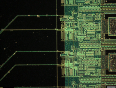
Reflected light microscopy: Reflected light microscopy or epi-illumination is the choice for illumination of opaque specimens like metals, ores, ceramics, polymers, semiconductors (e.g., unprocessed silicon, wafers, integrated circuits), slag, coal, plastics, and paint.
The system isn't perfect. The dome cannot take into account backlighting, and they don't do well with extreme side-lighting. For those conditions, I recommend taking both an incident reading and a reflected light reading and then averaging them. I have a tutorial that explains this method in more detail.
Still relevant today, Köhler illumination should be applied whenever suboptimal specimen illumination is preventing proper observation. Köhler illumination can provide even illumination of specimens in brightfield, darkfield, and all variations of phase contrast microscopy using either transmitted or reflected light pathways. Köhler illumination facilitates even illumination, high resolution, and good specimen contrast.
Focusing inmicroscope
Magnification system ofmicroscope
A handheld light meter is, I believe, the single most important accessory that a photographer can own. They provide more useful and accurate readings than the meters built in to cameras.
Function of light switch inmicroscope
Transmitted light microscopy: Köhler illumination requires several optical components of the microscope to function in between the light source and the specimen. These include the collector lens, field diaphragm, condenser diaphragm, and condenser lens. The collector lens acts to collect light from the light source and focus it on the plane of the condenser diaphragm. The condenser lens then projects this light through to the specimen.
Self-illuminated or trans-illuminated objects, such as stained glass windows and neon signs cannot be measured with an incident light meter. For those, a reflected light meter is required. Because an incident meter requires you to meter from the location of the subject, or in the same light as the subject, it cannot be used for inaccessible places like a theatre stage during a play or concert.
Criticalillumination
Rob Bellinger is a product applications manager for industrial microscopes at Evident. He has been part of Evident for more than 15 years. He currently provides application support for our industrial microscope systems in the US, Canada, and Latin America.
Critical illumination was the predominant technique in microscopy until August Köhler (1866–1948) developed a new method of illumination, now known as Köhler illumination. The problem with critical illumination was that the bright light lamp source created a filament image in the same plane as the specimen image. Visibility of the bulb filament in the final image caused uneven specimen illumination and introduced glare and shadowing artifacts. Köhler illumination resolved these artifacts by using a defocused light source image to evenly illuminate the specimen.
The 5 degree viewfinder turns the meter into a reflected light meter with a smaller angle of view, allowing you to take readings from different parts of the scene. An actual 1 degree spotmeter (discussed on Page 3) is better for this, though.
There are two types of exposure meters. Reflected light meters, and incident light meters. In addition, there are also Color Meters, which do not give exposure information; they tell you the white balance settings to use with your camera. If you want to know more about them, my Color Temperature Meter Tutorial dicusses their use.
Incident light meters are instantly recognizable because they have a white dome covering the meter's sensor. To use the meter, you stand close to your subject, so that the meter will be in the same light as your subject, and point the white dome directly toward the camera.
Darkfield microscopy: Well-suited for applications where contrast comes from light scattered by the specimen. Light not scattered by the specimen is not collected by the objective lens and not incorporated into the image. Consequently, the field around the specimen appears dark. The main limitation of darkfield microscopy is the low light levels seen in the final image. This is how Köhler illumination techniques contribute—the sample must be strongly illuminated. Raised features that are too smooth to cast shadows will not appear in brightfield images, but the light that reflects off the sides of the feature will be visible in the darkfield images.
The reason for the white dome over the sensor is that the three dimensional dome simulates a three dimensional subject. Light hits the subject from different directions, and the dome allows the meter to see light coming from the sides, from above or below, and from straight on.
If you have ever used a camera with a built-in light meter, you have already used a reflected light meter. All of the meters built in to cameras that read the light coming in through the lens are reflected light meters. There are also handheld reflected light meters.
What isilluminationinMicroscope
Phase contrast microscopy: An optical microscopy technique that generates sample contrast from interference of different path lengths of light reflected from the specimen. Amplitude and phase shifts occur based on properties of the specimen. These changes show as variations in brightness from the scattering and absorption of light. Phase contrast microscopy is particularly important in industrial microscopy as it reveals many features or structures of a specimen not visible in brightfield microscopy.
Terms Of Use | Privacy Notice | Cookies | Cookie Settings | About Us | Imprint | Careers | Careers | Sitemap
The knowledge that I am sharing took many years of study and practice to attain. If you find it valuable, please donate through my Paypal button below. My creative work is how I support myself and my son. Thank you!
Two reflected light meters. The Weston Master V is a classic meter from the 1960s, a development of the original handheld meters invented by Weston in the 1930s. The Gossen Ultra-Pro is mainly designed as a reflected light meter, but includes a small incident light dome that can be slid over the front of the sensor.
By adjusting the field diaphragm, the image of the field diaphragm aperture in the specimen plane is set to a size slightly larger than the imaged region of the specimen, which corresponds to the portion of the specimen image seen at the eyepiece field stop. As the field diaphragm, specimen, and eyepiece field stop all lie on the same conjugate image plane, this adjustment allows the illuminating rays to completely fill the eyepiece field of view, while minimizing the amount of extraneous light blocked by the eyepiece field stop.

To use a reflected light meter, you stand at the camera position and point the meter toward the subject. As the name implies, a reflected light meter measures the light that reflects off of the subject. This is a serious disadvantage because the meter has no way of knowing if you are photographing something that should be rendered as a light tone (like a white building) or as a dark tone, or a middle tone. Because of this, the meters are calibrated to assume that you are photographing an "average" subject; one whose various parts will average out to a middle-gray tone.
Differential interference contrast (DIC microscopy): A new phase contrast imaging method. DIC microscopy enhances contrast by creating artificial shadows, as if the object is illuminated from the side. To achieve DIC microscopy, polarized light is separated into two orthogonally polarized parts. The mutually coherent parts, which are spatially displaced (sheared) at the specimen plane, are then recombined before observation. The interference of the two parts at recombination is sensitive to their optical path differences, the product of their refractive index and geometric path length. The observed contrast is proportional to the path length gradient along the shear direction, giving the appearance of a three-dimensional physical relief.


 Ms.Cici
 8618319014500