
PRO'SKIT MT-4617 LED Light Intensity Meter - led meter light
A guide to light-sheetfluorescence microscopyfor multiscale imaging
N1 - Funding Information: The Ceratitis , Apis and Gryllus live imaging data were acquired by a cooperation of F.S. and E.H.K.S. with M. F. Schetelig (Justus-Liebig-Universität, Gießen, Germany), P. Siefert and B. Grünewald (Institut für Bienenkunde, Oberursel, Germany) and T. Mito (University of Tokushima, Japan), respectively. The human prostate biopsy images were kindly provided by A. K. Glaser and J. T. C. Liu (Department of Mechanical Engineering, University of Washington, USA) and N. P. Reder and L. D. True (Department of Pathology, University of Washington, USA). F.S. and E.H.K.S. thank S. Plath for his assistance in generating the computer-assisted design schemes. Publisher Copyright: © 2021, Springer Nature Limited.
We use cookies to help provide and enhance our service and tailor content. By continuing you agree to the use of cookies
light sheetmicroscopy中文
!!All content on this site: Copyright © 2024 University of Texas Southwestern Medical Center, its licensors, and contributors. All rights are reserved, including those for text and data mining, AI training, and similar technologies. For all open access content, the relevant licensing terms apply
Light sheet fluorescence microscopy
AB - Light sheet fluorescence microscopy (LSFM) uses a thin sheet of light to excite only fluorophores within the focal volume. Light sheet microscopes (LSMs) have a true optical sectioning capability and, hence, provide axial resolution, restrict photobleaching and phototoxicity to a fraction of the sample and use cameras to record tens to thousands of images per second. LSMs are used for in-depth analyses of large, optically cleared samples and long-term three-dimensional (3D) observations of live biological specimens at high spatio-temporal resolution. The independently operated illumination and detection trains and the canonical implementations, selective/single plane illumination microscope (SPIM) and digital scanned laser microscope (DSLM), are the basis for many LSM designs. In this Primer, we discuss various applications of LSFM for imaging multicellular specimens, developing vertebrate and invertebrate embryos, brain and heart function, 3D cell culture models, single cells, tissue sections, plants, organismic interaction and entire cleared brains. Further, we describe the combination of LSFM with other imaging approaches to allow for super-resolution or increased penetration depth and the use of sophisticated spatio-temporal manipulations to allow for observations along multiple directions. Finally, we anticipate developments of the field in the near future.

Lattice light-sheet microscopy: imaging molecules to embryos at high spatiotemporal resolution
Light sheet microscopy
Cage Code : 57235 We accept Local, State & Federal Government Purchase orders to both [email protected] and [email protected] to ensure receipt. We also accept Fortune 500 purchase orders at the above email, as well other corporate purchase orders, please include contact to call for payment information.
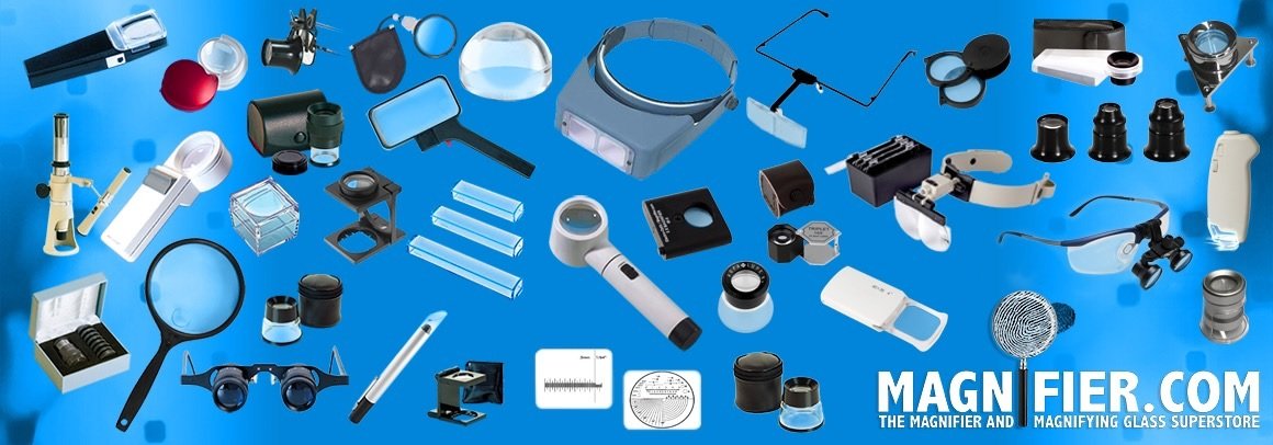
Magnifier.com Welcome to the Magnifier & Magnifying Glass Superstore! Shop one of the most extensive selections of magnifiers, loupes, low vision aids, and magnifying glasses. Magnifiers are specialized vision enhancement tools that use lenses and magnification to help the eye see more clearly. Our wide range of hundreds of magnifier and magnifying glass variations includes dome magnifiers, headband magnifiers, reading magnifiers, low vision aids, glass lens magnifiers, linen testers, loupes, and technical measuring magnifiers. Acquiring an effective magnifier for your specific need or task can be challenging, as specialized requirements & magnification often necessitate a particular type of magnifier. Whether you need augmented magnification for a visual task, detailed inspection, vision enhancement, or general reading, our online store offers a variety of magnifiers that will enhance your vision.

N2 - Light sheet fluorescence microscopy (LSFM) uses a thin sheet of light to excite only fluorophores within the focal volume. Light sheet microscopes (LSMs) have a true optical sectioning capability and, hence, provide axial resolution, restrict photobleaching and phototoxicity to a fraction of the sample and use cameras to record tens to thousands of images per second. LSMs are used for in-depth analyses of large, optically cleared samples and long-term three-dimensional (3D) observations of live biological specimens at high spatio-temporal resolution. The independently operated illumination and detection trains and the canonical implementations, selective/single plane illumination microscope (SPIM) and digital scanned laser microscope (DSLM), are the basis for many LSM designs. In this Primer, we discuss various applications of LSFM for imaging multicellular specimens, developing vertebrate and invertebrate embryos, brain and heart function, 3D cell culture models, single cells, tissue sections, plants, organismic interaction and entire cleared brains. Further, we describe the combination of LSFM with other imaging approaches to allow for super-resolution or increased penetration depth and the use of sophisticated spatio-temporal manipulations to allow for observations along multiple directions. Finally, we anticipate developments of the field in the near future.
JavaScript seems to be disabled in your browser. For the best experience on our site, be sure to turn on Javascript in your browser.
Light sheet fluorescence microscopy (LSFM) uses a thin sheet of light to excite only fluorophores within the focal volume. Light sheet microscopes (LSMs) have a true optical sectioning capability and, hence, provide axial resolution, restrict photobleaching and phototoxicity to a fraction of the sample and use cameras to record tens to thousands of images per second. LSMs are used for in-depth analyses of large, optically cleared samples and long-term three-dimensional (3D) observations of live biological specimens at high spatio-temporal resolution. The independently operated illumination and detection trains and the canonical implementations, selective/single plane illumination microscope (SPIM) and digital scanned laser microscope (DSLM), are the basis for many LSM designs. In this Primer, we discuss various applications of LSFM for imaging multicellular specimens, developing vertebrate and invertebrate embryos, brain and heart function, 3D cell culture models, single cells, tissue sections, plants, organismic interaction and entire cleared brains. Further, we describe the combination of LSFM with other imaging approaches to allow for super-resolution or increased penetration depth and the use of sophisticated spatio-temporal manipulations to allow for observations along multiple directions. Finally, we anticipate developments of the field in the near future.


 Ms.Cici
 8618319014500