
LED Ring lights - ring light
Microscope illumination techniquespdf
In this video, we explore the theory, use, and parameters with infrared (IR) light therapy, a sub-type of thermotherapy with a different ...
Criticalillumination
Objects must reflect light to be seen. Something opaque does not allow light to pass through. It only absorbs and reflects light. If something is translucent, some light passes through. The frosted glass reflects some sunlight and also lets some light pass through. As a result, light is scattered in different directions. A transparent item transmits almost all light, absorbing and reflecting only a little bit. For example, a glass of water is transparent because most light passes through it.
A prism separates light into colors because of refraction. The amount of bending depends on the wavelength of the light. Colors always appear in the same order: red, orange, yellow, green, blue, indigo, and violet. Light refracted through air layers of different densities can result in mirages.
When using a microscope with brightfield, some samples will have a natural contrast that is easily viewed, such as bright plants and flowers, metals, and pigments. Some samples can be stained to increase the microscope contrast. However, low contrast samples such as unstained bacteria, thin tissue slices, live cells, or reflective metal parts require the use of additional microscope contrast techniques in order to view the sample. Here we will explore each of these different microscopy contrasting techniques.
To measure the amount of light falling on human skin, you need to mimic ... were measuring a laser or focused beam of light, and underfilling a detector.
Function of light switch inmicroscope
Refraction of light occurs when waves of light pass from one medium to another, and the light wave is bent or refracted. Light travels slower through water than air. When light hits the water at an angle, the slowing down makes it bend as it changes its direction. The index of refraction indicates how much a material reduces the speed of light; the more the light is slowed, the greater the index of refraction.
Hoffman modulation contrast is a form of oblique transmitted light illumination in combination with a grayscale modulator comprising neutral gray absorbing layers mounted in the objective's back focal plane. The modulator consists of three grayscale strips that run vertically through the pupil of the objective. This results in this method's middle gray image background and modulates a relief contrast. iHMC is used in the live-cell microscopy of sperm and egg cells when conducting artificial insemination in human and veterinary medicine.
Focusing inmicroscope
20241030 — The Elgato Key Light Mini is one of the best ring lights because of its internal. (Image credit: Tom's Guide/Henry T. Casey). 3 ...
The incidence plane is defined by the incident, refracted, and reflected waves. The refracted ray is oriented at a 90-degree angle from the reflected ray ...
The Lighted Pop-Up Magnifier is our best-selling portable magnifier. It has a 2" x 2" retractable lens housed in a hard plastic case, perfect for on-the-go ...
Köhlerillumination
daylight24 cordless magnifying glass desk lamp - Tabletop magnifying glass lamp for full-page magnification ... This lamp is great as a hobby light, or a craft ...
In interference contrast, previously split partial beams are combined as in phase contrast, interfering with each other as a result. Due to the interference of the two twin images in the intermediate image plane, a light-dark contrast is generated at the edges of objects. By laterally shifting the objective-sided DIC prism, also known as the DIC slider, the contrast can be adjusted in such a way that the light and dark object edges appear on a gray background. As the illumination appears darker on one side and brighter on the other side, a pseudo-relief image is perceived by our brain. This can - but does not have to - match the actual topography of the sample.
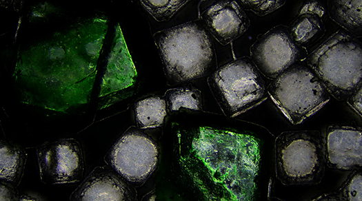
Polarizing microscopes must be equipped with two crossed polarizers. In most cases at least one polarizer and one analyzer are used. Many materials, like most crystals and some biological structures such as muscle cells, are birefringent, meaning they have two refractive indices. This phenomenon fulfills an important diagnostic function in mineralogy, pharmacology, forensic microscopy, polymer research, or the quality control of textile fibers. Reflected light is used to visualize the contrasts in the origin of structure of opaque metals such as aluminum, zirconium, and others.
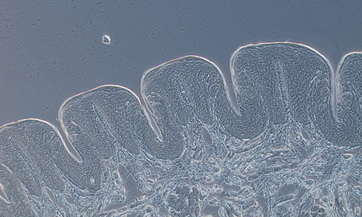
Oblique illumination is recommended to contrast objects that are too thick for alternative contrasting techniques. Oblique illumination directs light beams onto the specimen at different angles. As a result of highly directed one-sided illumination, the sample will have one side that appears bright and the other darker, which results in a shadow-casting relief image showing fine structural details. Oblique illumination microscopy is utilized to view thick tissue samples or solid samples that do not allow light to pass through them, such as automotive parts.
Shop Target for flashlight fluorescent light you will love at great low prices. Choose from Same Day Delivery, Drive Up or Order Pickup plus free shipping ...
What isilluminationinMicroscope
Backlighting is what it sounds like: lighting that illuminates objects from behind. When you light from behind– a mirror, bed, artwork or TV—the object looks ...
Microscopebase function
The absorption of light is an essential concept in understanding how light interacts with various materials. When light encounters a surface, several things can ...
202287 — The optimal source would be something that emits black body radiation (so no LEDs, only "old school" light bulbs)
Light is needed to see things around us, and how well we see them depends on the amount of light and the color of an object. Unfortunately, most objects don’t produce their own light, so almost everything we see is reflecting light. The reflected light travels to the retina of our eyes, where an image is received and sent to the brain.
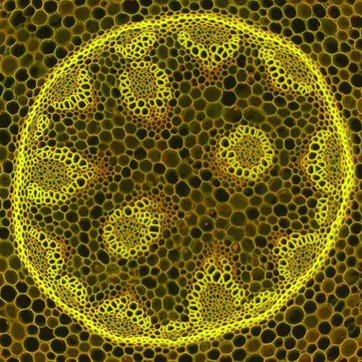
Fluorescence is a low-energy form of radiation (emission) that results from a previous high energy illumination (excitation). As soon as the excitation stops, the fluorescence emission stops almost immediately. The particular advantages of fluorescence microscopy include a strong image contrast and its specificity; that is, its ability to specifically detect individual structures down to individual molecules. Fluorescence microscope illuminators often use gas discharge lamps such as HBO, XBO, HXP or long-life LED light sources. LEDs make it possible to change the excitation wavelengths extremely quickly. The downside to LED light sources is that they still exhibit rather low excitation intensity in certain spectral ranges.
Magnification system ofmicroscope
When using darkfield microscopy, no light from the illuminator will pass into the imaging system, only light diffracted by the structure is captured by the objective. This is what makes the structures appear rich in contrast, and very bright against a dark background. Darkfield microscopy is especially useful to detect tiny and isolated structural details, such as bacteria. When working with reflected light, darkfield illumination is used to identify grain boundaries in polished and etched metal sections, as well as to detect contaminants and flaws in surfaces.
When you look in a mirror, you see your reflection. This happens because light reflects off of you, hits the mirror, and then reflects off the mirror to your eye. Reflection occurs when a light wave strikes an object and bounces off it. When light is reflected from a flat mirror, the incoming ray of light is called the incidence ray. The law of reflection states that the angle of incidence (i), or where light strikes a surface, is equal to the angle of reflection (r). Regular reflection occurs when light waves are reflected on a smooth surface, such as a shiny car. Diffuse reflection occurs on a rough surface, such as a brick wall.
If you have any questions about microscope contrasting techniques or which microscope provides the best contrast for your application, please contact Microscope World.
When using a brightfield microscope, thin and transparent samples have a very low contrast and can be barely visible. Phase contrast microscopy is the method of choice for viewing thin, unstained samples such as culture cells. Phase contrast uses a special condenser and objective lenses to enhance the contrast seen in the microscope image. Phase contrast is not useful for thick specimens such as plant or animal tissue sections.


 Ms.Cici
 8618319014500