
Why Is My Ring Doorbell Flashing Blue? Simple & Easy Fix - ring light blue
Fluorescence microscope
The patented Wendel LED Optical Contour Framing Projectors were designed by framing projector specialists with over 100 combined years of projector manufacturing and projector installation experience. Wendel LED Framing Projectors clear the mystery and simplify the path for a successful framing projector installation that can be performed by any qualified electrical contractor.
Type of microscope
The phase contrast method for microscopy was developed in the 1930s by the Dutch physicist Frits Zernike. After 1942, it became a widely used microscopy technique. In 1953, Zernike was awarded the Nobel Prize for Physics. For more details, refer to the articles: A Brief History of Light Microscopy – From the Medieval Reading Stone to Super-Resolution & Phase Contrast
Phase contrastmicroscopy
For forensic applications concerning the evidentiary investigation of paints, pigments, textiles, fibers, and human tissues, Leica microscopes offering phase contrast are very useful solutions.
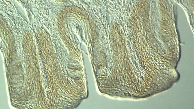
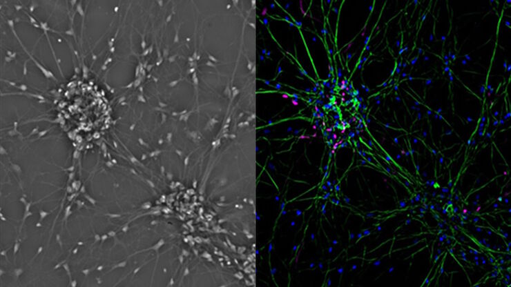
Most often biological specimens and tissues are observed with a phase contrast microscope. A large variety of biological specimens can be observed with phase contrast from fixed specimens to living cells and tissues. For examples, refer to the articles: Phase Contrast & Optical Contrast Methods
Our Lighting Modules use high efficacy 20 Watt LED Chips to drive a Compact Primary Optical System equivalent to 70 watts of halogen. The LED Framing Projector’s Objective Lens System delivers a uniform light with high lumen output. These Compact Optical LED Art Lights are the leading choice for art lighting specification.
Not all products or services are approved or offered in every market, and approved labelling and instructions may vary between countries. Please contact your local representative for further information.
Portions of the ring-shaped light are diffracted by optically dense structures of the specimen and experience a negative phase shift of about λ/4. This phase-shifted, diffracted light bypasses the λ/4 plate. In contrast, the portion of the ring-shaped light that passes directly through the specimen non-deviated will hit the phase plate which causes a positive λ/4 phase shift. As the total difference in phase shift between the light diffracted by the specimen’s structures and that which passes through phase plate will be about λ/2, destructive interference will occur. Consequently, more optically dense structures will appear darker than those that are less optically dense.
Darkfield microscopy
White High Color Rendering LED Light Chips were discriminately tested in art galleries to ensure art color enhancement. The 98 CRI High Color Rendering LED Chips include a High R9 Value to reveal true color in art for accurate color discrimination.
Lightfield microscopy
A phase contrast microscope is similar to a conventional brightfield microscope, except it uses an annular aperture in front of the light source and a quarter-wave phase plate after the objective lens. For more information, refer to the article: Phase Contrast
WENDEL® LED Framing Projectors are used for art accent lighting and they provide the art lighting designer and art lighting consultant with exposed and recessed LED Framing Projector options. They are fully adjustable framing projectors that use light masking shutters to shape light that is focused by optical lenses to frame light to the contour of art.
Differential interference contrastmicroscopy
Phase contrast is an optical contrast technique for microscopy which makes unstained structures in the cells of biological specimens visible. Cell structures that appear transparent with brightfield illumination can be viewed in high contrast and rich detail using phase contrast. Differences in optical density between structures in the cell can cause light that interacts with them to attain a phase shift. This phenomenon is the basis of phase contrast. As a result, more optically dense structures will look darker than less optically dense ones.
Wendelighting® Division of Jacksen International, Ltd 611 W. Huntington Dr., Suite A, Monrovia, CA 91016 800-528-0101 or 626-303-1142 Fax: 626-303-0046info@wendelighting.com
Confocalmicroscopy
The knowledge portal of Leica Microsystems offers scientific research and teaching material on the subjects of microscopy. The content is designed to support beginners, experienced practitioners and scientists alike in their everyday work and experiments.
Leica microscopes capable of phase contrast make a difference for the study of transparent and colorless minerals, crystals, and polymers.
Brightfield microscopy normally only provides a low-contrast image of many transparent biological specimens where few details are distinguished. One way to enhance contrast with brightfield microscopy is to use selective stains, but such stains are often toxic to living cells. A phase contrast light microscope offers a way to view the structures of many types of biological specimens in greater contrast without the need of stains. The contrast method exploits differences in optical density between structures of a specimen that lead to a phase shift of the light that interacts with the specimen and its structures.
Leica microscopes offer phase contrast for the study of cells or tissues concerning various life-science and forensic applications. Phase contrast can also be useful for certain material and earth-science applications.
A phase contrast microscope is similar to a conventional widefield microscope, except it uses an aperture in the shape of an annulus and a quarter-wave (λ/4) phase plate. The annular aperture is placed between the light source and condenser lens and the phase plate after the objective inside the microscope optics. Ring-shaped light that passes through the aperture is focused by the condenser onto the biological specimen to be observed.
Leica microscopes providing phase contrast are commonly used in life science research for the visualization, analysis, and documentation of biological structures and cellular processes.
What isbright-fieldmicroscopyused for

These LED Art Lights deliver optimum final adjustment control with light shaped to follow the contour of art. Illuminating art with a shaped beam of light, shaped to follow the contour of art, provides Fine LED Art Lighting focused on a subject for maximum visual impact. LED light is framed to follow the contour of paintings by adjusting shutters. Shutters are independently locked into position for the shaped light to continually conform to the contour of art. An art lighting specialist is not required for installation of a Wendel Shutter Framing Projector.
A Light Mask Kit is available for shaping light to any form, to follow non-rectilinear shaped art. The light mask kit requires field cutting a thin brass shim to any desired shape. A field cut light mask is easy when used in combination with our patent pending FADE IN® System. Fade In eliminates the need for an exact cut light mask and blends the shaped light border with a feathered low light perimeter that gradually increases in light intensity to a bright LED Art Illumination.
Wendel LED Framing Projectors include variable optics for illuminating art of any size or distance. Light beam spreads range from 12° to 90°. A 12° narrow beam objective lens focus tube is available for an incredibly tight LED spot of light for illuminating small art from long distances. The 90° objective lens focus tube is offered to illuminate large art or art positioned close to a ceiling.


 Ms.Cici
 8618319014500