
Wall lights - Wandleuchte DOT mint online kaufen - dot lights
Light microscopy does much what the name implies: visible light and magnifying lenses are used to view small objects. Light microscopes are the oldest form of higher quality imaging devices, dating back to the 1500s, and were the microscopes with which first cells were observed.
Govee TVBacklight
Your questions, but not your email details will be shared with OpenAI and retained for 30 days in accordance with their privacy principles.
In hospitals, quick examination of cells can be critical for doctors. In such situations, light microscopy can be used with tissues that have been frozen in carbon dioxide and sectioned using a microtome. This simpler method can be used urgently on patients who are in the operating room to guide the surgeon.
Registered members can chat with Azthena, request quotations, download pdf's, brochures and subscribe to our related newsletter content.
GoveeBacklight
News-Medical.Net provides this medical information service in accordance with these terms and conditions. Please note that medical information found on this website is designed to support, not to replace the relationship between patient and physician/doctor and the medical advice they may provide.
Ryding, Sara. 2023. Fluorescence Microscopy vs. Light Microscopy. News-Medical, viewed 19 December 2024, https://www.news-medical.net/life-sciences/Fluorescence-Microscopy-vs-Light-Microscopy.aspx.
LEDBacklightfor TV that changes with picture
Ryding, Sara. "Fluorescence Microscopy vs. Light Microscopy". News-Medical. https://www.news-medical.net/life-sciences/Fluorescence-Microscopy-vs-Light-Microscopy.aspx. (accessed December 19, 2024).
Ryding, Sara. (2023, July 21). Fluorescence Microscopy vs. Light Microscopy. News-Medical. Retrieved on December 19, 2024 from https://www.news-medical.net/life-sciences/Fluorescence-Microscopy-vs-Light-Microscopy.aspx.
Sara is a passionate life sciences writer who specializes in zoology and ornithology. She is currently completing a Ph.D. at Deakin University in Australia which focuses on how the beaks of birds change with global warming.
It takes a lot of costs to manufacture and transport Edge-lit LED panels. The technology within an edge-lit LED panel makes it expensive.
Backlit lighting photography
Ryding, Sara. "Fluorescence Microscopy vs. Light Microscopy". News-Medical. 19 December 2024. .
Furthermore, there are light microscopy techniques that can image both live and fixed samples, but there can be a tradeoff between signal-to-noise ratio and sample damage during the observation process. During fluorescence microscopy, cells undergo bleaching, in which the fluorescence diminishes during extended periods of observation. To conclude, there is flexibility in both microscopy groups.
PhilipsbacklightTV
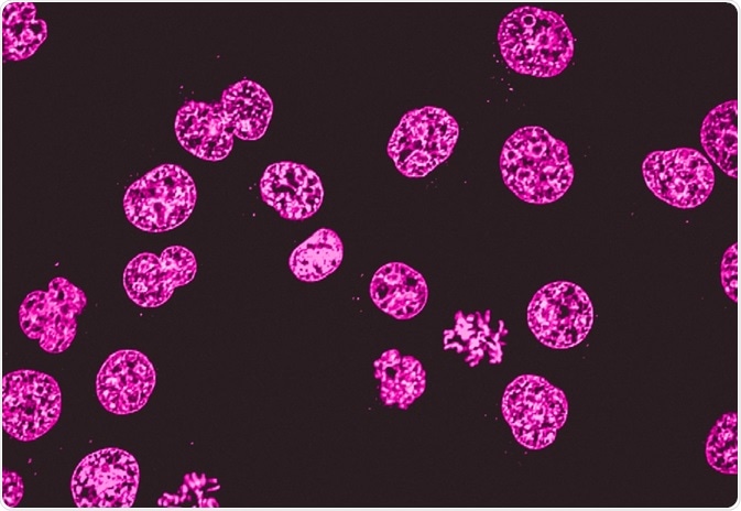
For example, a commonly used label is green fluorescent protein (GFP), which is excited with blue light and emits green light with a longer wavelength. Filters around the sample can separate the fluorescent light from the surrounding radiation.
LCDs need to be supported by additional lights because they don’t produce their light. To create this light, you can either use edge-lit LEDs or backlit LEDs. The primary difference between edge-lit and backlit LEDs is in the lighting is arranged. The light sources of Edge-lit displays are placed on a diffuser (usually thin) behind the LCD to create light dispersion.
Fluorescence microscopy is often applied in imaging cell structures or structural features, checking the viability of cells, imaging genetic material (both DNA and RNA), and imaging particular cells in a larger population.
The usefulness of traditional light microscopy is hampered by the fact that it uses visible light, as using visible light limits the resolution obtained from samples. On the other hand, fluorescence microscopy is not faced with this limitation, since it uses whatever light excites the fluorophores.
Backlit, as the name implies, is placed at the back of the frame instead of at the Edge. This position causes the LED to shine directly out, straight through the diffuser. In backlit LED, there is no light guard. As a result of this, the light shines directly outward and does not need to be directed. The straight direct light does not cause a glare because the backlit LED also has a diffuser which smoothens the light. The risk of yellowing of light guard is absent because there is no need for one. The directness of the LED light makes backlit more efficient than edge-lit. Unlike in edge-lit, where the light travels through the diffuser panel, light only passes through the thickness of the material in backlit.
Edge-lit could turn yellow due to the thinness of the panel, which allows for heating of the LED models. It makes edge-lit have a shorter life span.
Fluorescence microscopy can be used in conjunction with other types of light microscopy. Due to the fact that it creates images from the reflected light (rather than the direct light), it can be used with techniques such as phase contract microscopy.
As light microscopy developed, more forms using different techniques were invented. One of the types of microscopy within the broader light microscopy group is fluorescence microscopy. Fluorescence microscopy images cells or molecules that have been tagged with a fluorescent dye. The fluorescent substances are called fluorophores, which are integrated into the sample.
The significant advantage of using an Edge-lit flat panel lights LED display is the reduced thickness. As a result of this, the displays are placed on Edge and not directly behind the LCD. It leads to the thinness of the diffuser. Most thin diffusers are usually edge-lit and not backlit, and they transfer the majority of the components to a big stand.
Ledlight backlight
As mentioned, light microscopes that are used for light microscopy employ visible light to view the samples. This light is in the 400-700 nm range, whereas fluorescence microscopy uses light with much higher intensity.
TVBacklight
Switching from fluorescent troffer lights to LED troffer lights has its benefits, but the process could prove to be daunting and not so easy. You could either use Edge-lit panels or Backlit panels. Here are the significant differences between these panels.
Backlightfor TV repair
The fluorophores are excited by the light in the microscope, which causes them to give off light with lower energy and of longer wavelength. It is this light that produces the magnified view, rather than the original light source. This means that fluorescent microscopy uses reflected rather than transmitted light.
Both fluorescence microscopy and light microscopy represent specific imaging techniques to visualize cells or cellular components, albeit with somewhat different capabilities and uses. At its core, fluorescence microscopy is a form of light microscopy that uses many extra features to improve its capabilities.
The disadvantage of Backlit is the thickness of its diffuser. You cannot install it just anywhere. It has to be installed in a drop grid ceiling. Although its panel is thicker than Edge-lit, it is still easy to operate due to its lightweight around its surface.
A common method to visualize cells or tissue with light microscopes is to use dyes. Widely used ones might paint the main components, such as the dye combination of hematoxylin and eosin, which colors the nuclei violet and the cytoplasm pink. However, there are also more specialized dye techniques.
NewsMedical spoke with Hamilton Company about its syringe technology, custom syringe solutions, and syringe accessories for multi-industry use.
While we only use edited and approved content for Azthena answers, it may on occasions provide incorrect responses. Please confirm any data provided with the related suppliers or authors. We do not provide medical advice, if you search for medical information you must always consult a medical professional before acting on any information provided.
In this interview, Wirulda Pootakham, the Director of Thailand's National Omic Centre, talks to NewsMedical about genomic conservation in Thailand's mangroves.
Traditional light microscopes are widely used, and often require simpler dyes to visualize contrast which is not naturally visible. This is typically a simpler technique than fluorescence microscopy. Because of this, it is used in clinical settings, such as for immediate imaging of biopsied samples in hospitals and for cervical smears.
Concerning the location of the LEDs, Edge-lit light panels are placed on the Edge of the frame. The light that emits is guarded by a light guard panel that directs it sideways. The light board is designed for the smooth and evenly distribution of light in a given space. The sleek look of the diffusers is a result of the small depth of the panel. The installation of Edge-lit can be done in a standard grid ceiling. And the sleekness and thinness of the board make it possible to install Edge-lit LEDs on walls and ceilings using suspension box or cable installation kits.


 Ms.Cici
 8618319014500