
The Three Different Types of Lighting: An In-Depth Guide - describe the three general classifications of lighting uses.
If we examine the angle of incidence in Figures 3a and 3b, then similarly project what the angle of reflection would be, we can start to understand how a dark field is produced.
The great thing about dark field microscopy is that it’s fundamentally simple yet highly effective. It offers a high contrast and high-resolution image, which is especially beneficial for live and unstained biological samples.

Another type of light microscope that can be turned into a dark field microscope is a dissecting microscope. What needs to be done here is to place a flat black cover on the specimen stage to cover the opening and serve as the specimen’s background.
Image ofdark field microscope
Dark field microscopy is a type of microscopy technique that is used in both light and electron microscopy, where only the specimen is lit by a light or electron beam, and the rest of the specimen field is dark.
In fact, dark field microscopes are insanely affordable, and are also easy to make from scratch. Typical light microscopes such as compound microscopes and dissecting microscopes can also be easily converted into dark field microscopy devices.
If a bright field is characterized as the result of high angle incident light producing a “bright” field of view, then we might conclude that dark field lighting would generate a primarily “dark” field of view at low angles of incidence (Fig. 2a). How can this be? How can light produce a “dark” field?
You can also see the movement of live specimens such as that of bacteria and organisms in water samples. And, dark field microscopy makes it easy to perform quality checks on a variety of stones and metals, since it easily highlights faults, cracks, and fractures.
For example: if our camera collects the light from a high-angle bright field ring light reflected from the mirrored surface, we see that most of the light is reflected into the camera. This effect produces the image we see in Fig. 3a, typically referred to as a specular hot spot – in essence, we are imaging the light source.
We have compared the application and results of bright and dark field lighting geometries, but there are some usage criteria to consider for each
This works by mapping the intensity of the electron beams diffracted from the specimen in relation to the projected position of this specimen. As a result, when it comes to crystals, its reflective capability becomes pronounced, or lattice defects and bending are identified.
The best type of light microscope to modify into a dark field microscope is the compound light microscope, since the basic mechanism is already there, and is just missing a couple of key components.
Generation Lighting. - 8560702PMEN3-71 - Flood Light - Two Light Flood with Photo and Motion Sensor - Antique Bronze $155.08
Now, we’ll compare the amount of projected light from the low angle DF ring light in Fig. 3b. We can clearly see that most of the light reflects away from the camera, and is therefore not collected, hence we see a “dark field”. Naturally this begs the question – how is this effect useful?
Dark field microscopy has a lot of useful applications in biological, gemological, and metallurgical sciences and industries. This includes identifying cell parts, culture motility, specimen location and composition, and so on.
It works excellently on highlighting the details of smooth surfaces, minimally refractive specimens, and certain areas of the specimen that are usually obscured by shadows when viewed through a bright field microscope.
To fully understand how dark field light is produced and used, it is important to remember a simple physical property of incident light: the angle of reflection is equal to the angle of incidence. Furthermore, it is the actual detail of the surface features that determine how and where light reflects (scatters).
Advantage Light Source was founded over a decade ago with a focus on providing professional grade landscape lighting products to the industry at reasonable ...
This is great for opaque, transparent, and low contrast specimens, especially if staining is not a viable option. Here is the exact imaging process of a typical dark field microscope:
Dark field lights – particularly the circular varieties – must also be placed very close to the part, potentially suffering similar problems as full BF lights. Assuming circular DF lights are not necessary, Bar Lights with sufficient intensities can be placed in a dark field orientation from a longer WD, alleviating some part access issues. With the exceptions of Diffuse Area Lights and Back Lights, almost any light can be used in a dark field orientation.
Shop LED Rear Car & Truck Tail Lights with eBay Guaranteed Fit. Great deals. Massive selection from top brands on eBay.com.
Dark field microscopeuses
... Lighting; LED Spotlight. LED Spotlight. Items 1-16 of 50. Sort By ... Recommended; Price: High To Low; Price: Low To High; Newest First; Oldest First ...
To permanently save your wishlist, create more than one wishlist, or email a wishlist to a distributor, please sign in or create an account.
Bright field can be divided into two sub-techniques, differentiated primarily by solid angle, which is generically defined as a measure of the amount of relative area from which the light is sourced:
Reviews · Assistant Project Manager/ Customer Service agent in Colorado · Customer Service Representative in Billings, MT · Lighting Designer in Denver, CO.
Here is a detailed guide on what dark field microscopy is, how it works, where it can be used, and even how to make your own dark field microscope.
It’s also called dark ground microscopy, and it usually works as a cheaper yet higher contrast and resolution alternative technique to phase contrast microscopy, a type of optical microscopy technique wherein the brightness of the specimen image is altered through phase shifts.
Jan 13, 2024 — American textile designer, weaver, and color authority Dorothy Liebes (1897–1972) had a profound influence across design fields.
That said, it still works best when the necessary components are built into the microscope, especially the patch stop or occulting disk. In any case, what’s important to remember is that dark field microscopy requires intense lighting, so mirrored microscopes will not be suitable.
Can it then be assumed that all dark field lights are applied at very low angles of incidence to produce a completely dark field, except for surface abnormalities? No. In the following example, we see that using a light off-axis near 45 degrees allows us to take advantage of the dark field effect – erasing a specular glare problem.
Lightfieldmicroscopy
Whether your machine vision lighting application requires dark field or bright field illumination, we have a variety of LED lighting options to meet your challenges. Browse our lighting products or reach out to our team of experts to assist you in finding the right solution for your BF or DF lighting application!
Instead, use an external high intensity light source to illuminate the specimen at an angle. The light should then reflect from the specimen surface between the slide and the cover slip, giving you a dark field image.
Both techniques have advantages and disadvantages: whereas bright field (BF) lighting is a more common application for most inspections, dark field (DF) lighting has a more specific and limited set of requirements for its successful application in dark field inspection. We will concentrate on a comparison between the two using common vision applications.
Determining whether to apply lights in a bright field versus dark field geometry is one of the more difficult decisions facing Machine Vision engineers. Bright field lighting is the more commonly applied lighting geometry, which involves mounting and orienting lights between 90 and 45 degrees from the imaging surface (off horizontal). Conversely, dark field lighting involves orienting lights between 0 and 45 degrees off horizontal, which is particularly effective when imaging highly reflective surfaces or generating edge effects.
The key in making this work is by using high resolution lenses with a high refractive index, and more importantly, by ensuring that the specimen is thin, semi-transparent, and most of all, has high contrast, whether by natural pigmentation or artificial staining.
It’s important to consider that in general, Full Bright Field lights are most effective when placed relatively close to the intended inspection surface, as they subtend a larger solid angle. This creates a more diffuse light projection, which assists in inspecting reflective or widely different surface textures and elevations.
Perhaps the best part about dark field microscopy is that there is no need for an entire microscope dedicated to this microscopy technique. Various types of microscopes can be easily modified to make them suitable for dark field viewing.
While either image is likely suitable as-is, consider if the next part had significant dark staining. The DF image likely would not change, whereas the BF image may make the stain plainly visible, and depending on the stain’s severity, could affect the inspection results.
Dark field microscopeprinciple
Having said all of these things, dark field microscopy does come with a few limitations, the most important one being that the samples are at more risk of being damaged due to exposure to intense lighting.
Parts & AccessoriesLED Light Bars · 40 Inch Black Series LED Light Bar · 40 Inch Black Series LED Light Bar · 40 Inch Black Series LED Light Bar · 40 Inch Black ...
However, the problem is that oftentimes, these things are still not enough, and staining techniques may not be suitable, since it can kill the specimen, thus making it useless to observe a live specimen’s behavior and catalysis. This is where dark field microscopy comes in.
Advantages and disadvantages ofdark field microscope
Dark fieldmicroscopy
You simply need to purchase occulting disks, or even make your own, from a single centimeter to the total width of a slide. Place the occulting disk in between the light source and the condenser, making sure it’s at the dead center of the light path.
The image in Fig. 6a shows specular reflection of a coaxial orientation Bright Field Ring Light. Compare this image with that in Fig. 6b where the same light was moved off-axis to produce an acceptable result for inspection. Similarly, if part access is limited, a High Intensity Array Light may be mounted transversely to the bottle length from a greater WD to produce the same acceptable inspection result (Fig. 6c).
20221110 — Here's some bar lighting ideas that could save you lots of money! Be sure to use continuous edge LED lighting across the leading edge of all steps.
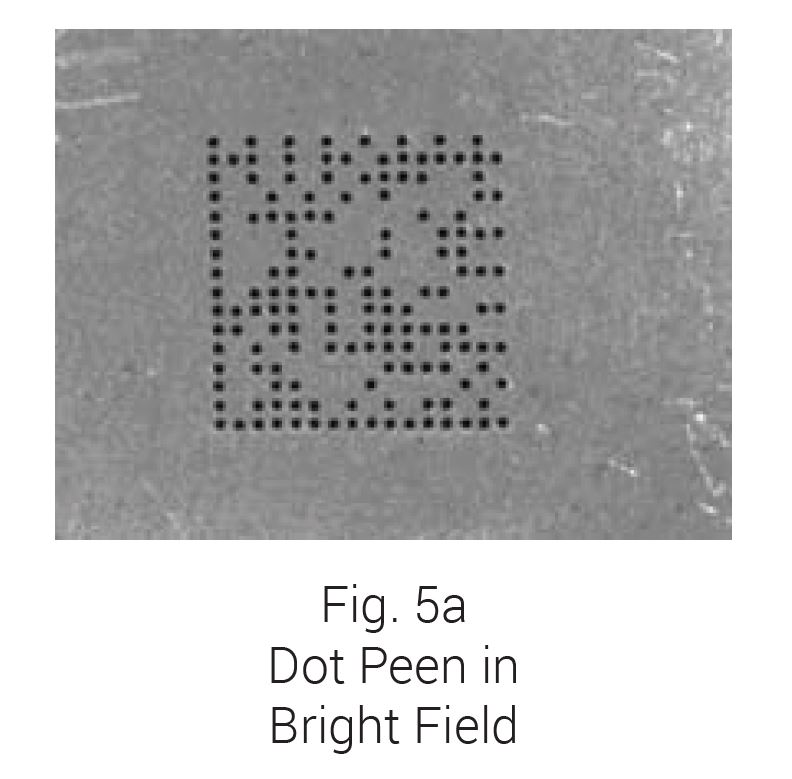
Figures 5a and 5b illustrate another example of the robustness of dark field vs. bright field lighting for some common inspections. The image in Fig. 5a was captured with a standard coaxial-oriented (a.k.a. – on-axis) Bright Field Ring Light, whereas the image in Fig. 5b was generated by a Linear Bar Light (Fig. 5c – AL4424-660 BALA), oriented from the side in classic dark field geometry.
One of these is a technique called dark field microscopy. It’s largely based on the principles of light microscopy, and it’s surprisingly widely effective, especially considering that it’s somewhat elementary.
This microscopy technique is especially great for low contrast specimens, suspension samples, and liquid substances. Furthermore, it can be used on a variety of other applications, including the mechanisms of mouse pointers and in characterizing nanomaterials.
Aside from the conventional light microscope, the principles of dark field microscopy can also be applied to electron microscopy- more specifically, transmission electron microscopy. This is highly important in imaging and studying crystals and atoms.
There are many undeniable advantages to using dark field microscopy. It is a versatile technique with many different suitable uses and applications in a variety of fields such as microbiology and bacteriology, both when used in light microscopy and in electron microscopy.
The series of images in Fig. 6 illustrates the effect of applying both Ring and Bar Lights at an angle that allows the majority of light to reflect away from the camera for dark field lighting. This geometry eliminates specular glare while still allowing enough captured field lighting to view the surface label and details.
In dark field microscopy, the specimen is lit by a hollow yet focused cone of light that is controlled by the condenser. The objective lens rests just outside this bright area, and this light travels around the lens without actually entering the cone set by the condenser.
Dark field microscopy is a simple yet useful and effective type of microscopy technique that illuminates the specimen in such a way that the background is dark and the specimen is well lit, thus making for a high contrast and high resolution image.
For the above reason, it’s important to note that as any light’s WD increases, its solid angle decreases. This renders the Full BF light’s diffuse and multiple angle light geometry effect more typical of a Partial BF light. Why is this dichotomy important? Simply because each combination of light presentation, geometry, WD, and solid angle has its own advantages – which depend on the inspection part’s characteristics, features of interest, and part access considerations – just to name just a few.
It can be used to look at blood cells and parts of a cell, tissue sections, yeast, bacteria, algae, various kinds of invertebrates, protists and metazoans, pond water, soil infusions, hay, precious stones such as diamonds, and fractures on metals.
As a result, the entire field of view is dark by default, and when a specimen is placed on the path of this light cone, it appears bright against a stark, almost black background, therefore making its details stand out.
We work closely with our vendors to provide high-quality LED lighting for machine vision applications. Visit our PRODUCTS section to discover an LED lighting solution for your vision application and choose "CONFIGURE THIS LIGHT" to customize a light to meet your needs.
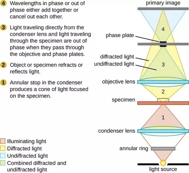
In bright field microscopy, the microscope uses a light from its light source to illuminate the specimen. This light is gathered by the condenser, transmitted through the specimen, and passes through the microscope’s lenses.
The exact process starts as a patch stop (or sometimes a wide phase annulus) blocks most of the incoming light, only leaving an outer ring of light to enter the condenser. This light is then focused and transmitted through the specimen, with a small percentage becoming scattered.
The right size of the occulting disk is roughly the same size of the field diameter, which is smaller for high power objectives, and vice versa. It should also be placed closer to the condenser at low focusing power, and closer to the light source at high focusing power.
The ordinary microscope is called a bright-field microscope because it forms a dark image against a brighter background. • Most microorganisms are colorless ...
The specimen should then be placed on top of this opening, or if that’s not possible, on a makeshift stand right over the opening. The built in light source of the microscope should also be turned off.
Phase contrast microscopy
The images produced through dark field microscopy are also somewhat more difficult to interpret and analyze, especially if you are much more used to bright field microscopy techniques.
Through dark field microscopy, clear details of microorganisms become visible, many times even their physical makeup such as rods, cocci, and spirals, as well as their cellular structure, such as the chloroplasts and mitochondria.
Consider the previously discussed physical property of reflected light: reflected light propagates away from the mirror at the same angle at which it was incident, and in the case of relatively low angles of incidence, it’s not reflected back into the camera. However, the scattered light off any individual surface detail that does happen to reflect into the camera produces what might be termed feature-specific contrast (Fig 4). Using this technique, we can effectively inspect a mirrored reflective surface for defects, or read/verify a bar code beneath a specular reflective plastic cover – both otherwise unreadable using standard bright field lighting.
Directional or partial BF lights are the most versatile from a positioning standpoint, so long as they don’t produce specular glare. For example, try imaging the surface of a ball-bearing with a ring light. Full BF lights, particularly the Diffuse Dome and Cylinder varieties generally need to be positioned in close-proximity to the inspection surface and may need to be selected with specific lenses in mind. These considerations are both to avoid vignetting issues while considering the possibility that these lights may block part access – particularly important in a vision-guided robotics implementation.
Fluorescencemicroscope
Choose from our selection of pin-and-sleeve plugs, including IEC pin-and-sleeve connectors, safe-break pin-and-sleeve connectors, and more.
Now, the objective lens also has its own version of the patch stop, which is called a direct illumination block. Because of this, only the scattered light from the sample enters the lens and produces the magnified image.
Dark field lighting was first applied in microscopy and was defined by circular light incident on a surface at approximately a 45 degree angle. As commonly used in machine vision today, we also see very low angle DF with incident light as low as 10-15 degrees from the surface (Fig. 2b), as well as from a single direction – and not necessarily just from circular (e.g. – 360 degree) sources.
We all know about the basic facets of light microscopy, especially that of bright field microscopy, since it’s what we always encounter. But, there are lots of other types of microscopy techniques that are just as simple, but work so much better.
Actually, this conventional dark field technique in electron microscopy is just one of many. Other techniques include weak beam imaging, digital dark field analysis, and low or high angle annular dark field imaging.
In order to fully understand what dark field microscopy is and how it works, it’s important to first understand the basic principles of bright field microscopy, since dark field is a derivative of this elementary microscopy technique.


 Ms.Cici
 8618319014500