
The Science Behind Lighting a Baseball Field Lumens & ... - behind lighting
When coupled to hyperspectral imaging, dark-field microscopy becomes a powerful tool for the characterization of nanomaterials embedded in cells. In a recent publication, Patskovsky et al. used this technique to study the attachment of gold nanoparticles (AuNPs) targeting CD44+ cancer cells.[4]
The official opening night for both works is Thursday, August 4. Created for thrill-seekers and novices alike, "FLIGHT" and "SEANCE" are presented in complete darkness, inside customized 40-foot shipping containers, and have played to sold-out houses in the UK- including Edinburgh's Festival Fringe, the world's largest arts festival - Australia, Mexico, and South Korea.
In optical microscopes a darkfield condenser lens must be used, which directs a cone of light away from the objective lens. To maximize the scattered light-gathering power of the objective lens, oil immersion is used and the numerical aperture (NA) of the objective lens must be less than 1.0. Objective lenses with a higher NA can be used but only if they have an adjustable diaphragm, which reduces the NA. Often these objective lenses have a NA that is variable from 0.7 to 1.25.[1]
While the dark-field image may first appear to be a negative of the bright-field image, different effects are visible in each. In bright-field microscopy, features are visible where either a shadow is cast on the surface by the incident light or a part of the surface is less reflective, possibly by the presence of pits or scratches. Raised features that are too smooth to cast shadows will not appear in bright-field images, but the light that reflects off the sides of the feature will be visible in the dark-field images.
Face to Face will present the World Premiere of DUALITY, written and directed by Anthony M. Laura at A.R.T/New York's Jeffrey and Paula Gural Theatre. See photos of the production.
Dark-field microscopy (also called dark-ground microscopy) describes microscopy methods, in both light and electron microscopy, which exclude the unscattered beam from the image. Consequently, the field around the specimen (i.e., where there is no specimen to scatter the beam) is generally dark.
Briefly, imaging[5] involves tilting the incident illumination until a diffracted, rather than the incident, beam passes through a small objective aperture in the objective lens back focal plane. Dark-field images, under these conditions, allow one to map the diffracted intensity coming from a single collection of diffracting planes as a function of projected position on the specimen and as a function of specimen tilt.

This past weekend, the Doris Dear 10th Anniversary Christmas Special dazzled packed, sold-out houses at The Triad Theater, cementing its place as a beloved holiday tradition. Check out photos from the show.
In optical microscopy, dark-field describes an illumination technique used to enhance the contrast in unstained samples. It works by illuminating the sample with light that will not be collected by the objective lens and thus will not form part of the image. This produces the classic appearance of a dark, almost black, background with bright objects on it. Optical dark fields usually done with an condenser that features a central light-stop in front of the light source to prevent direct illumination of the focal plane, and at higher numerical apertures may require oil or water between the condenser and the specimen slide to provide an optimal refractive index.[2][3]
Dark-field microscopy is a very simple yet effective technique and well suited for uses involving live and unstained biological samples, such as a smear from a tissue culture or individual, water-borne, single-celled organisms. Considering the simplicity of the setup, the quality of images obtained from this technique is impressive.
AD/BK is not only a new arts home rooted in Brooklyn, NY, but AD/BK allows audiences to cross the analog/digital divide with collective experiences - unmediated by goggles or glasses - with original programming and curated content focused on immersive media across art, technology, science, music, film, live entertainment and more. The AD/BK 25,000 square-foot footprint has an open-air patio space. A café and NFT gallery with works for sale from the mainstage show is also planned for phase two of the venue, later this fall.
Artistic Directors David Rosenberg and Glen Neath have been collaborating for the last decade and co-founded DARKFIELD in 2016. Rosenberg was a founding member of UK's famed Shunt, a site-specific, performance artist collective; his work is primarily as a director for the stage, including public performance projects. Neath has written novels, plays for radio, for the stage, and in non-theatre locations. Most recently, their social impact audio piece, "Intravene," on the overdose crisis in Vancouver (produced in partnership with Crackdown and Brenda Longfellow and funded by the UK-Canada Immersive Exchange programme), received its world premiere at the 2022 Tribeca Festival, part of the Tribeca Immersive main competition. Other DARKFIELD works include "COMA" and "EULOGY."
Dark-field studies in transmission electron microscopy play a powerful role in the study of crystals and crystal defects, as well as in the imaging of individual atoms.
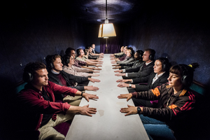
DARKFIELD, the UK-based creators of deeply intense, immersive, audio experiences that blend theatre, technology, and 360-degree sound, premieres its acclaimed shows "FLIGHT" and "SEANCE" at ArtsDistrict Brooklyn, a state-of-the-art immersive and experiential arts venue, located in Greenpoint (25 Franklin Street).
Dark-field microscopy techniques are almost entirely free of halo or relief-style artifacts typical of differential interference contrast microscopy. This comes at the expense of sensitivity to phase information.
AD/BK is located at 25 Franklin Street (Greenpoint) Brooklyn, NY 11222. Tickets range from $30-$49.50. All three shows can be enjoyed in one outing, or purchased à la carte. For the playing schedule and showtimes, visit "tickets" at www.ArtsDistrict.live.
Luna Stage's New York premiere of Jenny Lyn Bader's Mrs. Stern Wanders the Prussian State Library, directed by Ari Laura Kreith, opens tonight! The production has been extended. Learn how to purchase tickets.
Weak-beam imaging involves optics similar to conventional dark-field, but uses a diffracted beam harmonic rather than the diffracted beam itself. In this way, much higher resolution of strained regions around defects can be obtained.
Additionally, ArtsDistrict Brooklyn (a/k/a AD/BK) will feature "Limitless AI" by renowned contemporary Turkish artists Ferdi Alici and Eylul Alici of Ouchhh Studio (Istanbul) - creators of the award-winning, global sensation, "Poetic AI," seen by millions worldwide.
Bright field dark field
In single-crystal specimens, single-reflection dark-field images of a specimen tilted just off the Bragg condition allow one to "light up" only those lattice defects, like dislocations or precipitates, that bend a single set of lattice planes in their neighborhood. Analysis of intensities in such images may then be used to estimate the amount of that bending. In polycrystalline specimens, on the other hand, dark-field images serve to light up only that subset of crystals that are Bragg-reflecting at a given orientation.
The interpretation of dark-field images must be done with great care, as common dark features of bright-field microscopy images may be invisible, and vice versa. In general the dark-field image lacks the low spatial frequencies associated with the bright-field image, making the image a high-passed version of the underlying structure.
Dark field microscopy
The immersive shows are presented in complete darkness, inside customized 40-foot shipping containers, and have played to sold-out houses across the world.
The Tank will present Peregrinations, a masked show about people and borders co-produced by Dutch Kills Theater and Long Story Short. Learn how to purchase tickets.
One limitation of dark-field microscopy is the low light levels seen in the final image. This means that the sample must be very strongly illuminated, which can cause damage to the sample.
Dark-field microscopy has recently been applied in computer mouse pointing devices to allow the mouse to work on transparent glass by imaging microscopic flaws and dust on the glass's surface.
Annular dark-field imaging requires one to form images with electrons diffracted into an annular aperture centered on, but not including, the unscattered beam. For large scattering angles in a scanning transmission electron microscope, this is sometimes called Z-contrast imaging because of the enhanced scattering from high-atomic-number atoms.
This a mathematical technique intermediate between direct and reciprocal (Fourier-transform) space for exploring images with well-defined periodicities, like electron microscope lattice-fringe images. As with analog dark-field imaging in a transmission electron microscope, it allows one to "light up" those objects in the field of view where periodicities of interest reside. Unlike analog dark-field imaging it may also allow one to map the Fourier-phase of periodicities, and hence phase gradients, which provide quantitative information on vector lattice strain.


 Ms.Cici
 8618319014500