
Surgical Lights - illumination light
Dark fieldmicroscopy
I set this up on the crummiest little piece of garbage microscope I could find, and it looked very good. A relatively new student-model microscope should give a much better effect.
DIC microscopy
Dark field illumination is most readily set up at low magnifications (up to 100x), although it can be used with any dry objective lens. Any time you wish to view everything in a liquid sample, debris and all, dark field is best. Even tiny dust particles are obvious. Dark field is especially useful for finding cells in suspension. Dark field makes it easy to obtain the correct focal plane at low magnification for small, low contrast specimens. Use dark field for
You don't need sophisticated equipment to get a dark field effect, although the effect is most dramatic when the occulting disk is built into the condenser itself. You do need a higher intensity light, since you are seeing only reflected light. At low magnification (up to 100x) any decent optical instrument can be set up so that light is reflected toward the viewer rather than passing through the object directly toward the viewer.
Type ofmicroscope
A : Lens B : Light source C : Reflected light enters into the lens to cause glareD : Reflected light doesn’t reach the lens, creating shadows
Polarizingmicroscope
Suspensions of cells and samples of pond water look spectacular in dark field. While specimens may look washed out and lack detail in bright field, protists, metazoans, cell suspensions, algae, and other microscopic organisms are clearly distinguished and their details show up well. At 100x you can readily see bacteria, even distinguish some structure (rods, curved rods, spirals, or cocci) and movement. Non-motile bacteria look like vibrating bright dots against a dark background. Motile bacteria can be seen moving in a definite direction, sometimes remarkably fast. In pond water samples you may find Spirillum volutans, a very large (up to 0.5 mm) motile spiral bacterium.
To view a specimen in dark field, an opaque disc is placed underneath the condenser lens, so that only light that is scattered by objects on the slide can reach the eye (figure 2). Instead of coming up through the specimen, the light is reflected by particles on the slide. Everything is visible regardless of color, usually bright white against a dark background. Pigmented objects are often seen in "false colors," that is, the reflected light is of a color different than the color of the object. Better resolution can be obtained using dark field as opposed to bright field viewing.
Dark field optics are a low cost alternative to phase contrast optics. The contrast and resolution obtained with inexpensive dark field equipment may be superior to what you have with student grade phase contrast equipment. It is surprising that few manufacturers and vendors promote the use of dark field optics.
Phase contrast microscopy
KEYENCE supports customers from the selection process to line operations with on-site operating instructions and after-sales support.
Fluorescencemicroscope
I would start with the largest disk, sliding it around until it is directly in the center of the light path. Increasing the illumination should then produce a good dark field effect. To optimize, first try stopping down the field diaphragm to get the best contrast between background and specimen. Try to match the size of the occulting disk to the field diameter, so that the edge of the disk is just outside the field of veiw - smaller disks are appropriate for higher power objectives. Vertically, the disk should be a close to the condenser as possible, to make the contrast the greatest. On microscopes with built-in dark field equipment, the view is so impressive because the occulting disk is built into the condenser - very close and focused. After testing the set-up this way, a stand might be rigged to fit under the microscope, so the slide can be placed in position without holding it. Something that 'grabs' the condenser and supports the occulting disks would be ideal. The less the students have to mess with, the better.
To set up a dissecting microscope for "dark field" viewing, the specimen should be placed over an opening so that light reflects only from surfaces between cover slip and slide, not from a surface beneath the slide. You may need to make a stand to hold the slide. The surface beneath the opening should be a flat black. Turn off any built-in illuminator. Aim a high-intensity light source toward the specimen at an angle, from the top or side through a glass dish or jar.
With a compound microscope, dark field is obtained by placing an occulting disk in the light path between source and condenser. A cheap set of occulting disks can be prepared by cutting circular pieces of black electrical tape ranging from dime-size up to a diameter that equals the width of the slide, and sticking them to the slide in a row. The circles should be spaced well apart. A specimen is placed on the microscope stage as usual, and the illumination should be made as uniform as possible. If there is an aperture diaphragm in the condenser (contrast lever), it should be opened up wide. After focusing at low power, the slide with occulting disks is placed in the light path between source and condenser, bringing it as close to the bottom of the condenser as it will go.
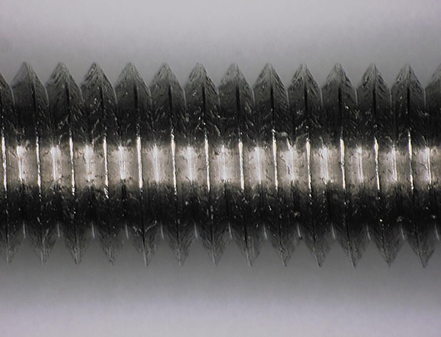
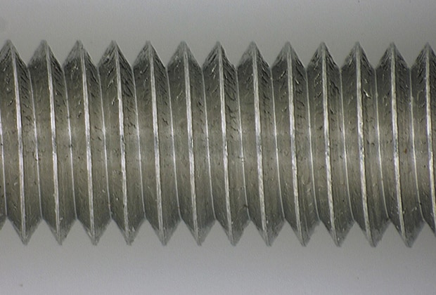
Some of the most difficult targets to properly illuminate are three-dimensional objects that are highly-reflective (e.g. medical devices, electronic components, etc).With typical lighting, images of these objects usually contain areas with extreme glare, while other parts of the target are dark.By using a diffuser attachment, light is uniformly spread over an area to provide the best possible illumination for clear observation of the target.
A dilute suspension of yeast cells makes a good practice specimen for dark field optics, particularly when cultured with living Paramecium.


 Ms.Cici
 8618319014500