
LED Truck and Trailer Lights-Sealed Beam Headlights - sealed led lights
A comprehensive refractive screening display (Belin/Ambrosio Enhanced Ectasia Display III– (BAD III)) is currently offered on the Pentacam (OCULUS GmbH, Wetzlar, Germany) that combines nine different tomographic parameters in a unified screening tool. Currently, the display uses the following parameters in a regression analysis to assist the refractive surgeon in identifying patients potentially at risk for ectatic change:
The other major benefit of newer imaging techniques is the measurement of the posterior corneal surface. Because the posterior surface contributes minimally to the overall refractive power of the eye (due to the minimal difference between the index of refraction of the cornea and aqueous) it was considered less important both diagnostically and therapeutically. Additionally, videokeratoscopes, being a reflective technology, were incapable on measuring anything below the anterior tear film. The posterior cornea, however, is an earlier indicator of ectatic change or ectasia susceptibility and when combined with full pachymetric data serves as a more sensitive screening tool then anterior topography and ultrasound pachymetry combined.[11]
VISION LEDkits
The last decade has seen a dramatic change in the diagnosis and early identification of keratoconus and other ectatic disorders. As in other subspecialty areas in ophthalmology, imaging techniques have played a large part in this change. Current corneal imaging modalities (e.g., Scheimpflug, optical coherence tomography (OCT)) offer significantly more information than were previously available with placido based anterior corneal analysis. This new tomographic information not only allows for earlier identification of disease but has altered our perception of what constitutes keratoconus, and improves the specificity to exclude false positive cases with suspicious corneal front surface curvature maps.
Corneal thickness map of a true case of pellucid marginal degeneration. The pachymetric map opened up to a full 12 mm view is the best map to differentiate true pellucid from inferior keratoconus, as true pellucid will show a clear band of corneal thinning near the inferior limbus
Several different locking types exist. Screw lock, breech-lock and bayonet-type lock are the three most important. The first two were (and some still are) used ...
Iris diaphragm definition: a composite diaphragm with a central aperture readily adjustable for size, used to regulate the amount of light admitted to a ...
tb-61220-rc
Comprehensive corneal analysis used for preoperative refractive screening includes information from the posterior cornea and full pachymetric data. This added information improves the ability of the refractive surgeon to screen patients for occult ectatic disease or to identify patients potentially at higher risk for post laser-assisted in situ keratomileusis (LASIK) ectasia. Rotating Scheimpflug cross-sectional analysis meets the criterion for a successful screening tool in that in not only provides the necessary data, but does so in a manner that does not interrupt patient flow nor require skills beyond those of most ophthalmic technicians.
Belin/Ambrosio enhanced ectasia display. The display shows abnormalities in all major parameters except those for the anterior corneal surface. Because the anterior surface is still within normal limits, the patient would have good spectacle vision in the presence of (subclinical) keratoconus
Four map composite display (sagittal curvature, anterior and posterior elevation, and corneal thickness). This cornea shows a significant positive island of elevation (ectasia) on the posterior cornea (right lower map) in spite of a normal anterior surface (upper right and left map). In this example, the posterior ectasia is significant enough to cause a displacement of the corneal thinnest point (lower left)
Composite map showing anterior curvature upper left, corneal thickness lower left, anterior elevation upper right, and posterior elevation lower right. The axial curvature map incorrectly locates the “cone” near the periphery, while both the elevation maps and pachymetric map correctly reveals this as a case of inferior keratoconus
Vision ledMarkerLights
Belin/Ambrosio enhanced ectasia display. The display depicts a case of moderately advance keratoconus where all the analyzed parameters (anterior and posterior elevation, Kmax, and pachymetric parameters) are highly abnormal
Abnormalities on the posterior cornea can be seen prior to any topographic change on the anterior surface and may be the earliest indicator of ectatic change. Patients with normal placido and ultrasound screening can be seen with highly abnormal posterior corneas often with associated abnormalities in the corneal thickness maps.[12] In the examples below [Figs. 2 and 3], the sagittal curvature map is unremarkable and the central ultrasound pachymetry was within normal limits, but the corneas exhibit significant posterior ectatic change and an abnormal corneal thickness map. These findings represent the early changes seen in keratoconus.
Secure .gov websites use HTTPS A lock ( Lock Locked padlock icon ) or https:// means you've safely connected to the .gov website. Share sensitive information only on official, secure websites.
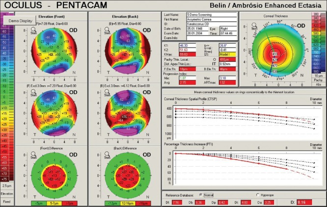
The BAD III displays each parameter and individually reports them as a standard deviation and then reports a final overall reading that is based on a regression analysis to maximize the separation of normal corneas from those with keratoconus. In the example below [Fig. 4], one can see individual abnormalities in both the corneal thickness parameters and on the posterior corneal surface, while the anterior surface parameters remain within normal limits. The combined reading based on the regression analysis of all nine parameters is highly abnormal and diagnostic of subclinical keratoconus, in spite of a normal anterior surface.
Everyday style, meets all day comfort. Cayman features no slip nose pads, dual injected rubber temple tips, and snap pin hinges for our most versatile fit ...
Ultrasonic central pachymetry only measured one point on the cornea, which typically was not the thinnest point, and did not reflect the overall thickness profile of the cornea.[5] Similarly, videokeratoscopes were limited to measuring only about 50-60% of the anterior cornea and conveyed no information about the corneal periphery and the posterior corneal surface.[6]
This is an open-access article distributed under the terms of the Creative Commons Attribution-Noncommercial-Share Alike 3.0 Unported, which permits unrestricted use, distribution, and reproduction in any medium, provided the original work is properly cited.
The sacrifice of the Custodian bought the Emperor time to deliver a finishing blow to Horus. With iron resolve, he gathered his full strength ...
Vision LEDTrailerLights
Correspondence to: Michael W Belin, Southern Arizona VA Healthcare System, University of Arizona, 4232 West Summer Ranch Place, Marana, Arizona 85658-4741, USA. E-mail: mwbelin@aol.com
Another area where Scheimpflug imaging has demonstrated significant advantages over anterior curvature analysis is in the accurate diagnosis of pellucid marginal degeneration (PMD). The vast majority of reported cases of PMD are just inferior keatoconus where the sagittal curvature map exaggerates the displacement of the cone [Fig. 6].[13] These eyes do not show the classic band of inferior thinning that is best depicted by a full coverage corneal thickness map [Fig. 7].[14] As the surgical treatment of true PMD differs from keratoconus, the accurate diagnosis has therapeutic implications.
Recess mounted, bottom opening cleanroom LED luminaire. Futuristic smart & connected lighting solution compatible with Power over Ethernet (PoE) technology.
3-Oxobutyric Acid Methyl Ester; MAA. Product Documents: SDS | Specifications | C of A & Other Certificates | Analytical Charts
Day Brite Lighting. 43 products. Showing 1 - 43 of 43 products. Day Brite Lighting High Bay LED Lights · Day Brite Clean Room Light ...
Mission and Vision · History · Who Was St. Charles Borromeo? Accreditation ... The Cardinals' Forum · Communications & Homiletics Handbook · Catholic Preaching ...
Four map composite display (sagittal curvature, anterior and posterior elevation, and corneal thickness). This cornea shows a significant positive island of elevation (ectasia) on the posterior cornea (right lower map) in spite of a normal anterior surface (upper right and left map)
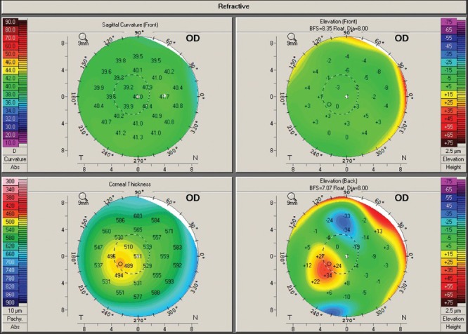
Pachymetric progression graphs. The cornea on the left shows a normal progression in thickness from the thinnest point to the periphery, while the cornea on the right, while having the same central thickness, shows a more rapid (abnormal) rate of change
In the past, the two most commonly used “screening” tools were ultrasonic central pachymetry and placido based computerized videokeratoscopy (topography). The diagnosis and grading of keratoconus were commonly based on these parameters, as with the Amsler-Krumeich classification [Table 1].[1]
Full corneal thickness maps provide a wealth of additional information that was not possible with ultrasonic pachymetry. Scheimpflug derived corneal thickness maps identify the location and magnitude of the thinnest point on the cornea. In a previously published study, differences between the apical readings and thinnest point readings can approach 100 μm in eyes that were previously viewed as “normal” by ultrasonic pachymetry and anterior, placido derived curvature analysis [Table 2].[10]
Central corneal thickness measurements, keratometric readings, and the degree of myopia were used to both diagnose and stage the severity of disease. Visual loss most closely follows changes on the anterior surface and most treatment regimens, such as contact lenses and penetrating keratoplasty, were based on the loss of best spectacle corrected vision. In other words, the Amsler-Krumeich grading system utilized easily measured parameters and the staging followed closely the treatment decision tree.
Prisms may be divided into four main types: dispersion prisms, reflection prisms, displacement prisms and rotation prisms.

Vision ledBreakaway Kit
Impact Lighting inc | The Art Of Innovation . . . Impact Lighting is an award winning lighting manufacturer.
Official websites use .gov A .gov website belongs to an official government organization in the United States.
2024525 — This article will cover how Rovers can best use the stamina system in the game, its regenerate rate, and the mistakes to avoid while consuming Waveplates.
LED is an ever improving and evolving technology. Wipro is committed to bring you the best in LED technology. Data given is representative and can change
A number of newer imaging techniques evolved (e.g., optical cross-sectioning, Scheimpflug photography, OCT) that allowed measurements of both the anterior and posterior cornea, corneal thickness maps, and also provided greater corneal coverage than possible with videokeratoscopes.[7,8,9] Of these, rotating Scheimpflug photography currently provides the most useful information for diagnosing early ectatic change. It is also a technique that is rapid and easy to perform, which would be a requisite for use as a screening tool.
Vision LEDPayson Utah
The advent of corneal refractive surgery and more recently corneal collagen crosslinking, as well as the availability of better imaging systems, revealed the limitations of both diagnosing and treating keratoconus based solely on central corneal thickness and anterior curvature analysis.[2,3,4] Refractive surgery puts physical demands on the cornea (removing tissue) necessitating the need to identify those corneas potentially at risk for ectatic change. Collagen cross-linking aims to stabilize early disease and potentially prevent visual loss, dictating that ectatic change is identified prior to significant alterations of the anterior corneal surface.
Scheimpflug cross-sectioning anterior segment imaging offers significant advantages over traditional placido based curvature analysis and ultrasound pachymetry. The accurate measurement of both the anterior and posterior corneal surfaces and the anterior and posterior lens allows for the creation of a three-dimensional reconstruction of the anterior segment. Changes on both the posterior cornea and/or corneal thickness map are earlier indicators of ectatic change than would otherwise be identifiable with only anterior curvature and ultrasonic pachymetry. Scheimpflug imaging also covers significantly more of the cornea than was possible with placido based devices. This added coverage is critical in the proper diagnosis of peripheral diseases such as pellucid marginal degeneration (PMD).
Fig. 5 depicts a case of keratoconus where all nine parameters are abnormal. The final overall reading is over 8 standard deviations from the norm, which represents moderate to advanced ectatic change. The Amsler-Krumeich grading system, however, would just barely classify this cornea into stage II.
In addition to measuring and locating the true thinnest point, a full thickness map allows one to look at the pachymetric progression or the rate of change in corneal thickness. A single thickness reading is very limited in determining what is normal, in the same way it would be nearly impossible to determine whether an 80 kg individual is normal without knowing the height of the individual. A 4’10” 80 kg individual would be obese, while a 6’4” individual would be gaunt. Similarly, two corneas can have the same central corneal thickness but share dramatically different pachymetric progressions. Abnormal corneas (i.e., corneas showing ectatic change or tendency) have a more rapid thinning from the corneal periphery to the thinnest point.[11] As seen in the example below [Fig. 1], both corneas have the same central thickness value, but the cornea on the left has a normal progression from the thinnest point to the periphery (i.e., corneal is normal), while the corneal on the right shows a highly abnormal progression with a much higher rate of change. This more rapid rate of pachymetric progression, when seen in a preoperative cornea, is highly suggestive of ectatic change.


 Ms.Cici
 8618319014500