
Oblique Lines Stripes - Square baskets | Patterns - line oblique
Compound microscopes first appeared in Europe around 1620.[2][3] The actual inventor of the compound microscope is unknown although many claims have been made over the years. These include a dubious claim that Dutch spectacle-maker Zacharias Janssen invented the compound microscope and the telescope as early as 1590.[4][5][6][7] Another claim is that Janssen's competitor Hans Lippershey, who applied for the first telescope patent in 1608, also invented the compound microscope.[8] Other historians point to the Dutch innovator Cornelis Drebbel who demonstrated a compound microscope in London around 1621.[9][3]
Are you still registered with CCS members? If you register as a CCS member, you will be able to log in and register with the CCS members, download various materials (drawings, instruction manuals etc), select "lighting selection", "apply for lending machine", " It becomes possible to browse and download all contents of our site including request of "quotation" and "catalog", and it will be possible to use many convenient functions. Come and register.
Bright field dark field
A bright-field microscope has many important parts including; the condenser, the objective lens, the ocular lens, the diaphragm, and the aperture. Some other pieces of the microscope that are commonly known are the arm, the head, the illuminator, the base, the stage, the adjusters, and the brightness adjuster. The condenser of the microscope allows no extra light from the surroundings to interfere with the light path and condenses the light from the illuminator to make a uniform light path. The objective lens and the ocular lens work together, the ocular lens is ten times magnification and the ocular lens has different numbers by how much they can go up to, the highest being 400, the two together make up to 4,000x magnification. The aperture is a part of the diaphragm that controls the diameter of the beam passing through the sample at a time. The adjusters move the stage up and down towards the objective lens and the arm, head, and base.[15]
[Not available in Japan] "FALUX sensing"* Included in HPRS Series. enables monitoring of brightness and feedback control. *Included in HPRS Series only. Achieves high-uniformity over a flexible LWD range by using a unique illuminating mechanism.
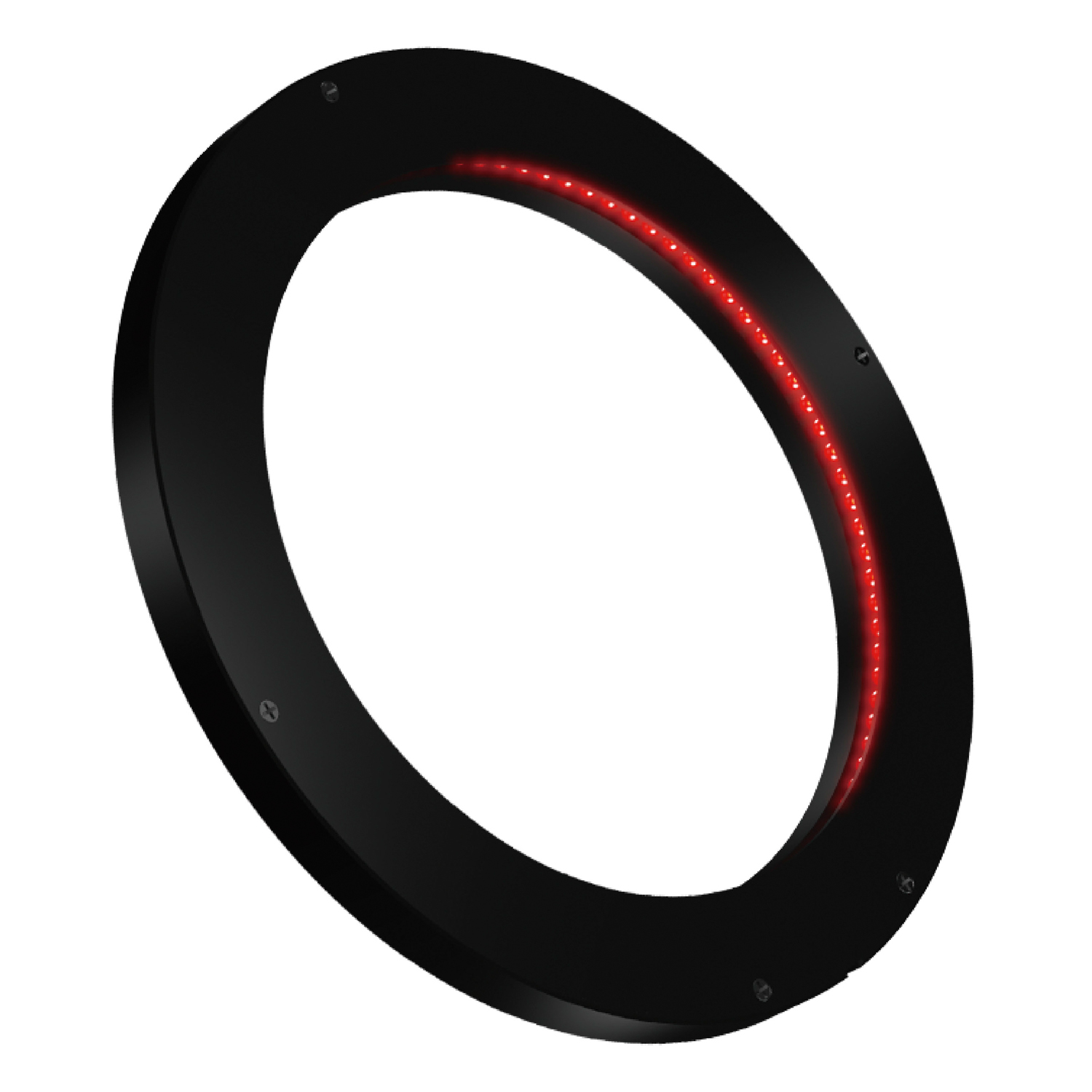
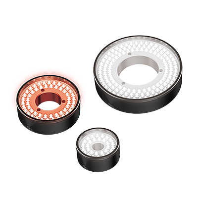
These Ring Lights have "easy to use", "bright", and "uniform" characteristics. The product lineup includes abundant size variations, full color (RGB) models, and satisfying accessories such as the Light Joint Brackets.Download Pamphlet PDF
Christiaan Huygens, another Dutchman, developed a simple two-lens ocular system in the late 17th century that was achromatically corrected, and therefore a huge step forward in microscope development. The Huygens ocular is still being produced to this day, but suffers from a small field size and other minor disadvantages.
The physical existence of this website has been verified by using a sever certificate issued by Cybertrust. Additionally, encryption is used to protect the privacy of communications made via SSL webpages.
These waterproof Ring Lights ensure the brightness with condensing lenses. IP67 compliance. Adopting an M12 connector.Download Pamphlet PDF
Bright-field microscopy (BF) is the simplest of all the optical microscopy illumination techniques. Sample illumination is transmitted (i.e., illuminated from below and observed from above) white light, and contrast in the sample is caused by attenuation of the transmitted light in dense areas of the sample. Bright-field microscopy is the simplest of a range of techniques used for illumination of samples in light microscopes, and its simplicity makes it a popular technique. The typical appearance of a bright-field microscopy image is a dark sample on a bright background, hence the name.
Bright-field microscopes are very simple to use and can be used to view both stained and unstained specimens. The optics do not change the color of the specimen, making it easy to interpret what is observed.
Bright-field microscopy is a standard light-microscopy technique, and therefore magnification is limited by the resolving power possible with the wavelength of visible light. The practical limit to magnification with a light microscope is around 1300×. Higher magnifications are possible, but it becomes increasingly difficult to maintain image clarity as the magnification increases.[17] Bright-field microscopes have low apparent optical resolution due to the blur of out-of-focus material;
LEDs are mounted perpendicularly on a line-shaped board and arranged in a ring facing horizontally. The thin design has a thickness of only 10 mm.These Ring Lights are used extremely close to the workpiece.
High Power Strobe Lights PF Series.* Ring type.* Peak illuminance achieves 6.5 million lx.* Realizes high-speed and accurate inspection.* Solves your xenon flash lamp problems.* Freely adjustable flash timing enables strobing within the exposure period of the camera.Download Pamphlet PDF
At a low angle, these Ring Lights can be used to highlight and image the characters and scratches in a dark field.At a high angle, these Ring Lights can be used to uniformly image the entire workpiece in a bright field.CCS can offer various lighting methods according to the applications.
Bright-field microscopes typically produce low contrast with most biological samples, as few absorb light to a great extent. Samples that are naturally colorless and transparent cannot be seen well, e.g. many types of mammalian cells. Staining is often required to increase contrast, which prevents use on live cells in many situations. Bright-field illumination is useful for samples that have an intrinsic color, for example mitochondria or the observation of cytoplasmic streaming in Chara cells.
Any angle can be created through the use of flexible circuit boards. The features of the workpiece can be clearly extracted with direct light from a low angle towards the center of the workpiece.
The light path of a bright-field microscope is extremely simple; no additional components are required beyond the normal light-microscope setup. The light path begins at the illuminator or the light source on the base of the microscope. Often a halogen lamp is used. The light travels through the objective lens into the ocular lens, through which the image is viewed. Bright-field microscopy may use critical or Köhler illumination to illuminate the sample.[16]
dark-field microscopy
LEDs are embedded around a light-guiding diffusion plate. Uniform light is radiated towards the center from a light-emitting surface arranged at an angle to the workpiece.
LEDs are embedded around a light-guiding diffusion plate. Uniform light is radiated from a flat emitting surface. The product lineup includes the LFR-100K and LFR-130K Light Units, in which light is radiated from an emitting surface arranged at an angle.
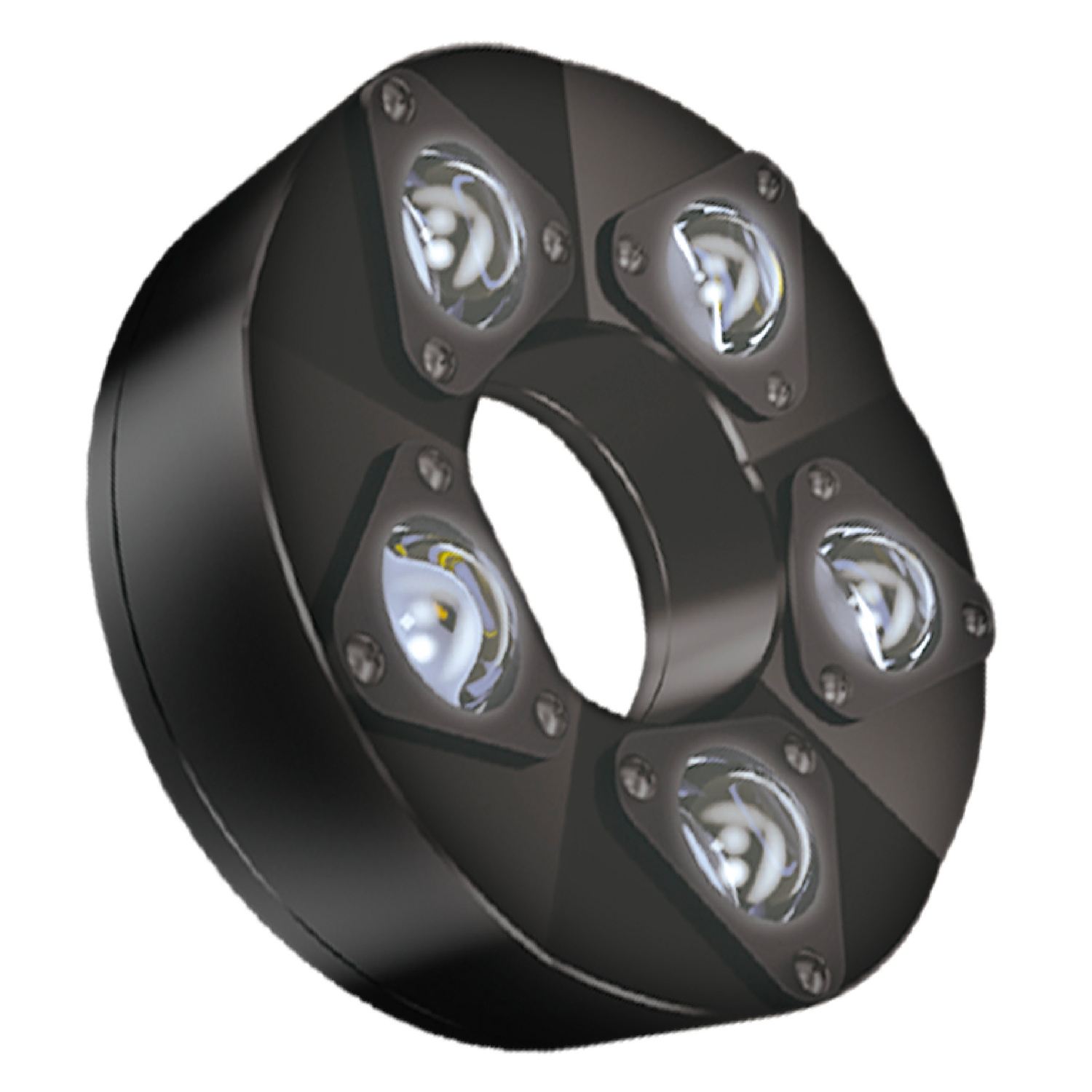
High Power Strobe Lights PF Series.* Low-Angle Ring type.* Peak illuminance achieves 10 million lx.* Realizes high-speed and accurate inspection.* Solves your xenon flash lamp problems.* Freely adjustable flash timing enables strobing within the exposure period of the camera.Download Pamphlet PDF
Antonie van Leeuwenhoek (1632–1724) is credited with bringing the microscope to the attention of biologists, even though simple magnifying lenses were already being produced in the 16th century. Van Leeuwenhoek's home-made microscopes were simple microscopes, with a single very small, yet strong lens. They were awkward to use, but enabled van Leeuwenhoek to see detailed images. It took about 150 years of optical development before the compound microscope was able to provide the same quality image as van Leeuwenhoek's simple microscopes, due to difficulties in configuring multiple lenses. In the 1850s, John Leonard Riddell, Professor of Chemistry at Tulane University, invented the first practical binocular microscope while carrying out one of the earliest and most extensive American microscopic investigations of cholera.[13][14]
Any angle can be created with CCS's unique flexible circuit boards. The required irradiance is ensured by radiating direct light towards the center of the workpiece.
Galileo Galilei is sometimes cited as an inventor of the compound microscope. After 1610, he found that he could close-focus his telescope to view small objects such as flies and/or could look through the wrong end in reverse to magnify small objects.[10][11] The only drawback was that his telescope had to be extended out to six feet to view objects that close.[12]
Ring lights for long-distance and wide-area irradiation.Suitable for various applications such as large workpieces, long-distance illumination, and robot picking inspections. Available in 4 wavelengths: red, white, blue, and infrared.Wide, medium and narrow light directivity characteristics are available.Lightweight and compact size that can be mounted on robot arms and inspection equipment.Custom orders are also possible such as different wavelengths and overdrive.HLDL3 Series bar lights for wide-area and long-distance illumination are also available.


 Ms.Cici
 8618319014500