
How the polarizing effect of social media is speeding up - polarizing
Dark fieldimmersive
Dark-field can be added to almost any light microscope for less than many other techniques. A simple method uses spider-stops (shown above) to create a cone of light by placing the spider-stops in a filter tray below the condenser or on top of the light source below the condenser. These metal filters can be purchased for a few dollars. Early on I used small coins like a penny, dime, nickel or quarter and placed them in the center of a light blue filter or on the light source under the condenser to achieve dark-field lighting. You will need different sized coins for different objectives and moving the condenser up or down also varies the light cone size. Spider-stops and coins generally only work with low power objectives but are easy to try though results will vary. Another method to add dark-field requires a desk lamp or fiber optic lamp and lighting the specimen from above and removing the condenser (R. Vossen, 2004). I have used this “top-lighting” method successfully with larger aquatic arthropods and low magnification objectives 2.5X and 5X. Some phase condensers have a dark-field option that can be used with low power objectives (10 to 40X) and if there isn’t a dark-field option sometimes you can achieve good dark-field by using the wrong phase ring with some objectives.
Dark-field microscopy is ideal for specimens with smooth reflective surfaces. Specimens with a different refractive index or refractive index gradients from their surrounding solution bend the light into the objective. Some of this is light is also diffracted entering the objective and can undergo interference. Under appropriate conditions dark-field microscopy can detect particles or fibers significantly smaller than the resolution limit of a normal light microscope (0.2 microns or 200 nm). This is possible due to diffraction disks provided the distance between the particles or fibers is greater than the resolving power of the objective. For this reason a dark-field microscope can detect suspended particles down to 40 nm in size and even bacteria flagella which is approximately 20 nm in width.
Dark FieldDenver
Unlike cones, cube and cuboid, a cylinder does not have any vertices, since the cylinder has a curved shape and no straight lines. It has two circular faces.
Dark-field is a technique that can be added to almost any light microscope and is economical in cost. Sometimes spider-disks or even coins can be used with low magnification objectives. The use of sliders or multipurpose condensers can produce better results for low power objectives (10, 20 and 40X). High magnification dark-field requires special condensers that have a numerical aperture greater than the high magnification objectives which have an iris diaphragms to reduce the objectives NA below that of the condenser. Dark-field has the ability to detect specimens below the limit of a normal light microscope resolution (200 nm) so that particles and fibers (20-40 nm) can be detected. This makes dark-field useful in the study of nanoparticles and microbes but it can also create beautiful images of aquatic-microorganisms.
DARKFIELD RADIO
The ultra-microscope was the first dark-field microscope. Richard Zsigmondy studied nanoparticles and developed the first ultra-microscope with Siedenkopf. Zsigmondy received the Nobel Prize in 1925 for his work on nanoparticles. Today there is a renewed interest in nanoparticles and dark-field microscopy has regained importance and popularity. I use dark-field microscopy to study and photograph mainly aquatic micro-organisms and find that it complements other forms of microscopy.
The downsides of dark-field microscopy are that it is not useful with thick specimens and it is less useful in identifying internal details. Also dirt, dust and particles in water show up as bright spots. I often need to clean background spots in images using image editing software like Photoshop.
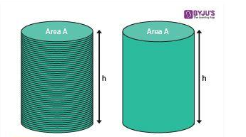
The area of the curved surface of the cylinder which is contained between the two parallel circular bases. It is also stated as a lateral surface area. The formula for it is given by:
Cylinder is one of the basic 3d shapes, in geometry, which has two parallel circular bases at a distance. The two circular bases are joined by a curved surface, at a fixed distance from the center. The line segment joining the center of two circular bases is the axis of the cylinder. The distance between the two circular bases is called the height of the cylinder. LPG gas-cylinder is one of the real-life examples of cylinders.
High magnification dark-field microscopy requires special dark-field condensers and the objectives for use with these possess an iris diaphragm which can be used to reduce the numerical aperture of the objective. Numerical aperture (NA) is commonly used in microscopy to describe the light acceptance cone of an objective. Objectives with larger numerical apertures offer more light gathering power and higher resolving power. The NA of the objective is written on each objective. More expensive objectives have higher numerical apertures and generally higher magnification objectives have greater NA’s then lower power objectives. For high resolution dark-field the NA of the condenser must be larger than the NA of the objective lens in order to prevent direct light from entering the objective. A 100X objective with NA = 1.25 requires a condenser with NA 1.4.
Dark fieldFLIGHT
Most high magnification dark-field condensers require oil immersion fluid between the condenser and microscope slide because the angle of incidence of light leaving the top of condenser is greater than the critical angle for glass to air; thus no light emerges from the condenser until it has the immersion oil applied to the condenser surface (Bagnell 2012). High magnification objectives designed for dark-field microscopy also have a built in iris diaphragm that permits the NA of the objective to be reduced.
Panthera C2 is a new world-class level cased in a revolutionary technical future-orientated solution, now accessible for life sciences.
The BA310 is designed for the daily routine work in universities, clinics, laboratories, and life sciences or medical applications.
Oblique illumination was the first step toward dark-field microscopy sometimes referred to as dark-ground microscopy. Dark-field microscopy illuminates specimens with oblique light in the form of a hollow cone. Oblique light from the cone is focused on the specimen but only light that is reflected, refracted or diffracted enters the objective. The specimens are generally highly refractile and must be spaced apart. Dark-field microscopy works primarily by increasing the contrast of the specimen. It does not work well with objects that are crowded or too thick and it can be used to study biological sections if unstained or if covered with silver or gold particles. Other good specimens for dark-field include: cell cultures, microbes, plankton, foods, fibers, crystals, colloids, arthropods, autoradiographs or tissues labelled with metal particles. The subjects appear bright against a black background and can produce striking images. The addition of coloured filters or Rheinberg filters can also be used with the technique though the colours do not provide additional information (R. Berdan 2017). Dark-field microscopy today is also used to examine pathogenic bacteria and in live blood analysis (see article on blood on this web site).
In mathematics, a cylinder is a three-dimensional solid that holds two parallel bases joined by a curved surface, at a fixed distance. These bases are normally circular in shape (like a circle) and the center of the two bases are joined by a line segment, which is called the axis. The perpendicular distance between the bases is the height, “h” and the distance from the axis to the outer surface is the radius “r” of the cylinder.
Since, the cylinder is a three-dimensional shape, therefore it has two major properties, i.e., surface area and volume. The total surface area of the cylinder is equal to the sum of its curved surface area and area of the two circular bases. The space occupied by a cylinder in three dimensions is called its volume.
Syphilis is a sexually transmitted disease caused by Treponema pallidum, a bacterium classified under the Spirochaetes phylum. Schaudinn and Hoffmann discovered the bacteria Treponema pallidum in tissue of patients with syphilis in 1905. In 1906, Landsteiner (he also discovered different AB0 blood types) introduced the use of dark-field microscopy for the detection of the spirochete causing syphilis which in turn increased the popularity of dark-field microscopy (Tampa et al. 2014).
Dark fieldexperience
A cylinder is a three-dimensional shape consisting of two parallel circular bases, joined by a curved surface. The center of the circular bases overlaps each other to form a right cylinder. The line segment joining the two centers is the axis, that denotes the height of the cylinder.
Each shape has some properties that differentiate one shape from another. Therefore, cylinders also have its characteristics.
Dark fieldmicroscopy
The total surface area of a cylinder is the sum of curved surafce area and the area of two circular bases. TSA = Curved surface + Area of Circular bases TSA = 2πrh + 2πr2 We can see, from the above expression, 2πr is common. Therefore,
V = 550 cm3 Frequently Asked Questions – FAQsQ1 What are the characteristics of cylinder?A cylinder has two parallel circular bases and a curved surface. The perpendicular distance between the two bases is the height of cylinder. The line segment joining the center of two bases is the axis of cylinder.Q2 How many vertices does a cylinder have?A cylinder has two faces that are circular in shape and parallel to each other. But it does not have any vertices, since it is a curved shape.Q3 What are the real life examples of cylinder?The real life examples of cylinder are: Gas cylinder, fire extinguisher, cans, pipes, etc.Q4 What are the formulas of cylinder?The formulas of cylinder are: Total surface area = 2πr(r+h) square units Volume of cylinder = πr2h cubic unitsQ5 How many bases does the cylinder have?A cylinder is having two circular bases.Q6 How to find total surface area of cylinder?First find the curved surface of cylinder which is equal to 2πrh, where r is the radius and h is the height of cylinder. Then add this curved surface area with the areas of two circular bases of cylinder to get the total surface area. Total surface area = Curved Surface area + Two circular base areas
Every three dimensional shape or a solid has volume that occupies some space. The volume of the cylinder is the space occupied by it in any three-dimensional plane. The amount of water that could be immersed in a cylinder is described by its volume. The formula for the volume of cylinder is given by:

Here we will learn about its definition, formulas, properties of cylinder and will solve some examples based on them. Apart from this figure, we have concepts of Sphere, Cone, Cuboid, Cube, etc. which we learn in Solid Geometry.


 Ms.Cici
 8618319014500