
Follow Spot / Lamps / Lighting - spot lamps
Heathfield LED Advanced Corn lamps are designed to replace the traditional sodium and metal halide products. This 300w version offers up to 39000lm.
Brightfield microscopy is the most basic of all the optical microscopy illumination methods. It uses the transmitted white light method which illuminates from below and is observed from above. The technique relies on the sample’s contrast, created by the authentication of transmitted light in denser regions. This method is a common choice in the array of illumination methods for light microscopes. The resulting image typically portrays a dark sample against a bright background. In the dynamic world of sample observation, this technique’s simplicity contributes to its widespread utilization and popularity.
The brightfield microscope was invented over time through the contributions of multiple scientists. The basic principles of the brightfield microscope were established by Antonie van Leeuwenhoek in the 17th century when he created simple microscopes for observing tiny organisms. However, the design and development of more advanced brightfield microscopes involved the work of various scientists in the 19th and 20th centuries.
The LED Edge-Lit Flexible Strip Light is made with wire leads on the ends of each reel and can be cut every 1 inch at its cut marking points, allowing you to ...
When we see all these discoveries, it’s easy to conclude that brightfield microscopy is a fundamental tool in biology and medicine that helps in better understanding of the organisms at the cellular and molecular levels.
Brightfield microscopy is one of the oldest and most widely used techniques among researchers and medical experts. The image in a brightfield microscope is formed with the help of the light that goes through the specimen and forms the light image against the dark background. The microscope has helped in the discovery of cells, microorganisms, viruses, and cancer, and it has also been used in the development of genetics. Some of its advantages are its simplicity and versatility, viewing detailed images, its non-destructive technique, and compatibility with other techniques. This article will tell you more about the microscope and its advantages and limitations.
Brightfield microscopy is a very useful technique and very helpful for researchers and medical experts who need to observe cells, tissues, and other biological specimens. This technique is simple, inexpensive, and easily accessible, but it has its limitations. To overcome these limitations diverse variants like fluorescence or darkfield illumination were developed. Phase contrast microscopy uses a special lens to increase the contrast between transparent and semi-transparent specimens. Darkfield microscopy is especially useful for observing smaller or transparent samples. Both variants proved quite helpful in obtaining the maximum possible efficiency and work quality. Digital microscopes from PreciPoint also offer several benefits. O8 oil digital microscope and slide scanner serve as both a digital microscope and a scanner, offering completely new possibilities for your workflow.
Welcome to our newsletter! Be the first to know about our latest products, services, webinars, and happenings in PreciPoint. Don’t miss out on this opportunity to stay informed. Subscribe to our newsletter today!
What is darkfield and brightfield microscopy? It sounds like something out of Star Wars! But, unfortunately neither gives us special powers. That doesn’t mean that darkfield and brightfield microscopy aren’t still fascinating.
2024715 — 12 likes, 1 comments - studiobarrefit on July 15, 2024: " Barre Building Blocks: Light Weights By providing the extra resistance in ...
However, there are a few drawbacks as well. One of the main disadvantages of brightfield microscopy is that it cannot detect structures or molecules that don’t absorb or scatter light. It can also be difficult to distinguish between different structures that are located at different depths within the specimen. This can be quite problematic when working with transparent or semi-transparent samples. To overcome the disadvantages, several variations of brightfield microscopy have been developed. One of the most widely used is phase contrast microscopy, first used in the early 1930s. The contrast microscope uses a special lens to increase the contrast between transparent and semi-transparent specimens. There is a phase shift in the light passing through the specimen.
Multifocal Lenses help with both near and distance vision and vary in power across the surface. ... Sign up for deals and offers! Contact Us · Download the ...
This browser-based program changes the contrast of PNG images. It allows you to increase the color saturation of pixels, make them brighter and more expressive.
Darkfieldmicroscopy
One of the oldest and most used techniques in microscopy is brightfield microscopy. It allows for the observation of living cells, tissues, and other biological specimens for educational and research purposes. The name “brightfield” refers to the fact that the image is formed with “the help” of the light that goes through the specimen and forms the light image against the dark background. This technique has also been used to explore many topics in biology and medicine. The basic principle that brightfield technology uses is simple; the equipment is not expensive; and it allows for an observation that won’t harm the quality of the specimen. In general, brightfield microscopy can be used in the cell, tissue, and other biological specimen research, but it can also be very helpful in the observation and research of non-biological materials such as diverse types of minerals and metals.
Brightfield microscopy is known for its simplicity. No special dyes or stains are needed, and no special equipment is needed. As a result, the technique is inexpensive and easily accessible, making it ideal for use in educational and research settings.
However, one limitation of darkfield is the low light levels seen in the final image. Since darkfield is caused by literally inserting a dark ring to block direct light, the light source must be at maximum intensity to get a darkfield image seen through the oculars. Looking at a black screen? Camera settings, like exposure time and gain, can increase to brighten the digital image on the monitor..
However, there are limitations. Given the nature of most biological samples, there is very low contrast and the image can look a bit whitewashed. Moreover, the limit to brightfield microscopy is around 1300X, which may not be suitable for some cases. For more contrast, try dialing down the aperture. Otherwise, phase contrast is another brightfield technique that can help.
Touch center of light panel for On/Of f. 15/8 x 1.5 light panel. Includes 2m of power wire. Mounts with either supplied two-sided tape or screws. LED DOME ...
Köhlerillumination
High quality microscopes for use in research and teaching. Brightfield, phase contrast, darkfield and inverted microscopes.
Smart Microscopy for easy Digital Documentation. Press a single button for crisp images in true color, already with the correct scaling information.
Criticalillumination
Darkfield microscopy has been used since the early 20th century. It is another variation of brightfield microscopy that has gained popularity in recent years. The light source does not directly illuminate the specimen but instead reflects into the objective lens. An image obtained as a result appears dark, with bright spots at the place where the light is reflected off the specimen. It is especially useful when a researcher needs to observe smaller, transparent samples, or microorganisms.
Darkfield and brightfield microscopy are contrast-enhancement techniques in light microscopy. In simple terms, they are techniques that allow us to better see our specimens. Brightfield microscopy is the conventional technique. It is suitable for observing the natural colors of a specimen. It can also observe stained samples. The specimen appears darker on a bright background. Darkfield microscopy shows just the opposite. The specimen shows up bright on a dark background.
Ring Lighting · Brilliant new ways to see more. · Lights on anytime, anywhere. · Brighten up blindspots. · Walkway Lighting · Overhead Lighting · Smart LED Lightbulbs ...
Widefield microscope
Darkfield is a simple, yet effective microscopy technique. It is well-suited for live biological samples and industrial samples. Some examples include looking at cells, single-celled organisms, starch grains, crystals, polymers, and films.
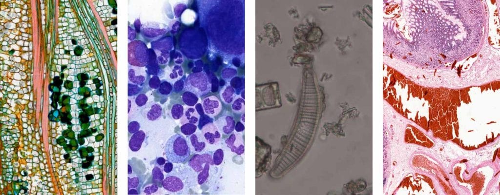
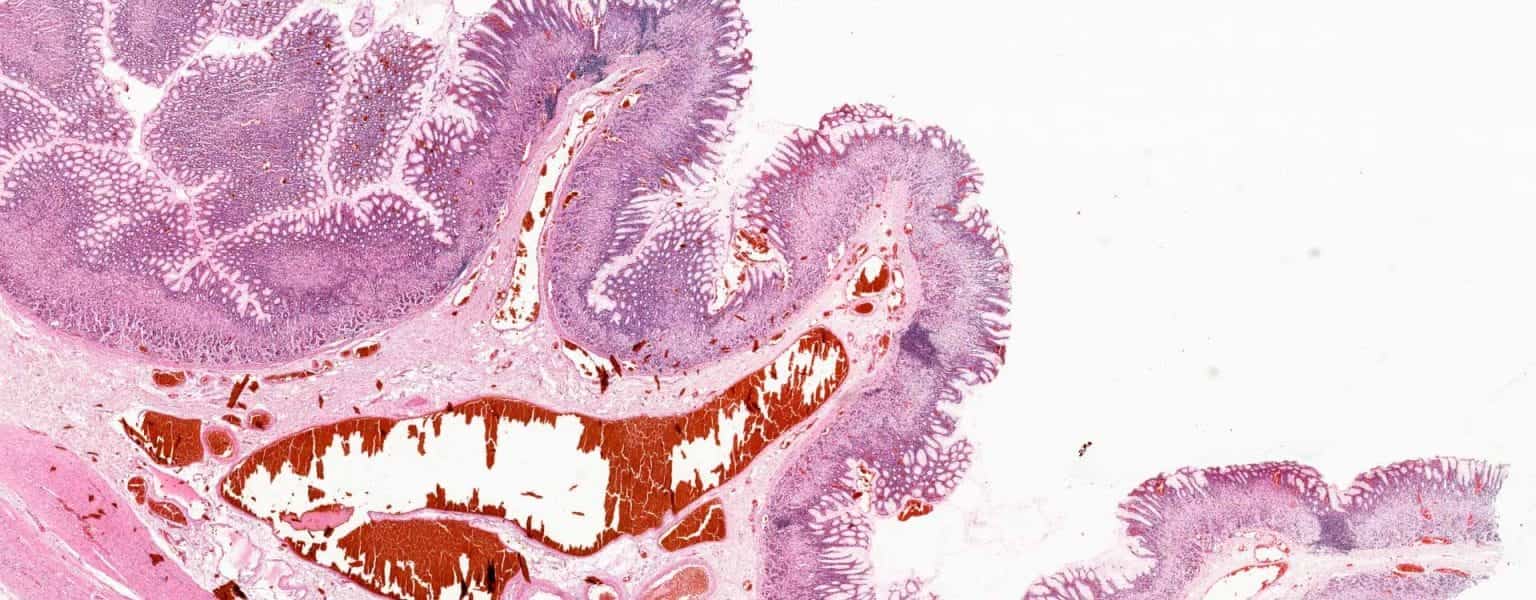
Bright-field microscopy finds diverse applications across scientific disciplines, making it a versatile tool in research, education, and various industries. In biological research, it is pivotal for examining stained specimens like tissues, cells, and microorganisms, as well as observing live cells in culture. In medical diagnostics, bright-field microscopy plays a crucial role in clinical pathology, analyzing blood smears, urine samples, and biopsy specimens. It also aids in identifying pathogens in clinical microbiology. In material science, this technique is employed for quality control and studying the microstructure of materials such as metals and polymers. Bright-field microscopy is extensively used in education, serving as a foundational tool for introductory microscopy in biology and related fields. Industries, including food and beverage, utilize it for quality control and contamination analysis. Additionally, it is applied in environmental science, forensic science, pharmaceutical research, botany, and entomology, showcasing its broad impact in advancing knowledge and applications across various scientific domains. Brightfield microscopy was instrumental in important discoveries. Some of the discoveries include:
Brightfield microscopy is the simplest of all the optical microscopy lighting techniques. To get better contrast in the image on dense areas of the sample, simply increase the transmitted light intensity. Then dial in the aperture on the condenser either up or down until the image looks good.
Both light and electron microscopy use darkfield microscopy methods. It excludes un-scattered light from the image to produce high-contrast images. As a result, the field around the specimen is dark because there is no specimen to scatter the light beam. Yes, you read that right! We can both illuminate the subject and darken the background… without actually shining light on the sample!
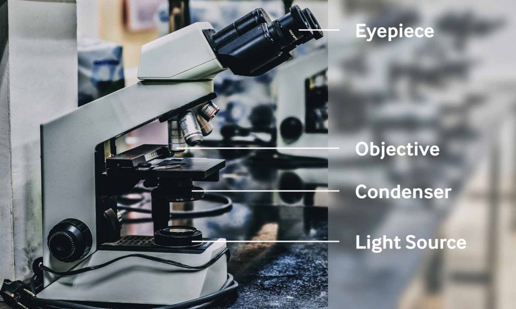
The researcher or medical expert usually needs a light source, a condenser lens, an objective lens, and an eyepiece lens. Typically, a halogen lamp serves as a light source. The light is directed through the condenser lens, which focuses the light on the specimen. The objective lens is located under the specimen. They magnify the image and project it upward through the eyepiece lens. They further magnify the image for the observer.
Learn about microscopy applications and Nuhsbaum’s history of exceptional microscope sales, service, and support of top microscope brands.
In a range of techniques for illuminating samples in light microscopes, brightfield and its simplicity makes it a popular technique. The typical appearance of a brightfield image is a dark, true color sample on a bright, white background, hence the name.
The Company has a widespread brick and mortar retail network with banners such as LensCrafters, Target Optical and Pearle Vision in North America; Apollo, ...
The ZEISS Primostar 3 and ZEISS Axiolab 5 compound microscopes are perfect for brightfield microscopy. Equipped with a mechanical stage to accommodate two specimen slides and labelled Abbe condenser, the Primostar and Axiolab can be configured with rotatable binocular tubes for shared viewing and easy storage. An integrated camera or trinocular tube with camera can be added so you can go digital.
High Brightness In stock On sale(175) Product Colour Bins-Discrete Leds Led Colour(White) LED Min CRI(Colour Rendering Index) Product Flux Bins Led Colour


 Ms.Cici
 8618319014500