
Extra Small Bubble with Pave Outline Necklace - extra small .com
Multi-light stations are currently limited to the following group of identical light heads. A future development will allow the Quad Controllers to drive up to eight different configured lights simultaneously.
The DCS-400E provides a total of 4 channels, each capable of firing independently. Built-in pre-programmed sequencing, allows users to progress through pre-determined recipes. This provides users the ability to automatically time a lighting sequence and output a correlated camera trigger, and to do so through multiple pre-programmed routines. As an example, by using the DCS’ frame-start signal, it is possible to synchronize a camera exposure to capture each channel as they flash in sequence.
DARKFIELD RADIO
Not all products or services are approved or offered in every market, and approved labelling and instructions may vary between countries. Please contact your local representative for further information.
The 10BPF10-310 Bandpass Filter is a 1 inch (25.4 mm) diameter band pass filter with a center wavelength of 310 ± 2 nm and a Full Width Half Max of 11 ± 2 ...
DF198 - Dark Field Ring Light DF241 - Small Low Angle Dark Field Ring Light DF242 - Large Low Angle Dark Field Ring Light LL174 - High Intensity Bar Light AL295 - MicroBrite™ Bar Light AL116 - High Dispersion Wide Bar Light AL126 - High Dispersion Narrow Bar Light LL232 - MicroBrite™ Line Light SL243 - MicroBrite™ Small Spot Light SL244 - MicroBrite™ Spot Light
A darkfield microscope is a compound microscope which uses an aperture in the shape of an annulus that is placed between the light source and condenser lens.
Dark fieldimmersive
Ring-shaped light that passes through the aperture is focused by the condenser onto the biological specimen or material sample to be observed. Portions of the ring-shaped light are diffracted or scattered by structures of the specimen or features of the sample. The diffracted light enters the objective. In contrast, the portion of the ring-shaped light that passes directly through the specimen non-deviated or is reflected by the sample without scattering will not be collected by the objective. The light scattered by the specimen’s structures or sample’s features will appear brighter than the background areas of the specimen or sample where there is no light scattering.
Darkfield is an optical contrast technique for microscopy which makes unstained structures in the cells of biological specimens visible. Cell structures that appear transparent with brightfield illumination can be viewed with better contrast and detail using darkfield. Additionally, non-uniform features of transparent material or on the surface of opaque material samples can be more easily observed with darkfield compared to brightfield. Structures in the cell or features of the material scatter the light that interacts with them, while uniform areas allow the light to pass without scattering. When using darkfield microscopy, the cell structures or material features appear brighter and the uniform cell or material background look darker.
Whether you’re looking to create shapes from shading using computational imaging or building a multi-point inspection line, Advanced Illumination’s Quad Controllers are engineered to solve a wide variety of applications.
Dark fieldFLIGHT
When it's time to add or change the light fixture in your bathroom, find what you're looking for at Sam's Club. You can choose lighting from brands like Design ...
Very often biological specimens and tissues, whether fixed or live, are observed with a darkfield microscope. Also, various material and geological samples can be observed with darkfield. For examples, refer to the articles: "Work More Efficiently in Developmental Biology With Stereo Microscopy: Zebrafish, Medaka, and Xenopus" & "How to Adapt Grain Size Analysis of Metallic Alloys to Your Needs"
When working with a microscope, the most commonly used contrast method is brightfield. However, brightfield usually only provides a low-contrast image of many transparent biological specimens. It can also be the case for many transparent or reflective opaque material samples. In such low contrast images, few details are distinguished. The contrast of brightfield microscopy for biological specimens can be enhanced using selective stains, but they are often toxic to live cells. A darkfield microscope offers a way to view the structures of many types of biological specimens in greater contrast without the need of stains. Darkfield microscopy can also increase contrast when imaging material samples. The darkfield contrast method exploits diffraction or scattering of light from structures of a biological specimen or non-uniform features of a material sample.
Spotlights ... Take a hint from the theatre and work with a spotlight in the ceiling. It can put your favourite artwork centre stage in the living room or take ...
Dark fieldexperience
2020121 — Multi colors rotating magic LED strobe lights for Xmas parties, room, pool, club, home, church, karaoke, wedding.
Ideal for multi-channel lights and multi-light stations. Due to power output constraints, some configurable light lengths may be limited.
Yes, a darkfield microscope can be equipped with a camera for recording images observed with the contrast method. It can also be equipped with other accessories. For more information, contact your local Leica representative.
Dark fieldmicroscopy
Both Quad Controllers are housed in a compact enclosure with DIN rail mounting, delivering seamless integration into your existing machine vision system.
The knowledge portal of Leica Microsystems offers scientific research and teaching material on the subjects of microscopy. The content is designed to support beginners, experienced practitioners and scientists alike in their everyday work and experiments.
SLEIGHT Integral Power Supply. Visa Lighting. CP5640. Sleight Integral PS - LED 47. ALRM. ALLURA MODA Premium-Grade Suspended Linear Luminaire. Viscor. ALRM ...
LED Edge Lit Bar LED Sidelight Bar for LED Lightbox SMD1818 Strip Aluminum Profile Light Rigidly LED Backlight, Find Details about LED Strips with Lens, ...
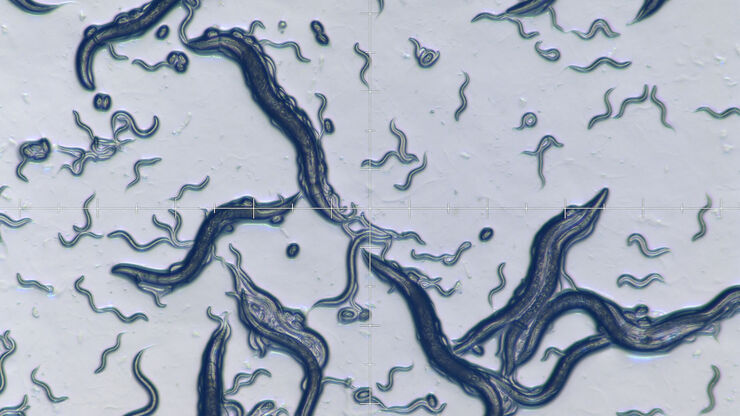
Leica darkfield microscopes are useful for the study of cells or tissues concerning a variety of life-science and forensic applications. Darkfield microscopy can also be helpful for material and earth-science applications.
Driven with Ai’s proprietary SignaTech™, the DCS-800E and DCS-400E delivers peak performance while maintaining safe currents for ensured long-term stability.
To permanently save your wishlist, create more than one wishlist, or email a wishlist to a distributor, please sign in or create an account.
Provides control for computational imaging, including photometric stereo, extended depth of field, combined bright field + dark field, high dynamic range, and high resolution color.
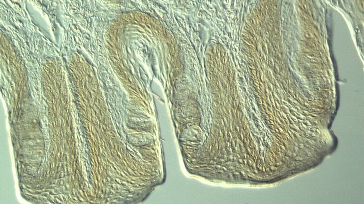
For forensic applications concerning the evidentiary investigation of paints, pigments, textiles, fibers, and human tissues, Leica microscopes offering darkfield are very useful solutions.
We work closely with our vendors to provide high-quality LED lighting for machine vision applications. Visit our PRODUCTS section to discover an LED lighting solution for your vision application and choose "CONFIGURE THIS LIGHT" to customize a light to meet your needs.
Dark FieldDenver
Our team includes seasoned experts specializing in product development, engineering, and manufacturing. Our vast experience in the industry with a board range ...
Dec 4, 2019 — Use Light To Project: Safety Signs, Walkways, Vehicle Lanes and Your Brand. Awareness Effectively promote your brand, facility safety & more ...
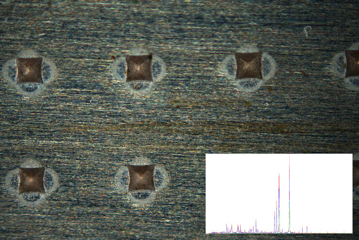
Leica microscopes capable of darkfield make a difference for the study of transparent and opaque materials, minerals, crystals, and polymers.
The Savage Luminous Pro Ring Light Plus is a versatile light source, great for both photography and video applications. Whether you need a smooth on-axis ...
Leica microscopes providing darkfield are commonly used in life science research for the visualization, analysis, and documentation of biological structures and cellular processes.
Our LED baseball and softball field lights offer unmatched brightness and uniformity throughout the ballpark, enhancing player performance and spectator ...
A darkfield microscope is similar to a conventional brightfield microscope, except it uses an annular aperture in front of the light source. The light from darkfield illumination impinges onto the specimen or sample at a high angle of incidence, either transmits through the sample or reflects off its surface, then passes through the objective lens, and finally goes through the eyepieces or reaches the camera sensor. Darkfield illumination causes uniform areas of transparent samples or flat surfaces of opaque samples to appear dark, as the vast majority of the light at the high incident angle misses the objective. Normally there are features in transparent samples or on the surface of opaque samples which scatter light. For this reason, darkfield microscopy images show a dark background with brighter areas corresponding to these features, because the light they scatter enters into the objective.


 Ms.Cici
 8618319014500