
dcs plus | Complete Travel Technology - d.cs
Getting the desired colors from C-41 film, whether by darkroom printing or scanning and editing, has always involved some level of manual adjustment. Photography is art, and as such it has always involved making subjective decisions about how images should look; all photos are interpretations of raw data and it’s up to the photographer to decide what interpretation looks best. The best starting point for editing is a scan that represents the image data stored in the dye layers of the film with minimal crosstalk.
By using a diffuser attachment, light is uniformly spread over an area to provide the best possible illumination for clear observation of the target.
Brightfield technique has been mated with cell imaging software to better perform tasks previously delegated to fluorescence microscopy. By using multiple focal levels the cell borders and nuclei can be located in cell populations.The benefit of using brightfield illumination for this task is that it frees fluorescent channels in microscopes and eliminates distortions caused by the overlapping of the color emissions of the stains and the excitation of the fluorescing materials.Here's a related article and interesting software for digital imaging applying digital colour brightness and true colour 3D.AdvantagesBrightfield microscopy is very simple to use with fewer adjustments needed to be made to view specimens.Some specimens can be viewed without staining and the optics used in the brightfield technique donât alter the color of the specimen.It is adaptable with new technology and optional pieces of equipment can be implemented with brightfield illumination to give versatility in the tasks it can perform.DisadvantagesCertain disadvantages are inherent in any optical imaging technique.By using an aperture diaphragm for contrast, past a certain point, greater contrast adds distortion. However, employing an iris diaphragm will help compensate for this problem.Brightfield microscopy canât be used to observe living specimens of bacteria, although when using fixed specimens, bacteria have an optimum viewing magnification of 1000x.Brightfield microscopy has very low contrast and most cells absolutely have to be stained to be seen; staining may introduce extraneous details into the specimen that should not be present.Also, the user will need to be knowledgeable in proper staining techniques.Lastly, this method requires a strong light source for high magnification applications and intense lighting can produce heat that will damage specimens or kill living microorganisms.Check out many more useful microscopy imaging techniques here. Great Microscopes to Consider...OMAX 40X-2500X Brighter Darkfield LED Trinocular Compound Microscope with 9MP Digital CameraAmScope T490A-PCT Compound Trinocular Microscope with Phase Contrast turretReturn from Brightfield Microscopy to Compound Light Microscope Return to Best Microscope HomeFind out how to advertise on MicroscopeMaster!FacebookTwitter
Although a basic method of microscopy, brightfield as a technique is well suited to mating with new technologies.Digital imaging systems can make high resolution images of properly stained microorganisms using this technique.Three-dimensional imaging accessories can be used with the brightfield method and newer technologies will allow real time viewing in 3D.Also suited to video imaging, this enhancement will allow the user to view motile organisms interacting with their environment.Brightfield technique has been mated with cell imaging software to better perform tasks previously delegated to fluorescence microscopy. By using multiple focal levels the cell borders and nuclei can be located in cell populations.The benefit of using brightfield illumination for this task is that it frees fluorescent channels in microscopes and eliminates distortions caused by the overlapping of the color emissions of the stains and the excitation of the fluorescing materials.Here's a related article and interesting software for digital imaging applying digital colour brightness and true colour 3D.AdvantagesBrightfield microscopy is very simple to use with fewer adjustments needed to be made to view specimens.Some specimens can be viewed without staining and the optics used in the brightfield technique donât alter the color of the specimen.It is adaptable with new technology and optional pieces of equipment can be implemented with brightfield illumination to give versatility in the tasks it can perform.DisadvantagesCertain disadvantages are inherent in any optical imaging technique.By using an aperture diaphragm for contrast, past a certain point, greater contrast adds distortion. However, employing an iris diaphragm will help compensate for this problem.Brightfield microscopy canât be used to observe living specimens of bacteria, although when using fixed specimens, bacteria have an optimum viewing magnification of 1000x.Brightfield microscopy has very low contrast and most cells absolutely have to be stained to be seen; staining may introduce extraneous details into the specimen that should not be present.Also, the user will need to be knowledgeable in proper staining techniques.Lastly, this method requires a strong light source for high magnification applications and intense lighting can produce heat that will damage specimens or kill living microorganisms.Check out many more useful microscopy imaging techniques here. Great Microscopes to Consider...OMAX 40X-2500X Brighter Darkfield LED Trinocular Compound Microscope with 9MP Digital CameraAmScope T490A-PCT Compound Trinocular Microscope with Phase Contrast turretReturn from Brightfield Microscopy to Compound Light Microscope Return to Best Microscope HomeFind out how to advertise on MicroscopeMaster!FacebookTwitter
Brightfield microscopy is the most elementary form of microscope illumination techniques and is generally used with compound microscopes.The name "brightfield" is derived from the fact that the specimen is dark and contrasted by the surrounding bright viewing field. Simple light microscopes are sometimes referred to as brightfield microscopes.How it WorksIn brightfield microscopy a specimen is placed on the stage of the microscope and incandescent light from the microscopeâs light source is aimed at a lens beneath the specimen. This lens is called a condenser.Featured right: Algae under the microscope with visible cells using brightfield illumination.The condenser usually contains an aperture diaphragm to control and focus light on the specimen; light passes through the specimen and then is collected by an objective lens situated in a turret above the stage.The objective magnifies the light and transmits it to an oracular lens or eyepiece and into the userâs eyes. Some of the light is absorbed by stains, pigmentation, or dense areas of the sample and this contrast allows you to see the specimen.For good results with this microscopic technique, the microscope should have a light source that can provide intense illumination necessary at high magnifications and lower light levels for lower magnifications.Uses and AdvancementsTo some extent, brightfield microscopy is used in most disciplines requiring microscopic investigation.Because it is a simple method, this is the first type of microscopy students learn in schools.The life sciences, particularly microbiology and bacteriology, have always relied on the brightfield technique.This technique can be used to view fixed specimens or live cells. Since many organic specimens are transparent or opaque, staining is required to cause the contrast that allows them to be visible under the microscope.Different stains and staining techniques are used depending upon the type of specimen and cell structure being examined.For example:Fuchsin is used to stain smooth muscle cellsMethylene blue is used to stain cell nucleiGram stain is used on bacteria and gives rise to the name gram-negative or gram-positive bacteria based on the reaction of the bacteria to the stain. In fact, many scientific journals will not accept microbiological research for publication that is not supported by gram staining and brightfield illumination methodology. Most routine medical microscopic examination of blood and tissue is performed using this illumination technique.Different complimentary techniques can be used to augment brightfield microscopy. By using a polarizing filter this illumination technique can be used in geological microscopic research and will reveal details not visible using white light.Properly stained, microorganisms may be magnified to 1200x; utilizing an oil immersion objective will increase resolution at this high magnification.Digital Imaging OptionsAlthough a basic method of microscopy, brightfield as a technique is well suited to mating with new technologies.Digital imaging systems can make high resolution images of properly stained microorganisms using this technique.Three-dimensional imaging accessories can be used with the brightfield method and newer technologies will allow real time viewing in 3D.Also suited to video imaging, this enhancement will allow the user to view motile organisms interacting with their environment.Brightfield technique has been mated with cell imaging software to better perform tasks previously delegated to fluorescence microscopy. By using multiple focal levels the cell borders and nuclei can be located in cell populations.The benefit of using brightfield illumination for this task is that it frees fluorescent channels in microscopes and eliminates distortions caused by the overlapping of the color emissions of the stains and the excitation of the fluorescing materials.Here's a related article and interesting software for digital imaging applying digital colour brightness and true colour 3D.AdvantagesBrightfield microscopy is very simple to use with fewer adjustments needed to be made to view specimens.Some specimens can be viewed without staining and the optics used in the brightfield technique donât alter the color of the specimen.It is adaptable with new technology and optional pieces of equipment can be implemented with brightfield illumination to give versatility in the tasks it can perform.DisadvantagesCertain disadvantages are inherent in any optical imaging technique.By using an aperture diaphragm for contrast, past a certain point, greater contrast adds distortion. However, employing an iris diaphragm will help compensate for this problem.Brightfield microscopy canât be used to observe living specimens of bacteria, although when using fixed specimens, bacteria have an optimum viewing magnification of 1000x.Brightfield microscopy has very low contrast and most cells absolutely have to be stained to be seen; staining may introduce extraneous details into the specimen that should not be present.Also, the user will need to be knowledgeable in proper staining techniques.Lastly, this method requires a strong light source for high magnification applications and intense lighting can produce heat that will damage specimens or kill living microorganisms.Check out many more useful microscopy imaging techniques here. Great Microscopes to Consider...OMAX 40X-2500X Brighter Darkfield LED Trinocular Compound Microscope with 9MP Digital CameraAmScope T490A-PCT Compound Trinocular Microscope with Phase Contrast turretReturn from Brightfield Microscopy to Compound Light Microscope Return to Best Microscope HomeFind out how to advertise on MicroscopeMaster!FacebookTwitter
Because the overall light transmission of C-41 film is heavy biased towards yellow and orange wavelengths due to the orange mask, this means that a lot of light gets through that interferes with both the red and green channels in the camera sensor. The red channel of the scan will contain not just information from the red-blocking cyan dye, but also the green-blocking magenta dye. The green channel will contain information from all three dye layers.
Backlight scanonline
The light source is suitable to use with a variety of existing film carriers, but was designed with future expansion in mind. More open source 3D printable film scanning gear is coming soon :3
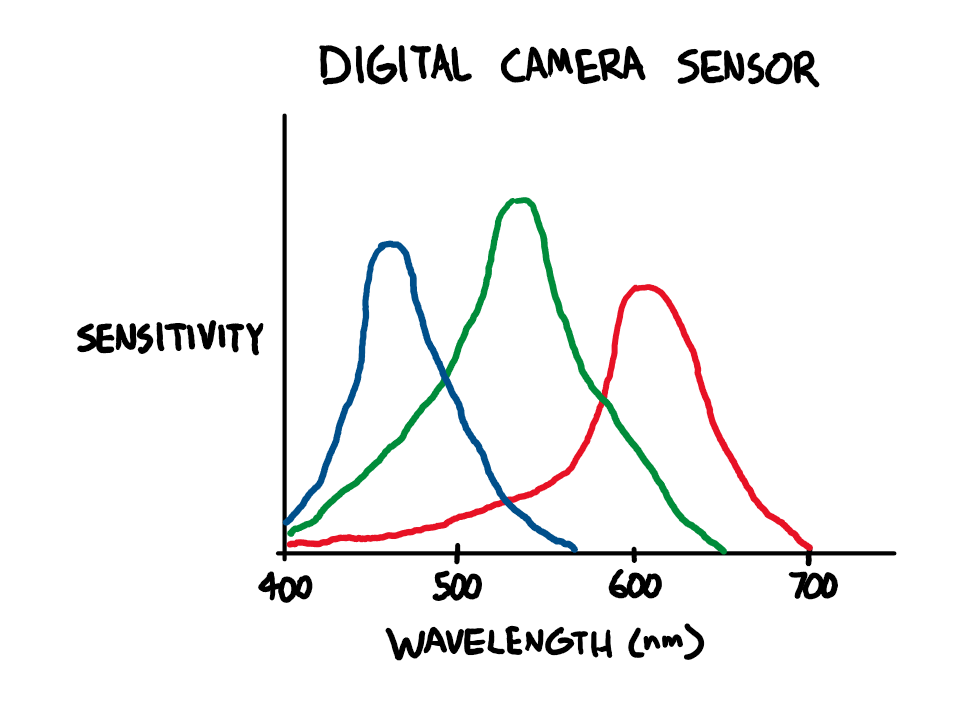
When considering the limitations of digital camera sensors, the ideal wavelengths are >650nm for red, 520-550nm for green, and <450nm for blue, as shown in the diagram above. However, to the best of my knowledge, there aren’t any commercially available light sources combining LEDs of these wavelengths, so the only way (for now) to get an ideal light source is to build one yourself, which is discussed in the next section of this article.
Properly stained, microorganisms may be magnified to 1200x; utilizing an oil immersion objective will increase resolution at this high magnification.Digital Imaging OptionsAlthough a basic method of microscopy, brightfield as a technique is well suited to mating with new technologies.Digital imaging systems can make high resolution images of properly stained microorganisms using this technique.Three-dimensional imaging accessories can be used with the brightfield method and newer technologies will allow real time viewing in 3D.Also suited to video imaging, this enhancement will allow the user to view motile organisms interacting with their environment.Brightfield technique has been mated with cell imaging software to better perform tasks previously delegated to fluorescence microscopy. By using multiple focal levels the cell borders and nuclei can be located in cell populations.The benefit of using brightfield illumination for this task is that it frees fluorescent channels in microscopes and eliminates distortions caused by the overlapping of the color emissions of the stains and the excitation of the fluorescing materials.Here's a related article and interesting software for digital imaging applying digital colour brightness and true colour 3D.AdvantagesBrightfield microscopy is very simple to use with fewer adjustments needed to be made to view specimens.Some specimens can be viewed without staining and the optics used in the brightfield technique donât alter the color of the specimen.It is adaptable with new technology and optional pieces of equipment can be implemented with brightfield illumination to give versatility in the tasks it can perform.DisadvantagesCertain disadvantages are inherent in any optical imaging technique.By using an aperture diaphragm for contrast, past a certain point, greater contrast adds distortion. However, employing an iris diaphragm will help compensate for this problem.Brightfield microscopy canât be used to observe living specimens of bacteria, although when using fixed specimens, bacteria have an optimum viewing magnification of 1000x.Brightfield microscopy has very low contrast and most cells absolutely have to be stained to be seen; staining may introduce extraneous details into the specimen that should not be present.Also, the user will need to be knowledgeable in proper staining techniques.Lastly, this method requires a strong light source for high magnification applications and intense lighting can produce heat that will damage specimens or kill living microorganisms.Check out many more useful microscopy imaging techniques here. Great Microscopes to Consider...OMAX 40X-2500X Brighter Darkfield LED Trinocular Compound Microscope with 9MP Digital CameraAmScope T490A-PCT Compound Trinocular Microscope with Phase Contrast turretReturn from Brightfield Microscopy to Compound Light Microscope Return to Best Microscope HomeFind out how to advertise on MicroscopeMaster!FacebookTwitter
Different stains and staining techniques are used depending upon the type of specimen and cell structure being examined.For example:Fuchsin is used to stain smooth muscle cellsMethylene blue is used to stain cell nucleiGram stain is used on bacteria and gives rise to the name gram-negative or gram-positive bacteria based on the reaction of the bacteria to the stain. In fact, many scientific journals will not accept microbiological research for publication that is not supported by gram staining and brightfield illumination methodology. Most routine medical microscopic examination of blood and tissue is performed using this illumination technique.Different complimentary techniques can be used to augment brightfield microscopy. By using a polarizing filter this illumination technique can be used in geological microscopic research and will reveal details not visible using white light.Properly stained, microorganisms may be magnified to 1200x; utilizing an oil immersion objective will increase resolution at this high magnification.Digital Imaging OptionsAlthough a basic method of microscopy, brightfield as a technique is well suited to mating with new technologies.Digital imaging systems can make high resolution images of properly stained microorganisms using this technique.Three-dimensional imaging accessories can be used with the brightfield method and newer technologies will allow real time viewing in 3D.Also suited to video imaging, this enhancement will allow the user to view motile organisms interacting with their environment.Brightfield technique has been mated with cell imaging software to better perform tasks previously delegated to fluorescence microscopy. By using multiple focal levels the cell borders and nuclei can be located in cell populations.The benefit of using brightfield illumination for this task is that it frees fluorescent channels in microscopes and eliminates distortions caused by the overlapping of the color emissions of the stains and the excitation of the fluorescing materials.Here's a related article and interesting software for digital imaging applying digital colour brightness and true colour 3D.AdvantagesBrightfield microscopy is very simple to use with fewer adjustments needed to be made to view specimens.Some specimens can be viewed without staining and the optics used in the brightfield technique donât alter the color of the specimen.It is adaptable with new technology and optional pieces of equipment can be implemented with brightfield illumination to give versatility in the tasks it can perform.DisadvantagesCertain disadvantages are inherent in any optical imaging technique.By using an aperture diaphragm for contrast, past a certain point, greater contrast adds distortion. However, employing an iris diaphragm will help compensate for this problem.Brightfield microscopy canât be used to observe living specimens of bacteria, although when using fixed specimens, bacteria have an optimum viewing magnification of 1000x.Brightfield microscopy has very low contrast and most cells absolutely have to be stained to be seen; staining may introduce extraneous details into the specimen that should not be present.Also, the user will need to be knowledgeable in proper staining techniques.Lastly, this method requires a strong light source for high magnification applications and intense lighting can produce heat that will damage specimens or kill living microorganisms.Check out many more useful microscopy imaging techniques here. Great Microscopes to Consider...OMAX 40X-2500X Brighter Darkfield LED Trinocular Compound Microscope with 9MP Digital CameraAmScope T490A-PCT Compound Trinocular Microscope with Phase Contrast turretReturn from Brightfield Microscopy to Compound Light Microscope Return to Best Microscope HomeFind out how to advertise on MicroscopeMaster!FacebookTwitter
Different complimentary techniques can be used to augment brightfield microscopy. By using a polarizing filter this illumination technique can be used in geological microscopic research and will reveal details not visible using white light.Properly stained, microorganisms may be magnified to 1200x; utilizing an oil immersion objective will increase resolution at this high magnification.Digital Imaging OptionsAlthough a basic method of microscopy, brightfield as a technique is well suited to mating with new technologies.Digital imaging systems can make high resolution images of properly stained microorganisms using this technique.Three-dimensional imaging accessories can be used with the brightfield method and newer technologies will allow real time viewing in 3D.Also suited to video imaging, this enhancement will allow the user to view motile organisms interacting with their environment.Brightfield technique has been mated with cell imaging software to better perform tasks previously delegated to fluorescence microscopy. By using multiple focal levels the cell borders and nuclei can be located in cell populations.The benefit of using brightfield illumination for this task is that it frees fluorescent channels in microscopes and eliminates distortions caused by the overlapping of the color emissions of the stains and the excitation of the fluorescing materials.Here's a related article and interesting software for digital imaging applying digital colour brightness and true colour 3D.AdvantagesBrightfield microscopy is very simple to use with fewer adjustments needed to be made to view specimens.Some specimens can be viewed without staining and the optics used in the brightfield technique donât alter the color of the specimen.It is adaptable with new technology and optional pieces of equipment can be implemented with brightfield illumination to give versatility in the tasks it can perform.DisadvantagesCertain disadvantages are inherent in any optical imaging technique.By using an aperture diaphragm for contrast, past a certain point, greater contrast adds distortion. However, employing an iris diaphragm will help compensate for this problem.Brightfield microscopy canât be used to observe living specimens of bacteria, although when using fixed specimens, bacteria have an optimum viewing magnification of 1000x.Brightfield microscopy has very low contrast and most cells absolutely have to be stained to be seen; staining may introduce extraneous details into the specimen that should not be present.Also, the user will need to be knowledgeable in proper staining techniques.Lastly, this method requires a strong light source for high magnification applications and intense lighting can produce heat that will damage specimens or kill living microorganisms.Check out many more useful microscopy imaging techniques here. Great Microscopes to Consider...OMAX 40X-2500X Brighter Darkfield LED Trinocular Compound Microscope with 9MP Digital CameraAmScope T490A-PCT Compound Trinocular Microscope with Phase Contrast turretReturn from Brightfield Microscopy to Compound Light Microscope Return to Best Microscope HomeFind out how to advertise on MicroscopeMaster!FacebookTwitter
The material on this page is not medical advice and is not to be used for diagnosis or treatment. Although care has been taken when preparing this page, its accuracy cannot be guaranteed. Scientific understanding changes over time.**  Be sure to take the utmost precaution and care when performing a microscope experiment.  MicroscopeMaster is not liable for your results or any personal issues resulting from performing the experiment. The MicroscopeMaster website is for educational purposes only.
Different complimentary techniques can be used to augment brightfield microscopy. By using a polarizing filter this illumination technique can be used in geological microscopic research and will reveal details not visible using white light.Properly stained, microorganisms may be magnified to 1200x; utilizing an oil immersion objective will increase resolution at this high magnification.Digital Imaging OptionsAlthough a basic method of microscopy, brightfield as a technique is well suited to mating with new technologies.Digital imaging systems can make high resolution images of properly stained microorganisms using this technique.Three-dimensional imaging accessories can be used with the brightfield method and newer technologies will allow real time viewing in 3D.Also suited to video imaging, this enhancement will allow the user to view motile organisms interacting with their environment.Brightfield technique has been mated with cell imaging software to better perform tasks previously delegated to fluorescence microscopy. By using multiple focal levels the cell borders and nuclei can be located in cell populations.The benefit of using brightfield illumination for this task is that it frees fluorescent channels in microscopes and eliminates distortions caused by the overlapping of the color emissions of the stains and the excitation of the fluorescing materials.Here's a related article and interesting software for digital imaging applying digital colour brightness and true colour 3D.AdvantagesBrightfield microscopy is very simple to use with fewer adjustments needed to be made to view specimens.Some specimens can be viewed without staining and the optics used in the brightfield technique donât alter the color of the specimen.It is adaptable with new technology and optional pieces of equipment can be implemented with brightfield illumination to give versatility in the tasks it can perform.DisadvantagesCertain disadvantages are inherent in any optical imaging technique.By using an aperture diaphragm for contrast, past a certain point, greater contrast adds distortion. However, employing an iris diaphragm will help compensate for this problem.Brightfield microscopy canât be used to observe living specimens of bacteria, although when using fixed specimens, bacteria have an optimum viewing magnification of 1000x.Brightfield microscopy has very low contrast and most cells absolutely have to be stained to be seen; staining may introduce extraneous details into the specimen that should not be present.Also, the user will need to be knowledgeable in proper staining techniques.Lastly, this method requires a strong light source for high magnification applications and intense lighting can produce heat that will damage specimens or kill living microorganisms.Check out many more useful microscopy imaging techniques here. Great Microscopes to Consider...OMAX 40X-2500X Brighter Darkfield LED Trinocular Compound Microscope with 9MP Digital CameraAmScope T490A-PCT Compound Trinocular Microscope with Phase Contrast turretReturn from Brightfield Microscopy to Compound Light Microscope Return to Best Microscope HomeFind out how to advertise on MicroscopeMaster!FacebookTwitter
Apply these adjustments to all scans from the same roll of film. Manually fine-tune the black and white points, and if necessary, white balance or curves for all scans.
The PCB should be fabricated with black soldermask to prevent reflections off the soldermask or fluorescence of the substrate material from affecting the emitted light.
The PCB schematic, layout, and Gerber files and the 3D CAD files for this project are released under the CERN Open Hardware Licence Version 2 - Weakly Reciprocal (CERN-OHL-W V2). The text and illustrations in this article may not be reproduced without permission.
Here's a related article and interesting software for digital imaging applying digital colour brightness and true colour 3D.AdvantagesBrightfield microscopy is very simple to use with fewer adjustments needed to be made to view specimens.Some specimens can be viewed without staining and the optics used in the brightfield technique donât alter the color of the specimen.It is adaptable with new technology and optional pieces of equipment can be implemented with brightfield illumination to give versatility in the tasks it can perform.DisadvantagesCertain disadvantages are inherent in any optical imaging technique.By using an aperture diaphragm for contrast, past a certain point, greater contrast adds distortion. However, employing an iris diaphragm will help compensate for this problem.Brightfield microscopy canât be used to observe living specimens of bacteria, although when using fixed specimens, bacteria have an optimum viewing magnification of 1000x.Brightfield microscopy has very low contrast and most cells absolutely have to be stained to be seen; staining may introduce extraneous details into the specimen that should not be present.Also, the user will need to be knowledgeable in proper staining techniques.Lastly, this method requires a strong light source for high magnification applications and intense lighting can produce heat that will damage specimens or kill living microorganisms.Check out many more useful microscopy imaging techniques here. Great Microscopes to Consider...OMAX 40X-2500X Brighter Darkfield LED Trinocular Compound Microscope with 9MP Digital CameraAmScope T490A-PCT Compound Trinocular Microscope with Phase Contrast turretReturn from Brightfield Microscopy to Compound Light Microscope Return to Best Microscope HomeFind out how to advertise on MicroscopeMaster!FacebookTwitter
For example:Fuchsin is used to stain smooth muscle cellsMethylene blue is used to stain cell nucleiGram stain is used on bacteria and gives rise to the name gram-negative or gram-positive bacteria based on the reaction of the bacteria to the stain. In fact, many scientific journals will not accept microbiological research for publication that is not supported by gram staining and brightfield illumination methodology. Most routine medical microscopic examination of blood and tissue is performed using this illumination technique.Different complimentary techniques can be used to augment brightfield microscopy. By using a polarizing filter this illumination technique can be used in geological microscopic research and will reveal details not visible using white light.Properly stained, microorganisms may be magnified to 1200x; utilizing an oil immersion objective will increase resolution at this high magnification.Digital Imaging OptionsAlthough a basic method of microscopy, brightfield as a technique is well suited to mating with new technologies.Digital imaging systems can make high resolution images of properly stained microorganisms using this technique.Three-dimensional imaging accessories can be used with the brightfield method and newer technologies will allow real time viewing in 3D.Also suited to video imaging, this enhancement will allow the user to view motile organisms interacting with their environment.Brightfield technique has been mated with cell imaging software to better perform tasks previously delegated to fluorescence microscopy. By using multiple focal levels the cell borders and nuclei can be located in cell populations.The benefit of using brightfield illumination for this task is that it frees fluorescent channels in microscopes and eliminates distortions caused by the overlapping of the color emissions of the stains and the excitation of the fluorescing materials.Here's a related article and interesting software for digital imaging applying digital colour brightness and true colour 3D.AdvantagesBrightfield microscopy is very simple to use with fewer adjustments needed to be made to view specimens.Some specimens can be viewed without staining and the optics used in the brightfield technique donât alter the color of the specimen.It is adaptable with new technology and optional pieces of equipment can be implemented with brightfield illumination to give versatility in the tasks it can perform.DisadvantagesCertain disadvantages are inherent in any optical imaging technique.By using an aperture diaphragm for contrast, past a certain point, greater contrast adds distortion. However, employing an iris diaphragm will help compensate for this problem.Brightfield microscopy canât be used to observe living specimens of bacteria, although when using fixed specimens, bacteria have an optimum viewing magnification of 1000x.Brightfield microscopy has very low contrast and most cells absolutely have to be stained to be seen; staining may introduce extraneous details into the specimen that should not be present.Also, the user will need to be knowledgeable in proper staining techniques.Lastly, this method requires a strong light source for high magnification applications and intense lighting can produce heat that will damage specimens or kill living microorganisms.Check out many more useful microscopy imaging techniques here. Great Microscopes to Consider...OMAX 40X-2500X Brighter Darkfield LED Trinocular Compound Microscope with 9MP Digital CameraAmScope T490A-PCT Compound Trinocular Microscope with Phase Contrast turretReturn from Brightfield Microscopy to Compound Light Microscope Return to Best Microscope HomeFind out how to advertise on MicroscopeMaster!FacebookTwitter
Because it is a simple method, this is the first type of microscopy students learn in schools.The life sciences, particularly microbiology and bacteriology, have always relied on the brightfield technique.This technique can be used to view fixed specimens or live cells. Since many organic specimens are transparent or opaque, staining is required to cause the contrast that allows them to be visible under the microscope.Different stains and staining techniques are used depending upon the type of specimen and cell structure being examined.For example:Fuchsin is used to stain smooth muscle cellsMethylene blue is used to stain cell nucleiGram stain is used on bacteria and gives rise to the name gram-negative or gram-positive bacteria based on the reaction of the bacteria to the stain. In fact, many scientific journals will not accept microbiological research for publication that is not supported by gram staining and brightfield illumination methodology. Most routine medical microscopic examination of blood and tissue is performed using this illumination technique.Different complimentary techniques can be used to augment brightfield microscopy. By using a polarizing filter this illumination technique can be used in geological microscopic research and will reveal details not visible using white light.Properly stained, microorganisms may be magnified to 1200x; utilizing an oil immersion objective will increase resolution at this high magnification.Digital Imaging OptionsAlthough a basic method of microscopy, brightfield as a technique is well suited to mating with new technologies.Digital imaging systems can make high resolution images of properly stained microorganisms using this technique.Three-dimensional imaging accessories can be used with the brightfield method and newer technologies will allow real time viewing in 3D.Also suited to video imaging, this enhancement will allow the user to view motile organisms interacting with their environment.Brightfield technique has been mated with cell imaging software to better perform tasks previously delegated to fluorescence microscopy. By using multiple focal levels the cell borders and nuclei can be located in cell populations.The benefit of using brightfield illumination for this task is that it frees fluorescent channels in microscopes and eliminates distortions caused by the overlapping of the color emissions of the stains and the excitation of the fluorescing materials.Here's a related article and interesting software for digital imaging applying digital colour brightness and true colour 3D.AdvantagesBrightfield microscopy is very simple to use with fewer adjustments needed to be made to view specimens.Some specimens can be viewed without staining and the optics used in the brightfield technique donât alter the color of the specimen.It is adaptable with new technology and optional pieces of equipment can be implemented with brightfield illumination to give versatility in the tasks it can perform.DisadvantagesCertain disadvantages are inherent in any optical imaging technique.By using an aperture diaphragm for contrast, past a certain point, greater contrast adds distortion. However, employing an iris diaphragm will help compensate for this problem.Brightfield microscopy canât be used to observe living specimens of bacteria, although when using fixed specimens, bacteria have an optimum viewing magnification of 1000x.Brightfield microscopy has very low contrast and most cells absolutely have to be stained to be seen; staining may introduce extraneous details into the specimen that should not be present.Also, the user will need to be knowledgeable in proper staining techniques.Lastly, this method requires a strong light source for high magnification applications and intense lighting can produce heat that will damage specimens or kill living microorganisms.Check out many more useful microscopy imaging techniques here. Great Microscopes to Consider...OMAX 40X-2500X Brighter Darkfield LED Trinocular Compound Microscope with 9MP Digital CameraAmScope T490A-PCT Compound Trinocular Microscope with Phase Contrast turretReturn from Brightfield Microscopy to Compound Light Microscope Return to Best Microscope HomeFind out how to advertise on MicroscopeMaster!FacebookTwitter
Invert the black and white points using the Levels or Curves tool and apply an appropriate curve to set the brightness of the output image.
The objective magnifies the light and transmits it to an oracular lens or eyepiece and into the userâs eyes. Some of the light is absorbed by stains, pigmentation, or dense areas of the sample and this contrast allows you to see the specimen.For good results with this microscopic technique, the microscope should have a light source that can provide intense illumination necessary at high magnifications and lower light levels for lower magnifications.Uses and AdvancementsTo some extent, brightfield microscopy is used in most disciplines requiring microscopic investigation.Because it is a simple method, this is the first type of microscopy students learn in schools.The life sciences, particularly microbiology and bacteriology, have always relied on the brightfield technique.This technique can be used to view fixed specimens or live cells. Since many organic specimens are transparent or opaque, staining is required to cause the contrast that allows them to be visible under the microscope.Different stains and staining techniques are used depending upon the type of specimen and cell structure being examined.For example:Fuchsin is used to stain smooth muscle cellsMethylene blue is used to stain cell nucleiGram stain is used on bacteria and gives rise to the name gram-negative or gram-positive bacteria based on the reaction of the bacteria to the stain. In fact, many scientific journals will not accept microbiological research for publication that is not supported by gram staining and brightfield illumination methodology. Most routine medical microscopic examination of blood and tissue is performed using this illumination technique.Different complimentary techniques can be used to augment brightfield microscopy. By using a polarizing filter this illumination technique can be used in geological microscopic research and will reveal details not visible using white light.Properly stained, microorganisms may be magnified to 1200x; utilizing an oil immersion objective will increase resolution at this high magnification.Digital Imaging OptionsAlthough a basic method of microscopy, brightfield as a technique is well suited to mating with new technologies.Digital imaging systems can make high resolution images of properly stained microorganisms using this technique.Three-dimensional imaging accessories can be used with the brightfield method and newer technologies will allow real time viewing in 3D.Also suited to video imaging, this enhancement will allow the user to view motile organisms interacting with their environment.Brightfield technique has been mated with cell imaging software to better perform tasks previously delegated to fluorescence microscopy. By using multiple focal levels the cell borders and nuclei can be located in cell populations.The benefit of using brightfield illumination for this task is that it frees fluorescent channels in microscopes and eliminates distortions caused by the overlapping of the color emissions of the stains and the excitation of the fluorescing materials.Here's a related article and interesting software for digital imaging applying digital colour brightness and true colour 3D.AdvantagesBrightfield microscopy is very simple to use with fewer adjustments needed to be made to view specimens.Some specimens can be viewed without staining and the optics used in the brightfield technique donât alter the color of the specimen.It is adaptable with new technology and optional pieces of equipment can be implemented with brightfield illumination to give versatility in the tasks it can perform.DisadvantagesCertain disadvantages are inherent in any optical imaging technique.By using an aperture diaphragm for contrast, past a certain point, greater contrast adds distortion. However, employing an iris diaphragm will help compensate for this problem.Brightfield microscopy canât be used to observe living specimens of bacteria, although when using fixed specimens, bacteria have an optimum viewing magnification of 1000x.Brightfield microscopy has very low contrast and most cells absolutely have to be stained to be seen; staining may introduce extraneous details into the specimen that should not be present.Also, the user will need to be knowledgeable in proper staining techniques.Lastly, this method requires a strong light source for high magnification applications and intense lighting can produce heat that will damage specimens or kill living microorganisms.Check out many more useful microscopy imaging techniques here. Great Microscopes to Consider...OMAX 40X-2500X Brighter Darkfield LED Trinocular Compound Microscope with 9MP Digital CameraAmScope T490A-PCT Compound Trinocular Microscope with Phase Contrast turretReturn from Brightfield Microscopy to Compound Light Microscope Return to Best Microscope HomeFind out how to advertise on MicroscopeMaster!FacebookTwitter
What about scanning film with a digital camera? Digital camera sensors are designed to capture light in a way that allows for a faithful reproduction of the colors that humans would perceive (this is a gross oversimplification.) C-41 film was not designed to be directly viewed by humans.
Import scans into image editing software. Use a linear RAW profile. Apply lens corrections if necessary. Optionally, use Lens Cast Calibration in Capture One or Flat-Field Correction in Lightroom to compensate for any unevenness in the light source.
Amazon and the Amazon logo are trademarks of Amazon.com, Inc. or its affiliatesImages are used with permission as required.
Neutralize the color of the minimum density (unexposed) areas of one negative, either by adjusting the white balance, adjusting the red, green, and blue channel max levels, or both.
When scanning film with a narrowband light source, it’s easy to get good results without using any specialized software. Using software designed for processing white light scans to process RGB scans may give suboptimal results.
Read MoreBetaproteobacteria â Examples, Characteristics and FunctionOct 25, 22 03:44 PMBetaproteobacteria is a heterogeneous group in the phylum Proteobacteria whose members can be found in a range of habitats from wastewater and hot springs to the Antarctic. Read more here.Read More
To get the best possible results, I designed my own custom RGB light source. All design files can be downloaded from the GitHub repository.
Backlightscreen
ULTRASEAL® XP is a composite membrane consisting of an XP technology layer that is integrally bonded to a high-strength geomembrane.
Scan all frames as RAW using fixed white balance and exposure. Exposure should be set such that none of the color channels are clipping.
MicroscopeMaster.com is a participant in the Amazon Services LLC Associates Program, an affiliate advertising program designed to provide a means to earn fees by linking to Amazon.com and affiliated sites.
Featured right: Algae under the microscope with visible cells using brightfield illumination.The condenser usually contains an aperture diaphragm to control and focus light on the specimen; light passes through the specimen and then is collected by an objective lens situated in a turret above the stage.The objective magnifies the light and transmits it to an oracular lens or eyepiece and into the userâs eyes. Some of the light is absorbed by stains, pigmentation, or dense areas of the sample and this contrast allows you to see the specimen.For good results with this microscopic technique, the microscope should have a light source that can provide intense illumination necessary at high magnifications and lower light levels for lower magnifications.Uses and AdvancementsTo some extent, brightfield microscopy is used in most disciplines requiring microscopic investigation.Because it is a simple method, this is the first type of microscopy students learn in schools.The life sciences, particularly microbiology and bacteriology, have always relied on the brightfield technique.This technique can be used to view fixed specimens or live cells. Since many organic specimens are transparent or opaque, staining is required to cause the contrast that allows them to be visible under the microscope.Different stains and staining techniques are used depending upon the type of specimen and cell structure being examined.For example:Fuchsin is used to stain smooth muscle cellsMethylene blue is used to stain cell nucleiGram stain is used on bacteria and gives rise to the name gram-negative or gram-positive bacteria based on the reaction of the bacteria to the stain. In fact, many scientific journals will not accept microbiological research for publication that is not supported by gram staining and brightfield illumination methodology. Most routine medical microscopic examination of blood and tissue is performed using this illumination technique.Different complimentary techniques can be used to augment brightfield microscopy. By using a polarizing filter this illumination technique can be used in geological microscopic research and will reveal details not visible using white light.Properly stained, microorganisms may be magnified to 1200x; utilizing an oil immersion objective will increase resolution at this high magnification.Digital Imaging OptionsAlthough a basic method of microscopy, brightfield as a technique is well suited to mating with new technologies.Digital imaging systems can make high resolution images of properly stained microorganisms using this technique.Three-dimensional imaging accessories can be used with the brightfield method and newer technologies will allow real time viewing in 3D.Also suited to video imaging, this enhancement will allow the user to view motile organisms interacting with their environment.Brightfield technique has been mated with cell imaging software to better perform tasks previously delegated to fluorescence microscopy. By using multiple focal levels the cell borders and nuclei can be located in cell populations.The benefit of using brightfield illumination for this task is that it frees fluorescent channels in microscopes and eliminates distortions caused by the overlapping of the color emissions of the stains and the excitation of the fluorescing materials.Here's a related article and interesting software for digital imaging applying digital colour brightness and true colour 3D.AdvantagesBrightfield microscopy is very simple to use with fewer adjustments needed to be made to view specimens.Some specimens can be viewed without staining and the optics used in the brightfield technique donât alter the color of the specimen.It is adaptable with new technology and optional pieces of equipment can be implemented with brightfield illumination to give versatility in the tasks it can perform.DisadvantagesCertain disadvantages are inherent in any optical imaging technique.By using an aperture diaphragm for contrast, past a certain point, greater contrast adds distortion. However, employing an iris diaphragm will help compensate for this problem.Brightfield microscopy canât be used to observe living specimens of bacteria, although when using fixed specimens, bacteria have an optimum viewing magnification of 1000x.Brightfield microscopy has very low contrast and most cells absolutely have to be stained to be seen; staining may introduce extraneous details into the specimen that should not be present.Also, the user will need to be knowledgeable in proper staining techniques.Lastly, this method requires a strong light source for high magnification applications and intense lighting can produce heat that will damage specimens or kill living microorganisms.Check out many more useful microscopy imaging techniques here. Great Microscopes to Consider...OMAX 40X-2500X Brighter Darkfield LED Trinocular Compound Microscope with 9MP Digital CameraAmScope T490A-PCT Compound Trinocular Microscope with Phase Contrast turretReturn from Brightfield Microscopy to Compound Light Microscope Return to Best Microscope HomeFind out how to advertise on MicroscopeMaster!FacebookTwitter
For example:Fuchsin is used to stain smooth muscle cellsMethylene blue is used to stain cell nucleiGram stain is used on bacteria and gives rise to the name gram-negative or gram-positive bacteria based on the reaction of the bacteria to the stain. In fact, many scientific journals will not accept microbiological research for publication that is not supported by gram staining and brightfield illumination methodology. Most routine medical microscopic examination of blood and tissue is performed using this illumination technique.Different complimentary techniques can be used to augment brightfield microscopy. By using a polarizing filter this illumination technique can be used in geological microscopic research and will reveal details not visible using white light.Properly stained, microorganisms may be magnified to 1200x; utilizing an oil immersion objective will increase resolution at this high magnification.Digital Imaging OptionsAlthough a basic method of microscopy, brightfield as a technique is well suited to mating with new technologies.Digital imaging systems can make high resolution images of properly stained microorganisms using this technique.Three-dimensional imaging accessories can be used with the brightfield method and newer technologies will allow real time viewing in 3D.Also suited to video imaging, this enhancement will allow the user to view motile organisms interacting with their environment.Brightfield technique has been mated with cell imaging software to better perform tasks previously delegated to fluorescence microscopy. By using multiple focal levels the cell borders and nuclei can be located in cell populations.The benefit of using brightfield illumination for this task is that it frees fluorescent channels in microscopes and eliminates distortions caused by the overlapping of the color emissions of the stains and the excitation of the fluorescing materials.Here's a related article and interesting software for digital imaging applying digital colour brightness and true colour 3D.AdvantagesBrightfield microscopy is very simple to use with fewer adjustments needed to be made to view specimens.Some specimens can be viewed without staining and the optics used in the brightfield technique donât alter the color of the specimen.It is adaptable with new technology and optional pieces of equipment can be implemented with brightfield illumination to give versatility in the tasks it can perform.DisadvantagesCertain disadvantages are inherent in any optical imaging technique.By using an aperture diaphragm for contrast, past a certain point, greater contrast adds distortion. However, employing an iris diaphragm will help compensate for this problem.Brightfield microscopy canât be used to observe living specimens of bacteria, although when using fixed specimens, bacteria have an optimum viewing magnification of 1000x.Brightfield microscopy has very low contrast and most cells absolutely have to be stained to be seen; staining may introduce extraneous details into the specimen that should not be present.Also, the user will need to be knowledgeable in proper staining techniques.Lastly, this method requires a strong light source for high magnification applications and intense lighting can produce heat that will damage specimens or kill living microorganisms.Check out many more useful microscopy imaging techniques here. Great Microscopes to Consider...OMAX 40X-2500X Brighter Darkfield LED Trinocular Compound Microscope with 9MP Digital CameraAmScope T490A-PCT Compound Trinocular Microscope with Phase Contrast turretReturn from Brightfield Microscopy to Compound Light Microscope Return to Best Microscope HomeFind out how to advertise on MicroscopeMaster!FacebookTwitter
Certain disadvantages are inherent in any optical imaging technique.By using an aperture diaphragm for contrast, past a certain point, greater contrast adds distortion. However, employing an iris diaphragm will help compensate for this problem.Brightfield microscopy canât be used to observe living specimens of bacteria, although when using fixed specimens, bacteria have an optimum viewing magnification of 1000x.Brightfield microscopy has very low contrast and most cells absolutely have to be stained to be seen; staining may introduce extraneous details into the specimen that should not be present.Also, the user will need to be knowledgeable in proper staining techniques.Lastly, this method requires a strong light source for high magnification applications and intense lighting can produce heat that will damage specimens or kill living microorganisms.Check out many more useful microscopy imaging techniques here. Great Microscopes to Consider...OMAX 40X-2500X Brighter Darkfield LED Trinocular Compound Microscope with 9MP Digital CameraAmScope T490A-PCT Compound Trinocular Microscope with Phase Contrast turretReturn from Brightfield Microscopy to Compound Light Microscope Return to Best Microscope HomeFind out how to advertise on MicroscopeMaster!FacebookTwitter
To some extent, brightfield microscopy is used in most disciplines requiring microscopic investigation.Because it is a simple method, this is the first type of microscopy students learn in schools.The life sciences, particularly microbiology and bacteriology, have always relied on the brightfield technique.This technique can be used to view fixed specimens or live cells. Since many organic specimens are transparent or opaque, staining is required to cause the contrast that allows them to be visible under the microscope.Different stains and staining techniques are used depending upon the type of specimen and cell structure being examined.For example:Fuchsin is used to stain smooth muscle cellsMethylene blue is used to stain cell nucleiGram stain is used on bacteria and gives rise to the name gram-negative or gram-positive bacteria based on the reaction of the bacteria to the stain. In fact, many scientific journals will not accept microbiological research for publication that is not supported by gram staining and brightfield illumination methodology. Most routine medical microscopic examination of blood and tissue is performed using this illumination technique.Different complimentary techniques can be used to augment brightfield microscopy. By using a polarizing filter this illumination technique can be used in geological microscopic research and will reveal details not visible using white light.Properly stained, microorganisms may be magnified to 1200x; utilizing an oil immersion objective will increase resolution at this high magnification.Digital Imaging OptionsAlthough a basic method of microscopy, brightfield as a technique is well suited to mating with new technologies.Digital imaging systems can make high resolution images of properly stained microorganisms using this technique.Three-dimensional imaging accessories can be used with the brightfield method and newer technologies will allow real time viewing in 3D.Also suited to video imaging, this enhancement will allow the user to view motile organisms interacting with their environment.Brightfield technique has been mated with cell imaging software to better perform tasks previously delegated to fluorescence microscopy. By using multiple focal levels the cell borders and nuclei can be located in cell populations.The benefit of using brightfield illumination for this task is that it frees fluorescent channels in microscopes and eliminates distortions caused by the overlapping of the color emissions of the stains and the excitation of the fluorescing materials.Here's a related article and interesting software for digital imaging applying digital colour brightness and true colour 3D.AdvantagesBrightfield microscopy is very simple to use with fewer adjustments needed to be made to view specimens.Some specimens can be viewed without staining and the optics used in the brightfield technique donât alter the color of the specimen.It is adaptable with new technology and optional pieces of equipment can be implemented with brightfield illumination to give versatility in the tasks it can perform.DisadvantagesCertain disadvantages are inherent in any optical imaging technique.By using an aperture diaphragm for contrast, past a certain point, greater contrast adds distortion. However, employing an iris diaphragm will help compensate for this problem.Brightfield microscopy canât be used to observe living specimens of bacteria, although when using fixed specimens, bacteria have an optimum viewing magnification of 1000x.Brightfield microscopy has very low contrast and most cells absolutely have to be stained to be seen; staining may introduce extraneous details into the specimen that should not be present.Also, the user will need to be knowledgeable in proper staining techniques.Lastly, this method requires a strong light source for high magnification applications and intense lighting can produce heat that will damage specimens or kill living microorganisms.Check out many more useful microscopy imaging techniques here. Great Microscopes to Consider...OMAX 40X-2500X Brighter Darkfield LED Trinocular Compound Microscope with 9MP Digital CameraAmScope T490A-PCT Compound Trinocular Microscope with Phase Contrast turretReturn from Brightfield Microscopy to Compound Light Microscope Return to Best Microscope HomeFind out how to advertise on MicroscopeMaster!FacebookTwitter
Brightfield microscopy has very low contrast and most cells absolutely have to be stained to be seen; staining may introduce extraneous details into the specimen that should not be present.Also, the user will need to be knowledgeable in proper staining techniques.Lastly, this method requires a strong light source for high magnification applications and intense lighting can produce heat that will damage specimens or kill living microorganisms.Check out many more useful microscopy imaging techniques here. Great Microscopes to Consider...OMAX 40X-2500X Brighter Darkfield LED Trinocular Compound Microscope with 9MP Digital CameraAmScope T490A-PCT Compound Trinocular Microscope with Phase Contrast turretReturn from Brightfield Microscopy to Compound Light Microscope Return to Best Microscope HomeFind out how to advertise on MicroscopeMaster!FacebookTwitter
Backlightfor scanning slides
The light source uses six each of deep red (665nm), green (525nm), and royal blue (450nm) 2835 package LEDs, with adjustable brightness for each color channel. The LEDs are driven by Diodes Incorporated AL8860 constant current buck drivers.
The name "brightfield" is derived from the fact that the specimen is dark and contrasted by the surrounding bright viewing field. Simple light microscopes are sometimes referred to as brightfield microscopes.How it WorksIn brightfield microscopy a specimen is placed on the stage of the microscope and incandescent light from the microscopeâs light source is aimed at a lens beneath the specimen. This lens is called a condenser.Featured right: Algae under the microscope with visible cells using brightfield illumination.The condenser usually contains an aperture diaphragm to control and focus light on the specimen; light passes through the specimen and then is collected by an objective lens situated in a turret above the stage.The objective magnifies the light and transmits it to an oracular lens or eyepiece and into the userâs eyes. Some of the light is absorbed by stains, pigmentation, or dense areas of the sample and this contrast allows you to see the specimen.For good results with this microscopic technique, the microscope should have a light source that can provide intense illumination necessary at high magnifications and lower light levels for lower magnifications.Uses and AdvancementsTo some extent, brightfield microscopy is used in most disciplines requiring microscopic investigation.Because it is a simple method, this is the first type of microscopy students learn in schools.The life sciences, particularly microbiology and bacteriology, have always relied on the brightfield technique.This technique can be used to view fixed specimens or live cells. Since many organic specimens are transparent or opaque, staining is required to cause the contrast that allows them to be visible under the microscope.Different stains and staining techniques are used depending upon the type of specimen and cell structure being examined.For example:Fuchsin is used to stain smooth muscle cellsMethylene blue is used to stain cell nucleiGram stain is used on bacteria and gives rise to the name gram-negative or gram-positive bacteria based on the reaction of the bacteria to the stain. In fact, many scientific journals will not accept microbiological research for publication that is not supported by gram staining and brightfield illumination methodology. Most routine medical microscopic examination of blood and tissue is performed using this illumination technique.Different complimentary techniques can be used to augment brightfield microscopy. By using a polarizing filter this illumination technique can be used in geological microscopic research and will reveal details not visible using white light.Properly stained, microorganisms may be magnified to 1200x; utilizing an oil immersion objective will increase resolution at this high magnification.Digital Imaging OptionsAlthough a basic method of microscopy, brightfield as a technique is well suited to mating with new technologies.Digital imaging systems can make high resolution images of properly stained microorganisms using this technique.Three-dimensional imaging accessories can be used with the brightfield method and newer technologies will allow real time viewing in 3D.Also suited to video imaging, this enhancement will allow the user to view motile organisms interacting with their environment.Brightfield technique has been mated with cell imaging software to better perform tasks previously delegated to fluorescence microscopy. By using multiple focal levels the cell borders and nuclei can be located in cell populations.The benefit of using brightfield illumination for this task is that it frees fluorescent channels in microscopes and eliminates distortions caused by the overlapping of the color emissions of the stains and the excitation of the fluorescing materials.Here's a related article and interesting software for digital imaging applying digital colour brightness and true colour 3D.AdvantagesBrightfield microscopy is very simple to use with fewer adjustments needed to be made to view specimens.Some specimens can be viewed without staining and the optics used in the brightfield technique donât alter the color of the specimen.It is adaptable with new technology and optional pieces of equipment can be implemented with brightfield illumination to give versatility in the tasks it can perform.DisadvantagesCertain disadvantages are inherent in any optical imaging technique.By using an aperture diaphragm for contrast, past a certain point, greater contrast adds distortion. However, employing an iris diaphragm will help compensate for this problem.Brightfield microscopy canât be used to observe living specimens of bacteria, although when using fixed specimens, bacteria have an optimum viewing magnification of 1000x.Brightfield microscopy has very low contrast and most cells absolutely have to be stained to be seen; staining may introduce extraneous details into the specimen that should not be present.Also, the user will need to be knowledgeable in proper staining techniques.Lastly, this method requires a strong light source for high magnification applications and intense lighting can produce heat that will damage specimens or kill living microorganisms.Check out many more useful microscopy imaging techniques here. Great Microscopes to Consider...OMAX 40X-2500X Brighter Darkfield LED Trinocular Compound Microscope with 9MP Digital CameraAmScope T490A-PCT Compound Trinocular Microscope with Phase Contrast turretReturn from Brightfield Microscopy to Compound Light Microscope Return to Best Microscope HomeFind out how to advertise on MicroscopeMaster!FacebookTwitter
It is adaptable with new technology and optional pieces of equipment can be implemented with brightfield illumination to give versatility in the tasks it can perform.
The life sciences, particularly microbiology and bacteriology, have always relied on the brightfield technique.This technique can be used to view fixed specimens or live cells. Since many organic specimens are transparent or opaque, staining is required to cause the contrast that allows them to be visible under the microscope.Different stains and staining techniques are used depending upon the type of specimen and cell structure being examined.For example:Fuchsin is used to stain smooth muscle cellsMethylene blue is used to stain cell nucleiGram stain is used on bacteria and gives rise to the name gram-negative or gram-positive bacteria based on the reaction of the bacteria to the stain. In fact, many scientific journals will not accept microbiological research for publication that is not supported by gram staining and brightfield illumination methodology. Most routine medical microscopic examination of blood and tissue is performed using this illumination technique.Different complimentary techniques can be used to augment brightfield microscopy. By using a polarizing filter this illumination technique can be used in geological microscopic research and will reveal details not visible using white light.Properly stained, microorganisms may be magnified to 1200x; utilizing an oil immersion objective will increase resolution at this high magnification.Digital Imaging OptionsAlthough a basic method of microscopy, brightfield as a technique is well suited to mating with new technologies.Digital imaging systems can make high resolution images of properly stained microorganisms using this technique.Three-dimensional imaging accessories can be used with the brightfield method and newer technologies will allow real time viewing in 3D.Also suited to video imaging, this enhancement will allow the user to view motile organisms interacting with their environment.Brightfield technique has been mated with cell imaging software to better perform tasks previously delegated to fluorescence microscopy. By using multiple focal levels the cell borders and nuclei can be located in cell populations.The benefit of using brightfield illumination for this task is that it frees fluorescent channels in microscopes and eliminates distortions caused by the overlapping of the color emissions of the stains and the excitation of the fluorescing materials.Here's a related article and interesting software for digital imaging applying digital colour brightness and true colour 3D.AdvantagesBrightfield microscopy is very simple to use with fewer adjustments needed to be made to view specimens.Some specimens can be viewed without staining and the optics used in the brightfield technique donât alter the color of the specimen.It is adaptable with new technology and optional pieces of equipment can be implemented with brightfield illumination to give versatility in the tasks it can perform.DisadvantagesCertain disadvantages are inherent in any optical imaging technique.By using an aperture diaphragm for contrast, past a certain point, greater contrast adds distortion. However, employing an iris diaphragm will help compensate for this problem.Brightfield microscopy canât be used to observe living specimens of bacteria, although when using fixed specimens, bacteria have an optimum viewing magnification of 1000x.Brightfield microscopy has very low contrast and most cells absolutely have to be stained to be seen; staining may introduce extraneous details into the specimen that should not be present.Also, the user will need to be knowledgeable in proper staining techniques.Lastly, this method requires a strong light source for high magnification applications and intense lighting can produce heat that will damage specimens or kill living microorganisms.Check out many more useful microscopy imaging techniques here. Great Microscopes to Consider...OMAX 40X-2500X Brighter Darkfield LED Trinocular Compound Microscope with 9MP Digital CameraAmScope T490A-PCT Compound Trinocular Microscope with Phase Contrast turretReturn from Brightfield Microscopy to Compound Light Microscope Return to Best Microscope HomeFind out how to advertise on MicroscopeMaster!FacebookTwitter
Photomynebacklight
2015817 — Diffusion paper is a great option if you want to soften your lighting up just a bit. The paper is usually clamped to the barn doors of a light ...
You can put any text and image on the front panel and touchpad of your controller. No need to worry about the size, we are here to modify your logo to fit ...
Fuchsin is used to stain smooth muscle cellsMethylene blue is used to stain cell nucleiGram stain is used on bacteria and gives rise to the name gram-negative or gram-positive bacteria based on the reaction of the bacteria to the stain. In fact, many scientific journals will not accept microbiological research for publication that is not supported by gram staining and brightfield illumination methodology. Most routine medical microscopic examination of blood and tissue is performed using this illumination technique.Different complimentary techniques can be used to augment brightfield microscopy. By using a polarizing filter this illumination technique can be used in geological microscopic research and will reveal details not visible using white light.Properly stained, microorganisms may be magnified to 1200x; utilizing an oil immersion objective will increase resolution at this high magnification.Digital Imaging OptionsAlthough a basic method of microscopy, brightfield as a technique is well suited to mating with new technologies.Digital imaging systems can make high resolution images of properly stained microorganisms using this technique.Three-dimensional imaging accessories can be used with the brightfield method and newer technologies will allow real time viewing in 3D.Also suited to video imaging, this enhancement will allow the user to view motile organisms interacting with their environment.Brightfield technique has been mated with cell imaging software to better perform tasks previously delegated to fluorescence microscopy. By using multiple focal levels the cell borders and nuclei can be located in cell populations.The benefit of using brightfield illumination for this task is that it frees fluorescent channels in microscopes and eliminates distortions caused by the overlapping of the color emissions of the stains and the excitation of the fluorescing materials.Here's a related article and interesting software for digital imaging applying digital colour brightness and true colour 3D.AdvantagesBrightfield microscopy is very simple to use with fewer adjustments needed to be made to view specimens.Some specimens can be viewed without staining and the optics used in the brightfield technique donât alter the color of the specimen.It is adaptable with new technology and optional pieces of equipment can be implemented with brightfield illumination to give versatility in the tasks it can perform.DisadvantagesCertain disadvantages are inherent in any optical imaging technique.By using an aperture diaphragm for contrast, past a certain point, greater contrast adds distortion. However, employing an iris diaphragm will help compensate for this problem.Brightfield microscopy canât be used to observe living specimens of bacteria, although when using fixed specimens, bacteria have an optimum viewing magnification of 1000x.Brightfield microscopy has very low contrast and most cells absolutely have to be stained to be seen; staining may introduce extraneous details into the specimen that should not be present.Also, the user will need to be knowledgeable in proper staining techniques.Lastly, this method requires a strong light source for high magnification applications and intense lighting can produce heat that will damage specimens or kill living microorganisms.Check out many more useful microscopy imaging techniques here. Great Microscopes to Consider...OMAX 40X-2500X Brighter Darkfield LED Trinocular Compound Microscope with 9MP Digital CameraAmScope T490A-PCT Compound Trinocular Microscope with Phase Contrast turretReturn from Brightfield Microscopy to Compound Light Microscope Return to Best Microscope HomeFind out how to advertise on MicroscopeMaster!FacebookTwitter
If going this route, I would recommend placing an array of RGB LED strips or an off-the-shelf RGB video light panel behind a diffuser made from some combination of diffusing film and white or matte clear acrylic sheets to get the most even illumination possible.
Brightfield microscopy is the most elementary form of microscope illumination techniques and is generally used with compound microscopes.The name "brightfield" is derived from the fact that the specimen is dark and contrasted by the surrounding bright viewing field. Simple light microscopes are sometimes referred to as brightfield microscopes.How it WorksIn brightfield microscopy a specimen is placed on the stage of the microscope and incandescent light from the microscopeâs light source is aimed at a lens beneath the specimen. This lens is called a condenser.Featured right: Algae under the microscope with visible cells using brightfield illumination.The condenser usually contains an aperture diaphragm to control and focus light on the specimen; light passes through the specimen and then is collected by an objective lens situated in a turret above the stage.The objective magnifies the light and transmits it to an oracular lens or eyepiece and into the userâs eyes. Some of the light is absorbed by stains, pigmentation, or dense areas of the sample and this contrast allows you to see the specimen.For good results with this microscopic technique, the microscope should have a light source that can provide intense illumination necessary at high magnifications and lower light levels for lower magnifications.Uses and AdvancementsTo some extent, brightfield microscopy is used in most disciplines requiring microscopic investigation.Because it is a simple method, this is the first type of microscopy students learn in schools.The life sciences, particularly microbiology and bacteriology, have always relied on the brightfield technique.This technique can be used to view fixed specimens or live cells. Since many organic specimens are transparent or opaque, staining is required to cause the contrast that allows them to be visible under the microscope.Different stains and staining techniques are used depending upon the type of specimen and cell structure being examined.For example:Fuchsin is used to stain smooth muscle cellsMethylene blue is used to stain cell nucleiGram stain is used on bacteria and gives rise to the name gram-negative or gram-positive bacteria based on the reaction of the bacteria to the stain. In fact, many scientific journals will not accept microbiological research for publication that is not supported by gram staining and brightfield illumination methodology. Most routine medical microscopic examination of blood and tissue is performed using this illumination technique.Different complimentary techniques can be used to augment brightfield microscopy. By using a polarizing filter this illumination technique can be used in geological microscopic research and will reveal details not visible using white light.Properly stained, microorganisms may be magnified to 1200x; utilizing an oil immersion objective will increase resolution at this high magnification.Digital Imaging OptionsAlthough a basic method of microscopy, brightfield as a technique is well suited to mating with new technologies.Digital imaging systems can make high resolution images of properly stained microorganisms using this technique.Three-dimensional imaging accessories can be used with the brightfield method and newer technologies will allow real time viewing in 3D.Also suited to video imaging, this enhancement will allow the user to view motile organisms interacting with their environment.Brightfield technique has been mated with cell imaging software to better perform tasks previously delegated to fluorescence microscopy. By using multiple focal levels the cell borders and nuclei can be located in cell populations.The benefit of using brightfield illumination for this task is that it frees fluorescent channels in microscopes and eliminates distortions caused by the overlapping of the color emissions of the stains and the excitation of the fluorescing materials.Here's a related article and interesting software for digital imaging applying digital colour brightness and true colour 3D.AdvantagesBrightfield microscopy is very simple to use with fewer adjustments needed to be made to view specimens.Some specimens can be viewed without staining and the optics used in the brightfield technique donât alter the color of the specimen.It is adaptable with new technology and optional pieces of equipment can be implemented with brightfield illumination to give versatility in the tasks it can perform.DisadvantagesCertain disadvantages are inherent in any optical imaging technique.By using an aperture diaphragm for contrast, past a certain point, greater contrast adds distortion. However, employing an iris diaphragm will help compensate for this problem.Brightfield microscopy canât be used to observe living specimens of bacteria, although when using fixed specimens, bacteria have an optimum viewing magnification of 1000x.Brightfield microscopy has very low contrast and most cells absolutely have to be stained to be seen; staining may introduce extraneous details into the specimen that should not be present.Also, the user will need to be knowledgeable in proper staining techniques.Lastly, this method requires a strong light source for high magnification applications and intense lighting can produce heat that will damage specimens or kill living microorganisms.Check out many more useful microscopy imaging techniques here. Great Microscopes to Consider...OMAX 40X-2500X Brighter Darkfield LED Trinocular Compound Microscope with 9MP Digital CameraAmScope T490A-PCT Compound Trinocular Microscope with Phase Contrast turretReturn from Brightfield Microscopy to Compound Light Microscope Return to Best Microscope HomeFind out how to advertise on MicroscopeMaster!FacebookTwitter
FilmBoxbacklight
This technique can be used to view fixed specimens or live cells. Since many organic specimens are transparent or opaque, staining is required to cause the contrast that allows them to be visible under the microscope.Different stains and staining techniques are used depending upon the type of specimen and cell structure being examined.For example:Fuchsin is used to stain smooth muscle cellsMethylene blue is used to stain cell nucleiGram stain is used on bacteria and gives rise to the name gram-negative or gram-positive bacteria based on the reaction of the bacteria to the stain. In fact, many scientific journals will not accept microbiological research for publication that is not supported by gram staining and brightfield illumination methodology. Most routine medical microscopic examination of blood and tissue is performed using this illumination technique.Different complimentary techniques can be used to augment brightfield microscopy. By using a polarizing filter this illumination technique can be used in geological microscopic research and will reveal details not visible using white light.Properly stained, microorganisms may be magnified to 1200x; utilizing an oil immersion objective will increase resolution at this high magnification.Digital Imaging OptionsAlthough a basic method of microscopy, brightfield as a technique is well suited to mating with new technologies.Digital imaging systems can make high resolution images of properly stained microorganisms using this technique.Three-dimensional imaging accessories can be used with the brightfield method and newer technologies will allow real time viewing in 3D.Also suited to video imaging, this enhancement will allow the user to view motile organisms interacting with their environment.Brightfield technique has been mated with cell imaging software to better perform tasks previously delegated to fluorescence microscopy. By using multiple focal levels the cell borders and nuclei can be located in cell populations.The benefit of using brightfield illumination for this task is that it frees fluorescent channels in microscopes and eliminates distortions caused by the overlapping of the color emissions of the stains and the excitation of the fluorescing materials.Here's a related article and interesting software for digital imaging applying digital colour brightness and true colour 3D.AdvantagesBrightfield microscopy is very simple to use with fewer adjustments needed to be made to view specimens.Some specimens can be viewed without staining and the optics used in the brightfield technique donât alter the color of the specimen.It is adaptable with new technology and optional pieces of equipment can be implemented with brightfield illumination to give versatility in the tasks it can perform.DisadvantagesCertain disadvantages are inherent in any optical imaging technique.By using an aperture diaphragm for contrast, past a certain point, greater contrast adds distortion. However, employing an iris diaphragm will help compensate for this problem.Brightfield microscopy canât be used to observe living specimens of bacteria, although when using fixed specimens, bacteria have an optimum viewing magnification of 1000x.Brightfield microscopy has very low contrast and most cells absolutely have to be stained to be seen; staining may introduce extraneous details into the specimen that should not be present.Also, the user will need to be knowledgeable in proper staining techniques.Lastly, this method requires a strong light source for high magnification applications and intense lighting can produce heat that will damage specimens or kill living microorganisms.Check out many more useful microscopy imaging techniques here. Great Microscopes to Consider...OMAX 40X-2500X Brighter Darkfield LED Trinocular Compound Microscope with 9MP Digital CameraAmScope T490A-PCT Compound Trinocular Microscope with Phase Contrast turretReturn from Brightfield Microscopy to Compound Light Microscope Return to Best Microscope HomeFind out how to advertise on MicroscopeMaster!FacebookTwitter
Either a 19-24V barrel jack AC adapter or a 20V-capable USB PD supply can be used as a power source; if using an AC adapter, J6, U2, R6, R7, R8, and C4 do not need to be installed. At the time of writing, all components can be sourced from DigiKey except for the IP2721 USB Power Delivery IC.
So if you scan color negative film with white light, you can’t just invert the colors and expect a good result. You need to do some math to try to recover the individual dye layers of the film from ambiguous input data. This can’t be done in standard image editing software, which is why dedicated software for negative conversion exists. This process will require extensive tuning for every combination of camera and light source used, and the color depth of the resulting positive image will be relatively low because much of the information recorded by the camera is effectively useless yellow-orange light that has passed through the film without being significantly attenuated by any of the dyes.
Oct 26, 22 05:01 PMChemoorganotrophs also known as organotrophs, include organisms that obtain their energy from organic chemicals like glucose. This process is known as chemoorganotrophy. Read more here.Read MoreBetaproteobacteria â Examples, Characteristics and FunctionOct 25, 22 03:44 PMBetaproteobacteria is a heterogeneous group in the phylum Proteobacteria whose members can be found in a range of habitats from wastewater and hot springs to the Antarctic. Read more here.Read More
You can’t get the exact same colors as the RGB scan by adjusting the white balance or individual color channels of a white light scan; the comparison image above has been updated to emphasize this. In fact, this is physically impossible because the light interacted with the film in a fundamentally different way in the white light scan vs. the RGB scan before it was reduced to three bins of wavelengths approximately representing red, green, and blue by the camera sensor.
Color negative (C-41) film stores an image using cyan, magenta, and yellow dyes. Dyes appear a certain color because they absorb some wavelengths of light; for example, yellow dye mostly absorbs light in the 400-550nm range (which we perceive as violet through green), while allowing other wavelengths (yellow through red) to pass through the film. These dyes are not intended to produce a human-viewable image, but rather to attenuate certain wavelengths of light for making prints on photosensitive paper.
I think it’s safe to say that most photographers shooting film in 2024 are looking for aesthetically pleasing colors, not technically perfect colors. If you have a more technically accurate way to process your scans, then by all means use it (also, write an article about it or build open-source software to automate the process!); but using a narrowband light source instead of a white light source will still get you closer to the results from a professional scanner.
Read MoreChemoorganotrophs - Definition, and ExamplesOct 26, 22 05:01 PMChemoorganotrophs also known as organotrophs, include organisms that obtain their energy from organic chemicals like glucose. This process is known as chemoorganotrophy. Read more here.Read MoreBetaproteobacteria â Examples, Characteristics and FunctionOct 25, 22 03:44 PMBetaproteobacteria is a heterogeneous group in the phylum Proteobacteria whose members can be found in a range of habitats from wastewater and hot springs to the Antarctic. Read more here.Read More
Privacy Policy by Hayley Anderson at MicroscopeMaster.comAll rights reserved 2010-2021Amazon and the Amazon logo are trademarks of Amazon.com, Inc. or its affiliatesImages are used with permission as required.
The diffuser film and microprism brightness enhancing film may be difficult to source. At the time of writing, they are available at a reasonable price from a variety of sellers in China. Brightness enhancing film is also being sold by CineStill under the name CS-LITEBRITE. It is also possible to obtain these materials by disassembling an old LCD display.
Return from Brightfield Microscopy to Compound Light Microscope Return to Best Microscope HomeFind out how to advertise on MicroscopeMaster!FacebookTwitter
What is backlighting in photography? Backlighting refers to light that comes from behind the subject. In other words, backlighting hits the back of the subject ...
White light is still ideal for scanning positive (slide) film, which is intended to be projected with white light. However, a narrowband light source may still be helpful for recovering colors from faded slide film.
Backlight scanapp
The condenser usually contains an aperture diaphragm to control and focus light on the specimen; light passes through the specimen and then is collected by an objective lens situated in a turret above the stage.The objective magnifies the light and transmits it to an oracular lens or eyepiece and into the userâs eyes. Some of the light is absorbed by stains, pigmentation, or dense areas of the sample and this contrast allows you to see the specimen.For good results with this microscopic technique, the microscope should have a light source that can provide intense illumination necessary at high magnifications and lower light levels for lower magnifications.Uses and AdvancementsTo some extent, brightfield microscopy is used in most disciplines requiring microscopic investigation.Because it is a simple method, this is the first type of microscopy students learn in schools.The life sciences, particularly microbiology and bacteriology, have always relied on the brightfield technique.This technique can be used to view fixed specimens or live cells. Since many organic specimens are transparent or opaque, staining is required to cause the contrast that allows them to be visible under the microscope.Different stains and staining techniques are used depending upon the type of specimen and cell structure being examined.For example:Fuchsin is used to stain smooth muscle cellsMethylene blue is used to stain cell nucleiGram stain is used on bacteria and gives rise to the name gram-negative or gram-positive bacteria based on the reaction of the bacteria to the stain. In fact, many scientific journals will not accept microbiological research for publication that is not supported by gram staining and brightfield illumination methodology. Most routine medical microscopic examination of blood and tissue is performed using this illumination technique.Different complimentary techniques can be used to augment brightfield microscopy. By using a polarizing filter this illumination technique can be used in geological microscopic research and will reveal details not visible using white light.Properly stained, microorganisms may be magnified to 1200x; utilizing an oil immersion objective will increase resolution at this high magnification.Digital Imaging OptionsAlthough a basic method of microscopy, brightfield as a technique is well suited to mating with new technologies.Digital imaging systems can make high resolution images of properly stained microorganisms using this technique.Three-dimensional imaging accessories can be used with the brightfield method and newer technologies will allow real time viewing in 3D.Also suited to video imaging, this enhancement will allow the user to view motile organisms interacting with their environment.Brightfield technique has been mated with cell imaging software to better perform tasks previously delegated to fluorescence microscopy. By using multiple focal levels the cell borders and nuclei can be located in cell populations.The benefit of using brightfield illumination for this task is that it frees fluorescent channels in microscopes and eliminates distortions caused by the overlapping of the color emissions of the stains and the excitation of the fluorescing materials.Here's a related article and interesting software for digital imaging applying digital colour brightness and true colour 3D.AdvantagesBrightfield microscopy is very simple to use with fewer adjustments needed to be made to view specimens.Some specimens can be viewed without staining and the optics used in the brightfield technique donât alter the color of the specimen.It is adaptable with new technology and optional pieces of equipment can be implemented with brightfield illumination to give versatility in the tasks it can perform.DisadvantagesCertain disadvantages are inherent in any optical imaging technique.By using an aperture diaphragm for contrast, past a certain point, greater contrast adds distortion. However, employing an iris diaphragm will help compensate for this problem.Brightfield microscopy canât be used to observe living specimens of bacteria, although when using fixed specimens, bacteria have an optimum viewing magnification of 1000x.Brightfield microscopy has very low contrast and most cells absolutely have to be stained to be seen; staining may introduce extraneous details into the specimen that should not be present.Also, the user will need to be knowledgeable in proper staining techniques.Lastly, this method requires a strong light source for high magnification applications and intense lighting can produce heat that will damage specimens or kill living microorganisms.Check out many more useful microscopy imaging techniques here. Great Microscopes to Consider...OMAX 40X-2500X Brighter Darkfield LED Trinocular Compound Microscope with 9MP Digital CameraAmScope T490A-PCT Compound Trinocular Microscope with Phase Contrast turretReturn from Brightfield Microscopy to Compound Light Microscope Return to Best Microscope HomeFind out how to advertise on MicroscopeMaster!FacebookTwitter
Oct 25, 22 03:44 PMBetaproteobacteria is a heterogeneous group in the phylum Proteobacteria whose members can be found in a range of habitats from wastewater and hot springs to the Antarctic. Read more here.Read More
Be aware that not all 2835 LEDs, even ones from the same manufacturer, have the same polarity. If using other LEDs than the ones specified in the BOM, be sure to check the datasheet before installing.
Nov 01, 22 04:44 PMDeltaproteobacteria is a large group (Class) of Gram-negative bacteria within the Phylum Proteobacteria. It consists of ecologically and metabolically diverse members. Read more here.Read MoreChemoorganotrophs - Definition, and ExamplesOct 26, 22 05:01 PMChemoorganotrophs also known as organotrophs, include organisms that obtain their energy from organic chemicals like glucose. This process is known as chemoorganotrophy. Read more here.Read MoreBetaproteobacteria â Examples, Characteristics and FunctionOct 25, 22 03:44 PMBetaproteobacteria is a heterogeneous group in the phylum Proteobacteria whose members can be found in a range of habitats from wastewater and hot springs to the Antarctic. Read more here.Read More
Narrowband trichromatic (RGB) light sources are superior to broadband (white) light for scanning color negative film; there are very good reasons why all commercial film scanners use RGB. (film scanners were using blue LEDs at least a decade before they would be widely used in consumer products!) Scanning film with an RGB light source produces high quality results with minimal post-processing, without the need for specialized software.
White light sources emit light over a broad spectrum of wavelengths. When white light passes through C-41 film, some of these wavelengths are only slightly absorbed by two or all three of the dyes, which means that these wavelengths aren’t useful for recovering the information stored in the individual dye layers - for example, the 580-620nm band (yellow through red-orange light) is mostly transmitted through the film, with a small amount being absorbed by both the magenta and cyan dye layers.
note: the orange color of C-41 film is often misunderstood to be part of the film base, or an orange tint evenly applied over the whole image. It is actually a corrective mask made up of yellow and magenta positive images.
In brightfield microscopy a specimen is placed on the stage of the microscope and incandescent light from the microscopeâs light source is aimed at a lens beneath the specimen. This lens is called a condenser.Featured right: Algae under the microscope with visible cells using brightfield illumination.The condenser usually contains an aperture diaphragm to control and focus light on the specimen; light passes through the specimen and then is collected by an objective lens situated in a turret above the stage.The objective magnifies the light and transmits it to an oracular lens or eyepiece and into the userâs eyes. Some of the light is absorbed by stains, pigmentation, or dense areas of the sample and this contrast allows you to see the specimen.For good results with this microscopic technique, the microscope should have a light source that can provide intense illumination necessary at high magnifications and lower light levels for lower magnifications.Uses and AdvancementsTo some extent, brightfield microscopy is used in most disciplines requiring microscopic investigation.Because it is a simple method, this is the first type of microscopy students learn in schools.The life sciences, particularly microbiology and bacteriology, have always relied on the brightfield technique.This technique can be used to view fixed specimens or live cells. Since many organic specimens are transparent or opaque, staining is required to cause the contrast that allows them to be visible under the microscope.Different stains and staining techniques are used depending upon the type of specimen and cell structure being examined.For example:Fuchsin is used to stain smooth muscle cellsMethylene blue is used to stain cell nucleiGram stain is used on bacteria and gives rise to the name gram-negative or gram-positive bacteria based on the reaction of the bacteria to the stain. In fact, many scientific journals will not accept microbiological research for publication that is not supported by gram staining and brightfield illumination methodology. Most routine medical microscopic examination of blood and tissue is performed using this illumination technique.Different complimentary techniques can be used to augment brightfield microscopy. By using a polarizing filter this illumination technique can be used in geological microscopic research and will reveal details not visible using white light.Properly stained, microorganisms may be magnified to 1200x; utilizing an oil immersion objective will increase resolution at this high magnification.Digital Imaging OptionsAlthough a basic method of microscopy, brightfield as a technique is well suited to mating with new technologies.Digital imaging systems can make high resolution images of properly stained microorganisms using this technique.Three-dimensional imaging accessories can be used with the brightfield method and newer technologies will allow real time viewing in 3D.Also suited to video imaging, this enhancement will allow the user to view motile organisms interacting with their environment.Brightfield technique has been mated with cell imaging software to better perform tasks previously delegated to fluorescence microscopy. By using multiple focal levels the cell borders and nuclei can be located in cell populations.The benefit of using brightfield illumination for this task is that it frees fluorescent channels in microscopes and eliminates distortions caused by the overlapping of the color emissions of the stains and the excitation of the fluorescing materials.Here's a related article and interesting software for digital imaging applying digital colour brightness and true colour 3D.AdvantagesBrightfield microscopy is very simple to use with fewer adjustments needed to be made to view specimens.Some specimens can be viewed without staining and the optics used in the brightfield technique donât alter the color of the specimen.It is adaptable with new technology and optional pieces of equipment can be implemented with brightfield illumination to give versatility in the tasks it can perform.DisadvantagesCertain disadvantages are inherent in any optical imaging technique.By using an aperture diaphragm for contrast, past a certain point, greater contrast adds distortion. However, employing an iris diaphragm will help compensate for this problem.Brightfield microscopy canât be used to observe living specimens of bacteria, although when using fixed specimens, bacteria have an optimum viewing magnification of 1000x.Brightfield microscopy has very low contrast and most cells absolutely have to be stained to be seen; staining may introduce extraneous details into the specimen that should not be present.Also, the user will need to be knowledgeable in proper staining techniques.Lastly, this method requires a strong light source for high magnification applications and intense lighting can produce heat that will damage specimens or kill living microorganisms.Check out many more useful microscopy imaging techniques here. Great Microscopes to Consider...OMAX 40X-2500X Brighter Darkfield LED Trinocular Compound Microscope with 9MP Digital CameraAmScope T490A-PCT Compound Trinocular Microscope with Phase Contrast turretReturn from Brightfield Microscopy to Compound Light Microscope Return to Best Microscope HomeFind out how to advertise on MicroscopeMaster!FacebookTwitter
Brightfield microscopy is very simple to use with fewer adjustments needed to be made to view specimens.Some specimens can be viewed without staining and the optics used in the brightfield technique donât alter the color of the specimen.It is adaptable with new technology and optional pieces of equipment can be implemented with brightfield illumination to give versatility in the tasks it can perform.DisadvantagesCertain disadvantages are inherent in any optical imaging technique.By using an aperture diaphragm for contrast, past a certain point, greater contrast adds distortion. However, employing an iris diaphragm will help compensate for this problem.Brightfield microscopy canât be used to observe living specimens of bacteria, although when using fixed specimens, bacteria have an optimum viewing magnification of 1000x.Brightfield microscopy has very low contrast and most cells absolutely have to be stained to be seen; staining may introduce extraneous details into the specimen that should not be present.Also, the user will need to be knowledgeable in proper staining techniques.Lastly, this method requires a strong light source for high magnification applications and intense lighting can produce heat that will damage specimens or kill living microorganisms.Check out many more useful microscopy imaging techniques here. Great Microscopes to Consider...OMAX 40X-2500X Brighter Darkfield LED Trinocular Compound Microscope with 9MP Digital CameraAmScope T490A-PCT Compound Trinocular Microscope with Phase Contrast turretReturn from Brightfield Microscopy to Compound Light Microscope Return to Best Microscope HomeFind out how to advertise on MicroscopeMaster!FacebookTwitter
** Â Be sure to take the utmost precaution and care when performing a microscope experiment. Â MicroscopeMaster is not liable for your results or any personal issues resulting from performing the experiment. The MicroscopeMaster website is for educational purposes only.
LCD displays emit white light that has been filtered through red, green, and blue filters and recombined. The results will depend on the emission spectrum of the exact display used and should be better than a white light source and worse than RGB LEDs. Newer high-gamut LCDs use special backlight LEDs designed to emit in narrower bands than typical white LEDs, so they will work significantly better than older ones.
OLED displays do contain separate red, green, and blue emitters, but they typically have broader emission spectra than inorganic LEDs. They will produce results that are significantly better than a white light source, but not as good as RGB LEDs.
For good results with this microscopic technique, the microscope should have a light source that can provide intense illumination necessary at high magnifications and lower light levels for lower magnifications.Uses and AdvancementsTo some extent, brightfield microscopy is used in most disciplines requiring microscopic investigation.Because it is a simple method, this is the first type of microscopy students learn in schools.The life sciences, particularly microbiology and bacteriology, have always relied on the brightfield technique.This technique can be used to view fixed specimens or live cells. Since many organic specimens are transparent or opaque, staining is required to cause the contrast that allows them to be visible under the microscope.Different stains and staining techniques are used depending upon the type of specimen and cell structure being examined.For example:Fuchsin is used to stain smooth muscle cellsMethylene blue is used to stain cell nucleiGram stain is used on bacteria and gives rise to the name gram-negative or gram-positive bacteria based on the reaction of the bacteria to the stain. In fact, many scientific journals will not accept microbiological research for publication that is not supported by gram staining and brightfield illumination methodology. Most routine medical microscopic examination of blood and tissue is performed using this illumination technique.Different complimentary techniques can be used to augment brightfield microscopy. By using a polarizing filter this illumination technique can be used in geological microscopic research and will reveal details not visible using white light.Properly stained, microorganisms may be magnified to 1200x; utilizing an oil immersion objective will increase resolution at this high magnification.Digital Imaging OptionsAlthough a basic method of microscopy, brightfield as a technique is well suited to mating with new technologies.Digital imaging systems can make high resolution images of properly stained microorganisms using this technique.Three-dimensional imaging accessories can be used with the brightfield method and newer technologies will allow real time viewing in 3D.Also suited to video imaging, this enhancement will allow the user to view motile organisms interacting with their environment.Brightfield technique has been mated with cell imaging software to better perform tasks previously delegated to fluorescence microscopy. By using multiple focal levels the cell borders and nuclei can be located in cell populations.The benefit of using brightfield illumination for this task is that it frees fluorescent channels in microscopes and eliminates distortions caused by the overlapping of the color emissions of the stains and the excitation of the fluorescing materials.Here's a related article and interesting software for digital imaging applying digital colour brightness and true colour 3D.AdvantagesBrightfield microscopy is very simple to use with fewer adjustments needed to be made to view specimens.Some specimens can be viewed without staining and the optics used in the brightfield technique donât alter the color of the specimen.It is adaptable with new technology and optional pieces of equipment can be implemented with brightfield illumination to give versatility in the tasks it can perform.DisadvantagesCertain disadvantages are inherent in any optical imaging technique.By using an aperture diaphragm for contrast, past a certain point, greater contrast adds distortion. However, employing an iris diaphragm will help compensate for this problem.Brightfield microscopy canât be used to observe living specimens of bacteria, although when using fixed specimens, bacteria have an optimum viewing magnification of 1000x.Brightfield microscopy has very low contrast and most cells absolutely have to be stained to be seen; staining may introduce extraneous details into the specimen that should not be present.Also, the user will need to be knowledgeable in proper staining techniques.Lastly, this method requires a strong light source for high magnification applications and intense lighting can produce heat that will damage specimens or kill living microorganisms.Check out many more useful microscopy imaging techniques here. Great Microscopes to Consider...OMAX 40X-2500X Brighter Darkfield LED Trinocular Compound Microscope with 9MP Digital CameraAmScope T490A-PCT Compound Trinocular Microscope with Phase Contrast turretReturn from Brightfield Microscopy to Compound Light Microscope Return to Best Microscope HomeFind out how to advertise on MicroscopeMaster!FacebookTwitter
Backlightfor scanning negatives
All 3D printed parts should be printed in ABS, ASA, PETG, or a similarly heat-resistant material in a neutral color. Using white material or painting the inside of the diffuser housing white may improve optical performance. 4x M2x4mm and 4x M2x10mm socket head cap screws are required for assembly.
Not only is the camera sensitive to wavelengths of light that are effectively ignored by RA-4 paper, there is significant overlap between the color channel sensitivities of digital camera sensors: light in the ~580-600nm range is sensed by both the red and green channels, and light in the ~480-500nm range is sensed by both the green and blue channels.
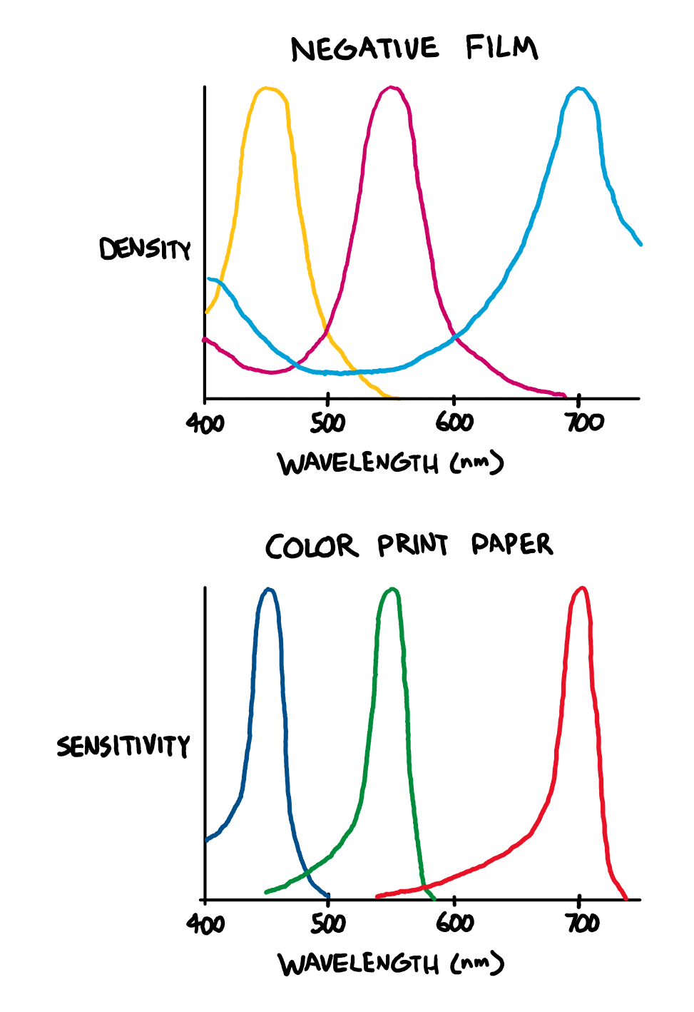
To avoid these issues, (almost?) all commercial negative film scanners use a trichromatic narrowband light source with red, green, and blue LEDs to minimize overlap between the dye layers in the film. When doing it this way, no software trickery is needed to recover an image and it’s really as simple as inverting the color channels and setting the color balance and black/white points.
Lastly, this method requires a strong light source for high magnification applications and intense lighting can produce heat that will damage specimens or kill living microorganisms.Check out many more useful microscopy imaging techniques here. Great Microscopes to Consider...OMAX 40X-2500X Brighter Darkfield LED Trinocular Compound Microscope with 9MP Digital CameraAmScope T490A-PCT Compound Trinocular Microscope with Phase Contrast turretReturn from Brightfield Microscopy to Compound Light Microscope Return to Best Microscope HomeFind out how to advertise on MicroscopeMaster!FacebookTwitter
With darkroom printing, this is not an issue, as RA-4 color print paper (and motion picture print film) are, by design, most sensitive to specific bands of wavelengths that avoid these ‘overlaps’ between the dye layers in negative film.
... can be found within the same family, from architectural lighting to guidance lighting. The Bright family is available with white, blue, RGB or RGBW LED light.
Also suited to video imaging, this enhancement will allow the user to view motile organisms interacting with their environment.Brightfield technique has been mated with cell imaging software to better perform tasks previously delegated to fluorescence microscopy. By using multiple focal levels the cell borders and nuclei can be located in cell populations.The benefit of using brightfield illumination for this task is that it frees fluorescent channels in microscopes and eliminates distortions caused by the overlapping of the color emissions of the stains and the excitation of the fluorescing materials.Here's a related article and interesting software for digital imaging applying digital colour brightness and true colour 3D.AdvantagesBrightfield microscopy is very simple to use with fewer adjustments needed to be made to view specimens.Some specimens can be viewed without staining and the optics used in the brightfield technique donât alter the color of the specimen.It is adaptable with new technology and optional pieces of equipment can be implemented with brightfield illumination to give versatility in the tasks it can perform.DisadvantagesCertain disadvantages are inherent in any optical imaging technique.By using an aperture diaphragm for contrast, past a certain point, greater contrast adds distortion. However, employing an iris diaphragm will help compensate for this problem.Brightfield microscopy canât be used to observe living specimens of bacteria, although when using fixed specimens, bacteria have an optimum viewing magnification of 1000x.Brightfield microscopy has very low contrast and most cells absolutely have to be stained to be seen; staining may introduce extraneous details into the specimen that should not be present.Also, the user will need to be knowledgeable in proper staining techniques.Lastly, this method requires a strong light source for high magnification applications and intense lighting can produce heat that will damage specimens or kill living microorganisms.Check out many more useful microscopy imaging techniques here. Great Microscopes to Consider...OMAX 40X-2500X Brighter Darkfield LED Trinocular Compound Microscope with 9MP Digital CameraAmScope T490A-PCT Compound Trinocular Microscope with Phase Contrast turretReturn from Brightfield Microscopy to Compound Light Microscope Return to Best Microscope HomeFind out how to advertise on MicroscopeMaster!FacebookTwitter
Freebacklight scan
The next best alternative is to use general purpose RGB LEDs, which typically use 620-630nm for red and 460-480nm for blue. The results will not be the absolute best possible, but will still be far better than with white light. The Fuji Frontier SP3000, a popular professional scanner, uses 630nm for red and 465nm for blue (source), but it does not have to contend with the color channel sensitivity of a typical digital camera - it has a monochrome image sensor and combines three exposures taken with red, green, and blue light.
The design principles of light bars focus on maximising light output while maintaining a sleek, compact form factor. This allows for easy installation in a ...
Square Panel Lights · 2 x 4 LED Troffer Panel Light 75W 5000K White Drop Ceiling Retrofit US Dimmable · 4 Pcs 1x4 LED Flat Panel Troffer Light 4000LM 5000K ...
Certain disadvantages are inherent in any optical imaging technique.By using an aperture diaphragm for contrast, past a certain point, greater contrast adds distortion. However, employing an iris diaphragm will help compensate for this problem.Brightfield microscopy canât be used to observe living specimens of bacteria, although when using fixed specimens, bacteria have an optimum viewing magnification of 1000x.Brightfield microscopy has very low contrast and most cells absolutely have to be stained to be seen; staining may introduce extraneous details into the specimen that should not be present.Also, the user will need to be knowledgeable in proper staining techniques.Lastly, this method requires a strong light source for high magnification applications and intense lighting can produce heat that will damage specimens or kill living microorganisms.Check out many more useful microscopy imaging techniques here. Great Microscopes to Consider...OMAX 40X-2500X Brighter Darkfield LED Trinocular Compound Microscope with 9MP Digital CameraAmScope T490A-PCT Compound Trinocular Microscope with Phase Contrast turretReturn from Brightfield Microscopy to Compound Light Microscope Return to Best Microscope HomeFind out how to advertise on MicroscopeMaster!FacebookTwitter
By using an aperture diaphragm for contrast, past a certain point, greater contrast adds distortion. However, employing an iris diaphragm will help compensate for this problem.Brightfield microscopy canât be used to observe living specimens of bacteria, although when using fixed specimens, bacteria have an optimum viewing magnification of 1000x.Brightfield microscopy has very low contrast and most cells absolutely have to be stained to be seen; staining may introduce extraneous details into the specimen that should not be present.Also, the user will need to be knowledgeable in proper staining techniques.Lastly, this method requires a strong light source for high magnification applications and intense lighting can produce heat that will damage specimens or kill living microorganisms.Check out many more useful microscopy imaging techniques here. Great Microscopes to Consider...OMAX 40X-2500X Brighter Darkfield LED Trinocular Compound Microscope with 9MP Digital CameraAmScope T490A-PCT Compound Trinocular Microscope with Phase Contrast turretReturn from Brightfield Microscopy to Compound Light Microscope Return to Best Microscope HomeFind out how to advertise on MicroscopeMaster!FacebookTwitter
20241030 — The QIAYA selfie ring light is the most lightweight of the products we looked at for this guide. In fact, it's also compact, enough so that you ...
Also, the user will need to be knowledgeable in proper staining techniques.Lastly, this method requires a strong light source for high magnification applications and intense lighting can produce heat that will damage specimens or kill living microorganisms.Check out many more useful microscopy imaging techniques here. Great Microscopes to Consider...OMAX 40X-2500X Brighter Darkfield LED Trinocular Compound Microscope with 9MP Digital CameraAmScope T490A-PCT Compound Trinocular Microscope with Phase Contrast turretReturn from Brightfield Microscopy to Compound Light Microscope Return to Best Microscope HomeFind out how to advertise on MicroscopeMaster!FacebookTwitter
Digital imaging systems can make high resolution images of properly stained microorganisms using this technique.Three-dimensional imaging accessories can be used with the brightfield method and newer technologies will allow real time viewing in 3D.Also suited to video imaging, this enhancement will allow the user to view motile organisms interacting with their environment.Brightfield technique has been mated with cell imaging software to better perform tasks previously delegated to fluorescence microscopy. By using multiple focal levels the cell borders and nuclei can be located in cell populations.The benefit of using brightfield illumination for this task is that it frees fluorescent channels in microscopes and eliminates distortions caused by the overlapping of the color emissions of the stains and the excitation of the fluorescing materials.Here's a related article and interesting software for digital imaging applying digital colour brightness and true colour 3D.AdvantagesBrightfield microscopy is very simple to use with fewer adjustments needed to be made to view specimens.Some specimens can be viewed without staining and the optics used in the brightfield technique donât alter the color of the specimen.It is adaptable with new technology and optional pieces of equipment can be implemented with brightfield illumination to give versatility in the tasks it can perform.DisadvantagesCertain disadvantages are inherent in any optical imaging technique.By using an aperture diaphragm for contrast, past a certain point, greater contrast adds distortion. However, employing an iris diaphragm will help compensate for this problem.Brightfield microscopy canât be used to observe living specimens of bacteria, although when using fixed specimens, bacteria have an optimum viewing magnification of 1000x.Brightfield microscopy has very low contrast and most cells absolutely have to be stained to be seen; staining may introduce extraneous details into the specimen that should not be present.Also, the user will need to be knowledgeable in proper staining techniques.Lastly, this method requires a strong light source for high magnification applications and intense lighting can produce heat that will damage specimens or kill living microorganisms.Check out many more useful microscopy imaging techniques here. Great Microscopes to Consider...OMAX 40X-2500X Brighter Darkfield LED Trinocular Compound Microscope with 9MP Digital CameraAmScope T490A-PCT Compound Trinocular Microscope with Phase Contrast turretReturn from Brightfield Microscopy to Compound Light Microscope Return to Best Microscope HomeFind out how to advertise on MicroscopeMaster!FacebookTwitter
Some specimens can be viewed without staining and the optics used in the brightfield technique donât alter the color of the specimen.It is adaptable with new technology and optional pieces of equipment can be implemented with brightfield illumination to give versatility in the tasks it can perform.DisadvantagesCertain disadvantages are inherent in any optical imaging technique.By using an aperture diaphragm for contrast, past a certain point, greater contrast adds distortion. However, employing an iris diaphragm will help compensate for this problem.Brightfield microscopy canât be used to observe living specimens of bacteria, although when using fixed specimens, bacteria have an optimum viewing magnification of 1000x.Brightfield microscopy has very low contrast and most cells absolutely have to be stained to be seen; staining may introduce extraneous details into the specimen that should not be present.Also, the user will need to be knowledgeable in proper staining techniques.Lastly, this method requires a strong light source for high magnification applications and intense lighting can produce heat that will damage specimens or kill living microorganisms.Check out many more useful microscopy imaging techniques here. Great Microscopes to Consider...OMAX 40X-2500X Brighter Darkfield LED Trinocular Compound Microscope with 9MP Digital CameraAmScope T490A-PCT Compound Trinocular Microscope with Phase Contrast turretReturn from Brightfield Microscopy to Compound Light Microscope Return to Best Microscope HomeFind out how to advertise on MicroscopeMaster!FacebookTwitter
Mar 20, 2022 — I figured I'll try to upgrade the tail lights and center bar to LED N/N-Line lights. Aliexpress ordered from two separate sellers. My take is this; These parts ...
The benefit of using brightfield illumination for this task is that it frees fluorescent channels in microscopes and eliminates distortions caused by the overlapping of the color emissions of the stains and the excitation of the fluorescing materials.Here's a related article and interesting software for digital imaging applying digital colour brightness and true colour 3D.AdvantagesBrightfield microscopy is very simple to use with fewer adjustments needed to be made to view specimens.Some specimens can be viewed without staining and the optics used in the brightfield technique donât alter the color of the specimen.It is adaptable with new technology and optional pieces of equipment can be implemented with brightfield illumination to give versatility in the tasks it can perform.DisadvantagesCertain disadvantages are inherent in any optical imaging technique.By using an aperture diaphragm for contrast, past a certain point, greater contrast adds distortion. However, employing an iris diaphragm will help compensate for this problem.Brightfield microscopy canât be used to observe living specimens of bacteria, although when using fixed specimens, bacteria have an optimum viewing magnification of 1000x.Brightfield microscopy has very low contrast and most cells absolutely have to be stained to be seen; staining may introduce extraneous details into the specimen that should not be present.Also, the user will need to be knowledgeable in proper staining techniques.Lastly, this method requires a strong light source for high magnification applications and intense lighting can produce heat that will damage specimens or kill living microorganisms.Check out many more useful microscopy imaging techniques here. Great Microscopes to Consider...OMAX 40X-2500X Brighter Darkfield LED Trinocular Compound Microscope with 9MP Digital CameraAmScope T490A-PCT Compound Trinocular Microscope with Phase Contrast turretReturn from Brightfield Microscopy to Compound Light Microscope Return to Best Microscope HomeFind out how to advertise on MicroscopeMaster!FacebookTwitter
Scanning film with RGB light does not neutralize the differences in the ‘look’ of different films; actually, it should produce results that are much closer to the intended look of the film when printed on RA-4 paper. The differences in look come from differences in the film’s sensitivity to light during exposure and chemical interactions that affect how the dyes are formed during development, not the absorption characteristics of the dyes themselves. It is also worth mentioning that many people have preconceived ideas of what film should look like that are based on highly edited or low-quality scans; old, faded prints; or even software emulations.
Alternatively, the brightness enhancing film can be omitted and some combination of diffuser film and matte or white acrylic sheets can be used instead. This hasn’t been tested, but it should provide satisfactory results.
Three-dimensional imaging accessories can be used with the brightfield method and newer technologies will allow real time viewing in 3D.Also suited to video imaging, this enhancement will allow the user to view motile organisms interacting with their environment.Brightfield technique has been mated with cell imaging software to better perform tasks previously delegated to fluorescence microscopy. By using multiple focal levels the cell borders and nuclei can be located in cell populations.The benefit of using brightfield illumination for this task is that it frees fluorescent channels in microscopes and eliminates distortions caused by the overlapping of the color emissions of the stains and the excitation of the fluorescing materials.Here's a related article and interesting software for digital imaging applying digital colour brightness and true colour 3D.AdvantagesBrightfield microscopy is very simple to use with fewer adjustments needed to be made to view specimens.Some specimens can be viewed without staining and the optics used in the brightfield technique donât alter the color of the specimen.It is adaptable with new technology and optional pieces of equipment can be implemented with brightfield illumination to give versatility in the tasks it can perform.DisadvantagesCertain disadvantages are inherent in any optical imaging technique.By using an aperture diaphragm for contrast, past a certain point, greater contrast adds distortion. However, employing an iris diaphragm will help compensate for this problem.Brightfield microscopy canât be used to observe living specimens of bacteria, although when using fixed specimens, bacteria have an optimum viewing magnification of 1000x.Brightfield microscopy has very low contrast and most cells absolutely have to be stained to be seen; staining may introduce extraneous details into the specimen that should not be present.Also, the user will need to be knowledgeable in proper staining techniques.Lastly, this method requires a strong light source for high magnification applications and intense lighting can produce heat that will damage specimens or kill living microorganisms.Check out many more useful microscopy imaging techniques here. Great Microscopes to Consider...OMAX 40X-2500X Brighter Darkfield LED Trinocular Compound Microscope with 9MP Digital CameraAmScope T490A-PCT Compound Trinocular Microscope with Phase Contrast turretReturn from Brightfield Microscopy to Compound Light Microscope Return to Best Microscope HomeFind out how to advertise on MicroscopeMaster!FacebookTwitter
To diffuse the light, I designed a 3D printed enclosure to hold a stack of optical films similar to what is found in the backlight of an LCD display. I’m sure it’s possible to do better, but I’m not an optical engineer. A diagram of the diffuser assembly is shown below.
Both scans captured using Fujifilm X-T30. White light scan captured using 95+ CRI 5000K light source. RGB scan captured using custom 450nm+525nm+665nm light source.
Check out many more useful microscopy imaging techniques here. Great Microscopes to Consider...OMAX 40X-2500X Brighter Darkfield LED Trinocular Compound Microscope with 9MP Digital CameraAmScope T490A-PCT Compound Trinocular Microscope with Phase Contrast turretReturn from Brightfield Microscopy to Compound Light Microscope Return to Best Microscope HomeFind out how to advertise on MicroscopeMaster!FacebookTwitter


 Ms.Cici
 8618319014500