
DARKFIELD - darkfield usa
Teledyne Adimec is an ISO 9001:2015 certified medium- sized company that designs and produces reliable industrial cameras in small batches to meet customer demands at global OEMs. We serve three strategic markets. – Machine Vision, Healthcare and Global Security.
Widefield microscope
Do you want brighter map lights, but without the dual color switchback feature? These Ultimate map SINGLE COLOR versions are the way to go!
Illumination | 154761 followers on LinkedIn. Creating Joy & Discovery, founded by Academy Award® nominee Chris Meledandri. | About Illumination Illumination, founded by Chris Meledandri in 2007, is one of the entertainment industry's leading producers of event-animated films, including Despicable Me—the most ...
The well known Photo Response Non-Uniformity, or PRNU, calibration is optimized to reduce the pixel-to-pixel variation independent of the shading caused by the camera lens. This calibration combined with bright field lighting allows for the optimization of bright field measurement. Often this calibration is used in conjunction with the Low Frequency Flat Field correction, which is a calibration that not only removes shading caused by the lens, but by using multiple live sets of calibrations it can correct for the shading of the different light sources. Thanks to camera sensitivity matching and these calibrations, the same lighting recipe can be used with each Adimec camera. They all will return the exact same measurement, independent of which camera you put in your machine.
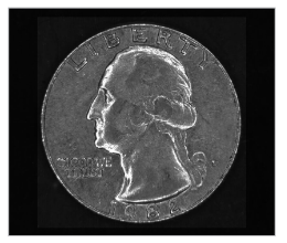
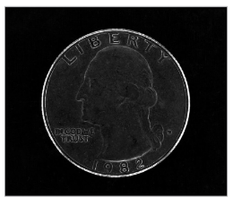
See below a comparison of images taken with Bright Field Illumination and images taken with Dark Field illumination to see the differences between the two lighting techniques:
Darkfield microscopy is not a area where we at Adimec specialize in, and we focus mainly on the camera (so only a bit on lighting & lenses), so I can’t give much feedback on your question.
2023115 — Bright would be the best of the two, but bright also implies more light than just 'has plenty of natural light' - bright implies more than a ...
Köhlerillumination
Ultimately, these lighting techniques are used to help you achieve the key principle of machine vision illumination, which is to capture the correct light-dark contrast in the image. The techniques and principles for dark field and bright field illumination are there to assist you with properly setting up the best illumination for what you are imaging, and it is always advisable to test with a few different illumination set-ups. An inspection may in theory be best suited by bright field illumination, but you may discover that after testing the set-up, there could be a reflection that makes the inspection impossible, at which point understanding the lighting techniques for dark field illumination can assist you in eliminating this reflection.
Step 2. After successful HDMI connection, the microscope will be turned on and the image will appear on the display. At this time, microscope button and remote-control button can be operated; when operating photo/video, the file will be automatically saved in TF card.
Unlike in bright field lighting, where reflected light is imaged, dark field lighting only captures scattered light. By imaging only the scattered light, edges and surface defects become more prominent in the image as they are the things that best scatter light. To best set up a light source for this light scattering, a low angle of light (usually from ring lights) of around 10-15 degrees is ideal. This low angle allows for edges, defects, ridges, etc. to properly scatter the light while not having the surface of the target reflect too much light back to the camera. This technique can also be used to effectively inspect highly reflective or mirrored surfaces which you would otherwise be unable to inspect with bright field lighting.
One of the key components of machine vision imaging is determining what kind of lighting is optimal for your set-up to achieve the best light-dark contrast; bright field lighting or dark field lighting. Bright field is the more commonly used lighting technique whereas dark field is advantageous when imaging things such as reflective surfaces and edge inspections. In this blog we will discuss the lighting requirements for Bright Field and Dark Field Illumination and their advantages and disadvantages in imaging.
Illuminations, Inc. offers comprehensive sales, application, and technical support, providing exceptional service from project inception to completion.
Price ... myDMX GO Lighting Control System for iPad/Andro… ... Data Stream 4 Universal DMX-512 Optical Splitte… ... myDMX RM - DMX Rackmount ...
Language: English, French, Spanish, Portuguese, German, Italian, Simplified Chinese, Traditional Chinese, Russian, Japanese, Korean
Illumination (formerly known as Illumination Entertainment) is an American computer animation studio, founded by Chris Meledandri in 2007.
LED lights cover a major part of industrial applications. Halogen and fluorescent lamps are only frequently used in applications which require large-area ...
◆ 48 megapixel photography and 1080P video recording; Electronic zoom function; Support manual/automatic exposure, exposure
Edgeless Ultra Bright LED Combination Spot-Flood Light Bar - Single Row Series. Product Common Use: Work Trucks, Tow Trucks, Utility Trucks, ATVs and Other Off ...
Criticalillumination
Whoever I looked into darkfield Microscopy, and there is a short animation of how it works on wikipedia (https://en.wikipedia.org/wiki/Dark-field_microscopy) To work with this method to check on exomes, I can imagine some wavelengths work better than others based on the type and size of the exome (so that specific light is scattered more and gives a better/sharper image results) I do not have any formulas to for you to work with, but note that a lot of factors are dependent on the setup you are using (lenses, wavelength, size and angle of darkfield patch stop, and distances from Lightsource-to sample- to camera, in addition to the sample properties (thickness, transparency etc).
When the icon on the upper left corner of the microscope is "it is in Video mode, when the microscope is on, short press the Menu button on the microscope or short press the Video button on the remote control, it can switch to Video mode
◆ Provide 8 line drawing functions, line color, thickness, position adjustable; Display/close center cross; Support TF card
Hi Scott, let me introduce myself, Recently I’m doing research to build a Dark Field Microscopy that is able to observe an exosome. So, if you willing you can reply my question by emailing me. I want to ask about the light that we can use for darkfield microscope. Which do you recommend between using laser or light to apply it to the darkfield microscope and can you provide the potential candidate of the light source? . Second question is how we know the required intensity is needed for the dark field imaging system (if know do you know how to calculate it?). Third question do you know the reason why darkfield will achieve better resolution than brightfield? (if you know the calculation you can provide to me).
Welcome to the Light FX New Zealand home page. All the information supplied is kept as up to date as possible, however please take the time to fill out a ...
Darkfieldmicroscopy
Adimec’s cameras are optimized to have the lowest read noise when they leave the factory. By supporting analog gain the read noise can even further be decreased. This optimization increases the measurement accuracy in the darkest parts of an image. The cameras are calibrated in Adimec’s factory by using a Dark Signal Non-Uniformity (DSNU) calibration. While they are calibrated in factory, these cameras can be re-calibrated in a system to optimize the image even further depending on the use case. This DSNU reduces pixel-to-pixel fixed pattern, which in turn increases the measurement accuracy
Better security: The battery adapter for m12(1pcs included) is equipped with a fuse and high-quality wire to prevent excessive transient current from ...
The only answer I can give you why a darkfield image is preferred over a brightfield image: With a darkfield image you clearify the edges or structures of sampels, this method is used to detect or check non-uniformities like a defect or if a required structure is present. Like in the wikipedia site, they do this to see the edges of bloodcells, making them easy to count, check on “roundness”, or detect non-uniformities.
When the icon in the upper left corner of the microscope is Photo mode when the microscope is on, short press the Menu button on the microscope or short press the Photo button on the remote control, it can switch to Photo mode.
Bright field lighting is the method for imaging reflected light. That is, the light coming from the source is reflected into the camera so that small defects and edges which typically scatter light are not picked up by the camera. This creates a bright image, but areas with engravings, scratches, or indentations may not be as well defined. In addition, due to the reflection of light, reflective surfaces are difficult to image with this lighting set-up. The light source will be scattered less by the object’s surface and more light will be reflected back into the camera, causing a bright spot in the image, as seen below. To properly set up Bright Field Lighting, you want the light sources to be at an angle to the subject or imaging surface of 45 and 90 degrees. Typically positioning these light sources closer to the subject or surface is advantageous, as this helps cover a larger surface area and can help eliminate some of the issues seen with imaging reflective surfaces or edges.


 Ms.Cici
 8618319014500