
Dark Field And Phase Contrast - Microscopes - how does dark field microscopy work
Keeping light out of the objective reveals the light that was scattered by samples on the slide itself. Instead of coming through the specimen and into the objective as is the case in bright field microscopy, light that is reflected by particles on the slide makes it to the observer.
When you shop for WAC Lighting Track Lighting with Lighting New York, you will enjoy our hassle-free buying experience where we simplify the process and take care of you every step of the way, ensuring that you will be proud of the spaces that you create. Guaranteed.
Registered members can chat with Azthena, request quotations, download pdf's, brochures and subscribe to our related newsletter content.
Track Lightingheads
As a high-contrast microscopy technique, dark-field microscopy is a cheaper alternative to more advanced phase-contrast optics developed in the 1940s by Dutch scientist Frits Zernike. Today, inexpensive dark field microscopy instrumentation delivers better contrast and resolution than student-grade phase-contrast equipment is capable of.
All orders over $49 ship for FREE in the contiguous United States. Contact a Lighting Expert for international shipping options.
While we only use edited and approved content for Azthena answers, it may on occasions provide incorrect responses. Please confirm any data provided with the related suppliers or authors. We do not provide medical advice, if you search for medical information you must always consult a medical professional before acting on any information provided.
Although more sophisticated high-contrast microscopy methods have been developed, dark-field microscopy is effective in some instances. It is a beneficial tool for studying biological and medical phenomena that cannot be easily separated from their immediate surroundings.
Track lightingtypes
Because of the way that it is manipulated in a dark field microscope, light passes through the condenser’s outer edge at a wide-angle, then strikes the sample at an oblique angle. This causes the light scattered by the sample to reach the objective lens, while other light that merely passes through the specimen misses the objective. The result is a brightly illuminated sample on a dark background.
Track lightingfor kitchen Ceiling
Beneath the opening, the surface should be a matt black. In-built illuminators must be switched off and replaced with a high-intensity light source such as a laser. Pointing this source to the specimen at an angle from the top or side through a glass dish or jar will generate the desired dark-field effect.
Dark-field microscopes were extremely popular in the first half of the 20th century, and many researchers focused on optimizing condenser systems and illuminators to make the technique as effective as possible. However, the introduction of phase-contrast, differential contrast, and Hoffman modulation contrast from the middle of the 20th century has caused dark-field techniques to fade from view.
A normal student microscope can be set up for dark-field microscopy by ensuring that light will only reflect from the surfaces between the coverslip and slide, but not from surfaces beneath the slide. Occulting discs can be easily prepared by cutting circles of black electrical tape in different sizes. These can be placed on the slide, evenly spaced in a row. Depending on the configuration of the microscope, a stand may be required to hold the slide in the correct position.
Dark-field microscopes are optical microscopes with an extra non-transparent disc placed beneath the condenser lens or a specially designed condenser lens that has a central area blacked out. With either technique, light from the source cannot directly enter the microscope’s objective.
Disclaimer: The views expressed here are those of the author expressed in their private capacity and do not necessarily represent the views of AZoM.com Limited T/A AZoNetwork the owner and operator of this website. This disclaimer forms part of the Terms and conditions of use of this website.
Pilkington, Ben. "What are Dark-Field Microscopes?". AZoOptics. 17 December 2024. .

Reuven Silverman of Ophir discusses the critical role of M2 measurements in laser technology for optimization and quality control in various industries.
Track lightingmodern
LIS Technologies is on the road to transforming nuclear fuel enrichment through advanced laser techniques, ensuring a sustainable and cost-effective approach to energy production.
Your questions, but not your email details will be shared with OpenAI and retained for 30 days in accordance with their privacy principles.
Ben Pilkington is a freelance writer who is interested in society and technology. He enjoys learning how the latest scientific developments can affect us and imagining what will be possible in the future. Since completing graduate studies at Oxford University in 2016, Ben has reported on developments in computer software, the UK technology industry, digital rights and privacy, industrial automation, IoT, AI, additive manufacturing, sustainability, and clean technology.
In this interview, Patrice Dionne, M.Sc, the product line manager for optical sensing at TeraXion, talks to AzoOptics about TeraXion's motivations and innovations in DFB laser technology and various real-world applications where their technology truly shines.
Dark-field effects can be obtained with relatively basic optical microscopy equipment, modified with an occulting disc. A reliable optical microscope can be modified to reflect light toward the viewer rather than pass directly through the sample to the viewer, particularly at low magnification (up to 100x). However, specialist dark field instrumentation with occulting discs that are built into the condenser itself yield much more dramatic dark-field imagery.
Caprette, D. R. (2012) Dark Field Viewing. [Online] Rice University. Available at: https://www.ruf.rice.edu/~bioslabs/methods/microscopy/dfield.html
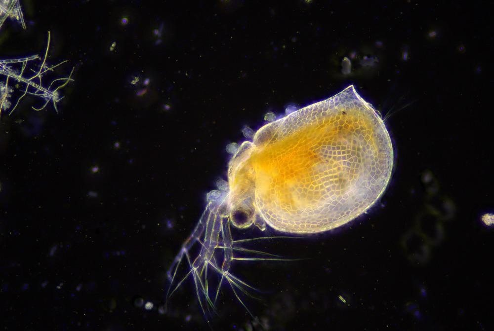
Dark-field microscopy has been used since the early 20th century to observe living, unstained cells and microorganisms. Specimens are brightly lit against a dark background to provide the contrast needed to study samples in solutions or natural aquatic environments such as pond water. This article looks at what dark-field microscopy is and its applications.
Zernike, F. (1955) How I Discovered Phase Contrast. Science. Vol 121, Issue 3141, pp. 345-349. https://doi.org/10.1126/science.121.3141.345.
Pilkington, Ben. "What are Dark-Field Microscopes?". AZoOptics. https://www.azooptics.com/Article.aspx?ArticleID=2125. (accessed December 17, 2024).
Dark-field microscopy is slightly more complicated in stereomicroscopy. A specialized stand that has a reflection mirror and a plate to shield light is required. This directs an inverted hollow cone of light toward the specimen at the correct oblique angles for a dark-field effect. Complex condenser systems with multiple lenses, or condensers with internal mirrors that reflect surfaces at specific geometries, are used in high-end stereo microscopes for dark-field microscopy.
Pilkington, Ben. 2022. What are Dark-Field Microscopes?. AZoOptics, viewed 17 December 2024, https://www.azooptics.com/Article.aspx?ArticleID=2125.
Track lighting has undergone many changes in recent years. The trend in track lighting has been toward smaller fixtures, which are much less noticeable in the space. Track lighting is excellent for its flexibility and can provide ambient, task or accent lighting. You can move, swivel, rotate and aim the individual fixtures in any direction along the track, giving you the versatility to change the lighting scheme when the need arises. With special attachments, you also can hang chandeliers and pendants from the track.
Pilkington, Ben. (2022, January 20). What are Dark-Field Microscopes?. AZoOptics. Retrieved on December 17, 2024 from https://www.azooptics.com/Article.aspx?ArticleID=2125.
Specimens are placed on the microscope stage to obtain a dark field optical image. The illumination must be as uniform as possible, and aperture diaphragm available in the condenser needs to be opened as wide as possible.
Any specimens in liquid samples can still be viewed with dark-field microscopy, particularly those that need to be seen with all the debris in the liquid.
AmazonTrack Lighting
LEDTrack lighting
The microscope operator focuses at low power, then places a slide with occulting discs between the light source and the condenser in the light’s path. The slide should be brought as close as possible to the bottom of the condenser.
Chambers, W., T. J. Fellers, and M. W. Davidson. Darkfield Illumination. [Online] Nikon Microscopy U. Available at: https://www.microscopyu.com/techniques/stereomicroscopy/darkfield-illumination

But there is still interest in developing and optimizing dark field microscopy techniques. Transmitted dark-field microscopy has recently received renewed interest as it can be used in combination with fluorescence microscopy. It is expected that newer high-contrast techniques, such as the oblique coherent contrast method developed by Nikon, will eventually displace dark field microscopy in research and industry settings.
Rijal, N. (2021) Dark-field Microscopy: Principle and Uses. [Online] Microbe Online. Available at: https://microbeonline.com/dark-field-microscopy/
Unless otherwise noted, we also offer free returns on regularly shipped items. We will accept returned freight orders, however, the customer is responsible for the return freight charges.


 Ms.Cici
 8618319014500