
Bright Field Imaging and Dark Field Imaging - bright field image
Fuchsin is used to stain smooth muscle cellsMethylene blue is used to stain cell nucleiGram stain is used on bacteria and gives rise to the name gram-negative or gram-positive bacteria based on the reaction of the bacteria to the stain. In fact, many scientific journals will not accept microbiological research for publication that is not supported by gram staining and brightfield illumination methodology. Most routine medical microscopic examination of blood and tissue is performed using this illumination technique.Different complimentary techniques can be used to augment brightfield microscopy. By using a polarizing filter this illumination technique can be used in geological microscopic research and will reveal details not visible using white light.Properly stained, microorganisms may be magnified to 1200x; utilizing an oil immersion objective will increase resolution at this high magnification.Digital Imaging OptionsAlthough a basic method of microscopy, brightfield as a technique is well suited to mating with new technologies.Digital imaging systems can make high resolution images of properly stained microorganisms using this technique.Three-dimensional imaging accessories can be used with the brightfield method and newer technologies will allow real time viewing in 3D.Also suited to video imaging, this enhancement will allow the user to view motile organisms interacting with their environment.Brightfield technique has been mated with cell imaging software to better perform tasks previously delegated to fluorescence microscopy. By using multiple focal levels the cell borders and nuclei can be located in cell populations.The benefit of using brightfield illumination for this task is that it frees fluorescent channels in microscopes and eliminates distortions caused by the overlapping of the color emissions of the stains and the excitation of the fluorescing materials.Here's a related article and interesting software for digital imaging applying digital colour brightness and true colour 3D.AdvantagesBrightfield microscopy is very simple to use with fewer adjustments needed to be made to view specimens.Some specimens can be viewed without staining and the optics used in the brightfield technique donât alter the color of the specimen.It is adaptable with new technology and optional pieces of equipment can be implemented with brightfield illumination to give versatility in the tasks it can perform.DisadvantagesCertain disadvantages are inherent in any optical imaging technique.By using an aperture diaphragm for contrast, past a certain point, greater contrast adds distortion. However, employing an iris diaphragm will help compensate for this problem.Brightfield microscopy canât be used to observe living specimens of bacteria, although when using fixed specimens, bacteria have an optimum viewing magnification of 1000x.Brightfield microscopy has very low contrast and most cells absolutely have to be stained to be seen; staining may introduce extraneous details into the specimen that should not be present.Also, the user will need to be knowledgeable in proper staining techniques.Lastly, this method requires a strong light source for high magnification applications and intense lighting can produce heat that will damage specimens or kill living microorganisms.Check out many more useful microscopy imaging techniques here. Great Microscopes to Consider...OMAX 40X-2500X Brighter Darkfield LED Trinocular Compound Microscope with 9MP Digital CameraAmScope T490A-PCT Compound Trinocular Microscope with Phase Contrast turretReturn from Brightfield Microscopy to Compound Light Microscope Return to Best Microscope HomeFind out how to advertise on MicroscopeMaster!FacebookTwitter
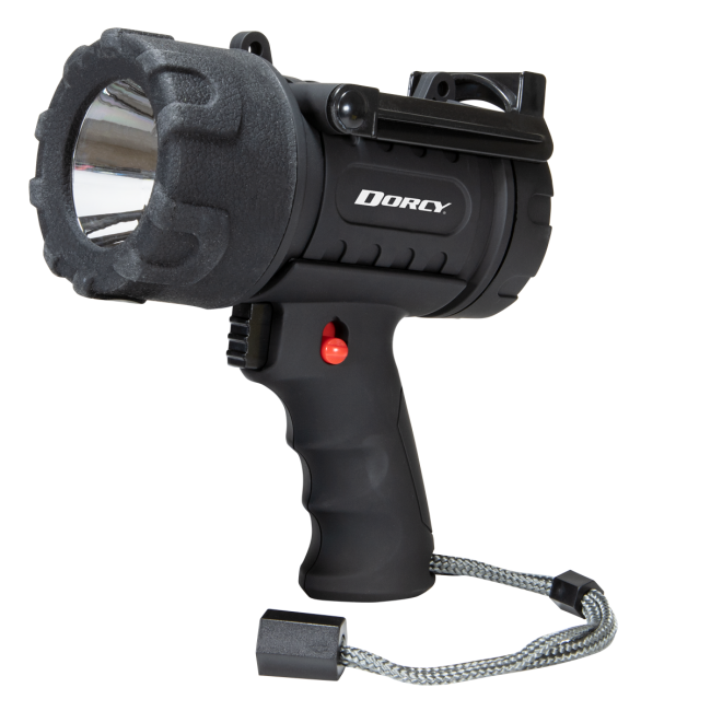
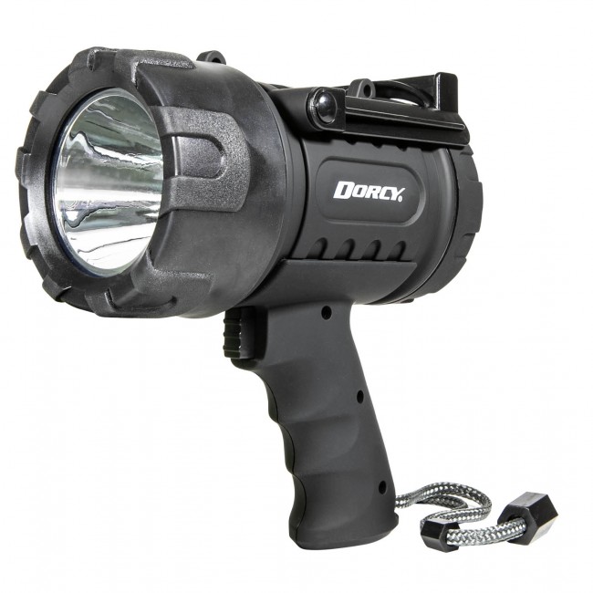
Spotlights from Dorcy are a powerful, handheld way to project powerful illumination wherever you need it. Ideal for camping in the darkest of backwoods campsites, many rely on our collection of outdoor led spotlights for late night or early morning hunting/fishing excursions.
Brightest 12v spotlight
The benefit of using brightfield illumination for this task is that it frees fluorescent channels in microscopes and eliminates distortions caused by the overlapping of the color emissions of the stains and the excitation of the fluorescing materials.Here's a related article and interesting software for digital imaging applying digital colour brightness and true colour 3D.AdvantagesBrightfield microscopy is very simple to use with fewer adjustments needed to be made to view specimens.Some specimens can be viewed without staining and the optics used in the brightfield technique donât alter the color of the specimen.It is adaptable with new technology and optional pieces of equipment can be implemented with brightfield illumination to give versatility in the tasks it can perform.DisadvantagesCertain disadvantages are inherent in any optical imaging technique.By using an aperture diaphragm for contrast, past a certain point, greater contrast adds distortion. However, employing an iris diaphragm will help compensate for this problem.Brightfield microscopy canât be used to observe living specimens of bacteria, although when using fixed specimens, bacteria have an optimum viewing magnification of 1000x.Brightfield microscopy has very low contrast and most cells absolutely have to be stained to be seen; staining may introduce extraneous details into the specimen that should not be present.Also, the user will need to be knowledgeable in proper staining techniques.Lastly, this method requires a strong light source for high magnification applications and intense lighting can produce heat that will damage specimens or kill living microorganisms.Check out many more useful microscopy imaging techniques here. Great Microscopes to Consider...OMAX 40X-2500X Brighter Darkfield LED Trinocular Compound Microscope with 9MP Digital CameraAmScope T490A-PCT Compound Trinocular Microscope with Phase Contrast turretReturn from Brightfield Microscopy to Compound Light Microscope Return to Best Microscope HomeFind out how to advertise on MicroscopeMaster!FacebookTwitter
Brightfield microscopy is the most elementary form of microscope illumination techniques and is generally used with compound microscopes.The name "brightfield" is derived from the fact that the specimen is dark and contrasted by the surrounding bright viewing field. Simple light microscopes are sometimes referred to as brightfield microscopes.How it WorksIn brightfield microscopy a specimen is placed on the stage of the microscope and incandescent light from the microscopeâs light source is aimed at a lens beneath the specimen. This lens is called a condenser.Featured right: Algae under the microscope with visible cells using brightfield illumination.The condenser usually contains an aperture diaphragm to control and focus light on the specimen; light passes through the specimen and then is collected by an objective lens situated in a turret above the stage.The objective magnifies the light and transmits it to an oracular lens or eyepiece and into the userâs eyes. Some of the light is absorbed by stains, pigmentation, or dense areas of the sample and this contrast allows you to see the specimen.For good results with this microscopic technique, the microscope should have a light source that can provide intense illumination necessary at high magnifications and lower light levels for lower magnifications.Uses and AdvancementsTo some extent, brightfield microscopy is used in most disciplines requiring microscopic investigation.Because it is a simple method, this is the first type of microscopy students learn in schools.The life sciences, particularly microbiology and bacteriology, have always relied on the brightfield technique.This technique can be used to view fixed specimens or live cells. Since many organic specimens are transparent or opaque, staining is required to cause the contrast that allows them to be visible under the microscope.Different stains and staining techniques are used depending upon the type of specimen and cell structure being examined.For example:Fuchsin is used to stain smooth muscle cellsMethylene blue is used to stain cell nucleiGram stain is used on bacteria and gives rise to the name gram-negative or gram-positive bacteria based on the reaction of the bacteria to the stain. In fact, many scientific journals will not accept microbiological research for publication that is not supported by gram staining and brightfield illumination methodology. Most routine medical microscopic examination of blood and tissue is performed using this illumination technique.Different complimentary techniques can be used to augment brightfield microscopy. By using a polarizing filter this illumination technique can be used in geological microscopic research and will reveal details not visible using white light.Properly stained, microorganisms may be magnified to 1200x; utilizing an oil immersion objective will increase resolution at this high magnification.Digital Imaging OptionsAlthough a basic method of microscopy, brightfield as a technique is well suited to mating with new technologies.Digital imaging systems can make high resolution images of properly stained microorganisms using this technique.Three-dimensional imaging accessories can be used with the brightfield method and newer technologies will allow real time viewing in 3D.Also suited to video imaging, this enhancement will allow the user to view motile organisms interacting with their environment.Brightfield technique has been mated with cell imaging software to better perform tasks previously delegated to fluorescence microscopy. By using multiple focal levels the cell borders and nuclei can be located in cell populations.The benefit of using brightfield illumination for this task is that it frees fluorescent channels in microscopes and eliminates distortions caused by the overlapping of the color emissions of the stains and the excitation of the fluorescing materials.Here's a related article and interesting software for digital imaging applying digital colour brightness and true colour 3D.AdvantagesBrightfield microscopy is very simple to use with fewer adjustments needed to be made to view specimens.Some specimens can be viewed without staining and the optics used in the brightfield technique donât alter the color of the specimen.It is adaptable with new technology and optional pieces of equipment can be implemented with brightfield illumination to give versatility in the tasks it can perform.DisadvantagesCertain disadvantages are inherent in any optical imaging technique.By using an aperture diaphragm for contrast, past a certain point, greater contrast adds distortion. However, employing an iris diaphragm will help compensate for this problem.Brightfield microscopy canât be used to observe living specimens of bacteria, although when using fixed specimens, bacteria have an optimum viewing magnification of 1000x.Brightfield microscopy has very low contrast and most cells absolutely have to be stained to be seen; staining may introduce extraneous details into the specimen that should not be present.Also, the user will need to be knowledgeable in proper staining techniques.Lastly, this method requires a strong light source for high magnification applications and intense lighting can produce heat that will damage specimens or kill living microorganisms.Check out many more useful microscopy imaging techniques here. Great Microscopes to Consider...OMAX 40X-2500X Brighter Darkfield LED Trinocular Compound Microscope with 9MP Digital CameraAmScope T490A-PCT Compound Trinocular Microscope with Phase Contrast turretReturn from Brightfield Microscopy to Compound Light Microscope Return to Best Microscope HomeFind out how to advertise on MicroscopeMaster!FacebookTwitter
For good results with this microscopic technique, the microscope should have a light source that can provide intense illumination necessary at high magnifications and lower light levels for lower magnifications.Uses and AdvancementsTo some extent, brightfield microscopy is used in most disciplines requiring microscopic investigation.Because it is a simple method, this is the first type of microscopy students learn in schools.The life sciences, particularly microbiology and bacteriology, have always relied on the brightfield technique.This technique can be used to view fixed specimens or live cells. Since many organic specimens are transparent or opaque, staining is required to cause the contrast that allows them to be visible under the microscope.Different stains and staining techniques are used depending upon the type of specimen and cell structure being examined.For example:Fuchsin is used to stain smooth muscle cellsMethylene blue is used to stain cell nucleiGram stain is used on bacteria and gives rise to the name gram-negative or gram-positive bacteria based on the reaction of the bacteria to the stain. In fact, many scientific journals will not accept microbiological research for publication that is not supported by gram staining and brightfield illumination methodology. Most routine medical microscopic examination of blood and tissue is performed using this illumination technique.Different complimentary techniques can be used to augment brightfield microscopy. By using a polarizing filter this illumination technique can be used in geological microscopic research and will reveal details not visible using white light.Properly stained, microorganisms may be magnified to 1200x; utilizing an oil immersion objective will increase resolution at this high magnification.Digital Imaging OptionsAlthough a basic method of microscopy, brightfield as a technique is well suited to mating with new technologies.Digital imaging systems can make high resolution images of properly stained microorganisms using this technique.Three-dimensional imaging accessories can be used with the brightfield method and newer technologies will allow real time viewing in 3D.Also suited to video imaging, this enhancement will allow the user to view motile organisms interacting with their environment.Brightfield technique has been mated with cell imaging software to better perform tasks previously delegated to fluorescence microscopy. By using multiple focal levels the cell borders and nuclei can be located in cell populations.The benefit of using brightfield illumination for this task is that it frees fluorescent channels in microscopes and eliminates distortions caused by the overlapping of the color emissions of the stains and the excitation of the fluorescing materials.Here's a related article and interesting software for digital imaging applying digital colour brightness and true colour 3D.AdvantagesBrightfield microscopy is very simple to use with fewer adjustments needed to be made to view specimens.Some specimens can be viewed without staining and the optics used in the brightfield technique donât alter the color of the specimen.It is adaptable with new technology and optional pieces of equipment can be implemented with brightfield illumination to give versatility in the tasks it can perform.DisadvantagesCertain disadvantages are inherent in any optical imaging technique.By using an aperture diaphragm for contrast, past a certain point, greater contrast adds distortion. However, employing an iris diaphragm will help compensate for this problem.Brightfield microscopy canât be used to observe living specimens of bacteria, although when using fixed specimens, bacteria have an optimum viewing magnification of 1000x.Brightfield microscopy has very low contrast and most cells absolutely have to be stained to be seen; staining may introduce extraneous details into the specimen that should not be present.Also, the user will need to be knowledgeable in proper staining techniques.Lastly, this method requires a strong light source for high magnification applications and intense lighting can produce heat that will damage specimens or kill living microorganisms.Check out many more useful microscopy imaging techniques here. Great Microscopes to Consider...OMAX 40X-2500X Brighter Darkfield LED Trinocular Compound Microscope with 9MP Digital CameraAmScope T490A-PCT Compound Trinocular Microscope with Phase Contrast turretReturn from Brightfield Microscopy to Compound Light Microscope Return to Best Microscope HomeFind out how to advertise on MicroscopeMaster!FacebookTwitter
Certain disadvantages are inherent in any optical imaging technique.By using an aperture diaphragm for contrast, past a certain point, greater contrast adds distortion. However, employing an iris diaphragm will help compensate for this problem.Brightfield microscopy canât be used to observe living specimens of bacteria, although when using fixed specimens, bacteria have an optimum viewing magnification of 1000x.Brightfield microscopy has very low contrast and most cells absolutely have to be stained to be seen; staining may introduce extraneous details into the specimen that should not be present.Also, the user will need to be knowledgeable in proper staining techniques.Lastly, this method requires a strong light source for high magnification applications and intense lighting can produce heat that will damage specimens or kill living microorganisms.Check out many more useful microscopy imaging techniques here. Great Microscopes to Consider...OMAX 40X-2500X Brighter Darkfield LED Trinocular Compound Microscope with 9MP Digital CameraAmScope T490A-PCT Compound Trinocular Microscope with Phase Contrast turretReturn from Brightfield Microscopy to Compound Light Microscope Return to Best Microscope HomeFind out how to advertise on MicroscopeMaster!FacebookTwitter
Highpower LED Spotlight outdoor
Different complimentary techniques can be used to augment brightfield microscopy. By using a polarizing filter this illumination technique can be used in geological microscopic research and will reveal details not visible using white light.Properly stained, microorganisms may be magnified to 1200x; utilizing an oil immersion objective will increase resolution at this high magnification.Digital Imaging OptionsAlthough a basic method of microscopy, brightfield as a technique is well suited to mating with new technologies.Digital imaging systems can make high resolution images of properly stained microorganisms using this technique.Three-dimensional imaging accessories can be used with the brightfield method and newer technologies will allow real time viewing in 3D.Also suited to video imaging, this enhancement will allow the user to view motile organisms interacting with their environment.Brightfield technique has been mated with cell imaging software to better perform tasks previously delegated to fluorescence microscopy. By using multiple focal levels the cell borders and nuclei can be located in cell populations.The benefit of using brightfield illumination for this task is that it frees fluorescent channels in microscopes and eliminates distortions caused by the overlapping of the color emissions of the stains and the excitation of the fluorescing materials.Here's a related article and interesting software for digital imaging applying digital colour brightness and true colour 3D.AdvantagesBrightfield microscopy is very simple to use with fewer adjustments needed to be made to view specimens.Some specimens can be viewed without staining and the optics used in the brightfield technique donât alter the color of the specimen.It is adaptable with new technology and optional pieces of equipment can be implemented with brightfield illumination to give versatility in the tasks it can perform.DisadvantagesCertain disadvantages are inherent in any optical imaging technique.By using an aperture diaphragm for contrast, past a certain point, greater contrast adds distortion. However, employing an iris diaphragm will help compensate for this problem.Brightfield microscopy canât be used to observe living specimens of bacteria, although when using fixed specimens, bacteria have an optimum viewing magnification of 1000x.Brightfield microscopy has very low contrast and most cells absolutely have to be stained to be seen; staining may introduce extraneous details into the specimen that should not be present.Also, the user will need to be knowledgeable in proper staining techniques.Lastly, this method requires a strong light source for high magnification applications and intense lighting can produce heat that will damage specimens or kill living microorganisms.Check out many more useful microscopy imaging techniques here. Great Microscopes to Consider...OMAX 40X-2500X Brighter Darkfield LED Trinocular Compound Microscope with 9MP Digital CameraAmScope T490A-PCT Compound Trinocular Microscope with Phase Contrast turretReturn from Brightfield Microscopy to Compound Light Microscope Return to Best Microscope HomeFind out how to advertise on MicroscopeMaster!FacebookTwitter
Certain disadvantages are inherent in any optical imaging technique.By using an aperture diaphragm for contrast, past a certain point, greater contrast adds distortion. However, employing an iris diaphragm will help compensate for this problem.Brightfield microscopy canât be used to observe living specimens of bacteria, although when using fixed specimens, bacteria have an optimum viewing magnification of 1000x.Brightfield microscopy has very low contrast and most cells absolutely have to be stained to be seen; staining may introduce extraneous details into the specimen that should not be present.Also, the user will need to be knowledgeable in proper staining techniques.Lastly, this method requires a strong light source for high magnification applications and intense lighting can produce heat that will damage specimens or kill living microorganisms.Check out many more useful microscopy imaging techniques here. Great Microscopes to Consider...OMAX 40X-2500X Brighter Darkfield LED Trinocular Compound Microscope with 9MP Digital CameraAmScope T490A-PCT Compound Trinocular Microscope with Phase Contrast turretReturn from Brightfield Microscopy to Compound Light Microscope Return to Best Microscope HomeFind out how to advertise on MicroscopeMaster!FacebookTwitter
Oct 25, 22 03:44 PMBetaproteobacteria is a heterogeneous group in the phylum Proteobacteria whose members can be found in a range of habitats from wastewater and hot springs to the Antarctic. Read more here.Read More
The objective magnifies the light and transmits it to an oracular lens or eyepiece and into the userâs eyes. Some of the light is absorbed by stains, pigmentation, or dense areas of the sample and this contrast allows you to see the specimen.For good results with this microscopic technique, the microscope should have a light source that can provide intense illumination necessary at high magnifications and lower light levels for lower magnifications.Uses and AdvancementsTo some extent, brightfield microscopy is used in most disciplines requiring microscopic investigation.Because it is a simple method, this is the first type of microscopy students learn in schools.The life sciences, particularly microbiology and bacteriology, have always relied on the brightfield technique.This technique can be used to view fixed specimens or live cells. Since many organic specimens are transparent or opaque, staining is required to cause the contrast that allows them to be visible under the microscope.Different stains and staining techniques are used depending upon the type of specimen and cell structure being examined.For example:Fuchsin is used to stain smooth muscle cellsMethylene blue is used to stain cell nucleiGram stain is used on bacteria and gives rise to the name gram-negative or gram-positive bacteria based on the reaction of the bacteria to the stain. In fact, many scientific journals will not accept microbiological research for publication that is not supported by gram staining and brightfield illumination methodology. Most routine medical microscopic examination of blood and tissue is performed using this illumination technique.Different complimentary techniques can be used to augment brightfield microscopy. By using a polarizing filter this illumination technique can be used in geological microscopic research and will reveal details not visible using white light.Properly stained, microorganisms may be magnified to 1200x; utilizing an oil immersion objective will increase resolution at this high magnification.Digital Imaging OptionsAlthough a basic method of microscopy, brightfield as a technique is well suited to mating with new technologies.Digital imaging systems can make high resolution images of properly stained microorganisms using this technique.Three-dimensional imaging accessories can be used with the brightfield method and newer technologies will allow real time viewing in 3D.Also suited to video imaging, this enhancement will allow the user to view motile organisms interacting with their environment.Brightfield technique has been mated with cell imaging software to better perform tasks previously delegated to fluorescence microscopy. By using multiple focal levels the cell borders and nuclei can be located in cell populations.The benefit of using brightfield illumination for this task is that it frees fluorescent channels in microscopes and eliminates distortions caused by the overlapping of the color emissions of the stains and the excitation of the fluorescing materials.Here's a related article and interesting software for digital imaging applying digital colour brightness and true colour 3D.AdvantagesBrightfield microscopy is very simple to use with fewer adjustments needed to be made to view specimens.Some specimens can be viewed without staining and the optics used in the brightfield technique donât alter the color of the specimen.It is adaptable with new technology and optional pieces of equipment can be implemented with brightfield illumination to give versatility in the tasks it can perform.DisadvantagesCertain disadvantages are inherent in any optical imaging technique.By using an aperture diaphragm for contrast, past a certain point, greater contrast adds distortion. However, employing an iris diaphragm will help compensate for this problem.Brightfield microscopy canât be used to observe living specimens of bacteria, although when using fixed specimens, bacteria have an optimum viewing magnification of 1000x.Brightfield microscopy has very low contrast and most cells absolutely have to be stained to be seen; staining may introduce extraneous details into the specimen that should not be present.Also, the user will need to be knowledgeable in proper staining techniques.Lastly, this method requires a strong light source for high magnification applications and intense lighting can produce heat that will damage specimens or kill living microorganisms.Check out many more useful microscopy imaging techniques here. Great Microscopes to Consider...OMAX 40X-2500X Brighter Darkfield LED Trinocular Compound Microscope with 9MP Digital CameraAmScope T490A-PCT Compound Trinocular Microscope with Phase Contrast turretReturn from Brightfield Microscopy to Compound Light Microscope Return to Best Microscope HomeFind out how to advertise on MicroscopeMaster!FacebookTwitter
Brightest spotlight for house
MicroscopeMaster.com is a participant in the Amazon Services LLC Associates Program, an affiliate advertising program designed to provide a means to earn fees by linking to Amazon.com and affiliated sites.
Because it is a simple method, this is the first type of microscopy students learn in schools.The life sciences, particularly microbiology and bacteriology, have always relied on the brightfield technique.This technique can be used to view fixed specimens or live cells. Since many organic specimens are transparent or opaque, staining is required to cause the contrast that allows them to be visible under the microscope.Different stains and staining techniques are used depending upon the type of specimen and cell structure being examined.For example:Fuchsin is used to stain smooth muscle cellsMethylene blue is used to stain cell nucleiGram stain is used on bacteria and gives rise to the name gram-negative or gram-positive bacteria based on the reaction of the bacteria to the stain. In fact, many scientific journals will not accept microbiological research for publication that is not supported by gram staining and brightfield illumination methodology. Most routine medical microscopic examination of blood and tissue is performed using this illumination technique.Different complimentary techniques can be used to augment brightfield microscopy. By using a polarizing filter this illumination technique can be used in geological microscopic research and will reveal details not visible using white light.Properly stained, microorganisms may be magnified to 1200x; utilizing an oil immersion objective will increase resolution at this high magnification.Digital Imaging OptionsAlthough a basic method of microscopy, brightfield as a technique is well suited to mating with new technologies.Digital imaging systems can make high resolution images of properly stained microorganisms using this technique.Three-dimensional imaging accessories can be used with the brightfield method and newer technologies will allow real time viewing in 3D.Also suited to video imaging, this enhancement will allow the user to view motile organisms interacting with their environment.Brightfield technique has been mated with cell imaging software to better perform tasks previously delegated to fluorescence microscopy. By using multiple focal levels the cell borders and nuclei can be located in cell populations.The benefit of using brightfield illumination for this task is that it frees fluorescent channels in microscopes and eliminates distortions caused by the overlapping of the color emissions of the stains and the excitation of the fluorescing materials.Here's a related article and interesting software for digital imaging applying digital colour brightness and true colour 3D.AdvantagesBrightfield microscopy is very simple to use with fewer adjustments needed to be made to view specimens.Some specimens can be viewed without staining and the optics used in the brightfield technique donât alter the color of the specimen.It is adaptable with new technology and optional pieces of equipment can be implemented with brightfield illumination to give versatility in the tasks it can perform.DisadvantagesCertain disadvantages are inherent in any optical imaging technique.By using an aperture diaphragm for contrast, past a certain point, greater contrast adds distortion. However, employing an iris diaphragm will help compensate for this problem.Brightfield microscopy canât be used to observe living specimens of bacteria, although when using fixed specimens, bacteria have an optimum viewing magnification of 1000x.Brightfield microscopy has very low contrast and most cells absolutely have to be stained to be seen; staining may introduce extraneous details into the specimen that should not be present.Also, the user will need to be knowledgeable in proper staining techniques.Lastly, this method requires a strong light source for high magnification applications and intense lighting can produce heat that will damage specimens or kill living microorganisms.Check out many more useful microscopy imaging techniques here. Great Microscopes to Consider...OMAX 40X-2500X Brighter Darkfield LED Trinocular Compound Microscope with 9MP Digital CameraAmScope T490A-PCT Compound Trinocular Microscope with Phase Contrast turretReturn from Brightfield Microscopy to Compound Light Microscope Return to Best Microscope HomeFind out how to advertise on MicroscopeMaster!FacebookTwitter
Bright Spotlight for backyard
Despite the impressive brightness of Dorcy's LED spotlights, it is possible to use them and not have to continually purchase new batteries for it! Our rechargeable LED spotlights are a great choice for outdoor enthusiasts who are looking to save money on consumables. Further, the portability of our handheld LED spotlights does not equate to sacrifices in durability.
Digital imaging systems can make high resolution images of properly stained microorganisms using this technique.Three-dimensional imaging accessories can be used with the brightfield method and newer technologies will allow real time viewing in 3D.Also suited to video imaging, this enhancement will allow the user to view motile organisms interacting with their environment.Brightfield technique has been mated with cell imaging software to better perform tasks previously delegated to fluorescence microscopy. By using multiple focal levels the cell borders and nuclei can be located in cell populations.The benefit of using brightfield illumination for this task is that it frees fluorescent channels in microscopes and eliminates distortions caused by the overlapping of the color emissions of the stains and the excitation of the fluorescing materials.Here's a related article and interesting software for digital imaging applying digital colour brightness and true colour 3D.AdvantagesBrightfield microscopy is very simple to use with fewer adjustments needed to be made to view specimens.Some specimens can be viewed without staining and the optics used in the brightfield technique donât alter the color of the specimen.It is adaptable with new technology and optional pieces of equipment can be implemented with brightfield illumination to give versatility in the tasks it can perform.DisadvantagesCertain disadvantages are inherent in any optical imaging technique.By using an aperture diaphragm for contrast, past a certain point, greater contrast adds distortion. However, employing an iris diaphragm will help compensate for this problem.Brightfield microscopy canât be used to observe living specimens of bacteria, although when using fixed specimens, bacteria have an optimum viewing magnification of 1000x.Brightfield microscopy has very low contrast and most cells absolutely have to be stained to be seen; staining may introduce extraneous details into the specimen that should not be present.Also, the user will need to be knowledgeable in proper staining techniques.Lastly, this method requires a strong light source for high magnification applications and intense lighting can produce heat that will damage specimens or kill living microorganisms.Check out many more useful microscopy imaging techniques here. Great Microscopes to Consider...OMAX 40X-2500X Brighter Darkfield LED Trinocular Compound Microscope with 9MP Digital CameraAmScope T490A-PCT Compound Trinocular Microscope with Phase Contrast turretReturn from Brightfield Microscopy to Compound Light Microscope Return to Best Microscope HomeFind out how to advertise on MicroscopeMaster!FacebookTwitter
Oct 26, 22 05:01 PMChemoorganotrophs also known as organotrophs, include organisms that obtain their energy from organic chemicals like glucose. This process is known as chemoorganotrophy. Read more here.Read MoreBetaproteobacteria â Examples, Characteristics and FunctionOct 25, 22 03:44 PMBetaproteobacteria is a heterogeneous group in the phylum Proteobacteria whose members can be found in a range of habitats from wastewater and hot springs to the Antarctic. Read more here.Read More
Read MoreChemoorganotrophs - Definition, and ExamplesOct 26, 22 05:01 PMChemoorganotrophs also known as organotrophs, include organisms that obtain their energy from organic chemicals like glucose. This process is known as chemoorganotrophy. Read more here.Read MoreBetaproteobacteria â Examples, Characteristics and FunctionOct 25, 22 03:44 PMBetaproteobacteria is a heterogeneous group in the phylum Proteobacteria whose members can be found in a range of habitats from wastewater and hot springs to the Antarctic. Read more here.Read More
For example:Fuchsin is used to stain smooth muscle cellsMethylene blue is used to stain cell nucleiGram stain is used on bacteria and gives rise to the name gram-negative or gram-positive bacteria based on the reaction of the bacteria to the stain. In fact, many scientific journals will not accept microbiological research for publication that is not supported by gram staining and brightfield illumination methodology. Most routine medical microscopic examination of blood and tissue is performed using this illumination technique.Different complimentary techniques can be used to augment brightfield microscopy. By using a polarizing filter this illumination technique can be used in geological microscopic research and will reveal details not visible using white light.Properly stained, microorganisms may be magnified to 1200x; utilizing an oil immersion objective will increase resolution at this high magnification.Digital Imaging OptionsAlthough a basic method of microscopy, brightfield as a technique is well suited to mating with new technologies.Digital imaging systems can make high resolution images of properly stained microorganisms using this technique.Three-dimensional imaging accessories can be used with the brightfield method and newer technologies will allow real time viewing in 3D.Also suited to video imaging, this enhancement will allow the user to view motile organisms interacting with their environment.Brightfield technique has been mated with cell imaging software to better perform tasks previously delegated to fluorescence microscopy. By using multiple focal levels the cell borders and nuclei can be located in cell populations.The benefit of using brightfield illumination for this task is that it frees fluorescent channels in microscopes and eliminates distortions caused by the overlapping of the color emissions of the stains and the excitation of the fluorescing materials.Here's a related article and interesting software for digital imaging applying digital colour brightness and true colour 3D.AdvantagesBrightfield microscopy is very simple to use with fewer adjustments needed to be made to view specimens.Some specimens can be viewed without staining and the optics used in the brightfield technique donât alter the color of the specimen.It is adaptable with new technology and optional pieces of equipment can be implemented with brightfield illumination to give versatility in the tasks it can perform.DisadvantagesCertain disadvantages are inherent in any optical imaging technique.By using an aperture diaphragm for contrast, past a certain point, greater contrast adds distortion. However, employing an iris diaphragm will help compensate for this problem.Brightfield microscopy canât be used to observe living specimens of bacteria, although when using fixed specimens, bacteria have an optimum viewing magnification of 1000x.Brightfield microscopy has very low contrast and most cells absolutely have to be stained to be seen; staining may introduce extraneous details into the specimen that should not be present.Also, the user will need to be knowledgeable in proper staining techniques.Lastly, this method requires a strong light source for high magnification applications and intense lighting can produce heat that will damage specimens or kill living microorganisms.Check out many more useful microscopy imaging techniques here. Great Microscopes to Consider...OMAX 40X-2500X Brighter Darkfield LED Trinocular Compound Microscope with 9MP Digital CameraAmScope T490A-PCT Compound Trinocular Microscope with Phase Contrast turretReturn from Brightfield Microscopy to Compound Light Microscope Return to Best Microscope HomeFind out how to advertise on MicroscopeMaster!FacebookTwitter

Although a basic method of microscopy, brightfield as a technique is well suited to mating with new technologies.Digital imaging systems can make high resolution images of properly stained microorganisms using this technique.Three-dimensional imaging accessories can be used with the brightfield method and newer technologies will allow real time viewing in 3D.Also suited to video imaging, this enhancement will allow the user to view motile organisms interacting with their environment.Brightfield technique has been mated with cell imaging software to better perform tasks previously delegated to fluorescence microscopy. By using multiple focal levels the cell borders and nuclei can be located in cell populations.The benefit of using brightfield illumination for this task is that it frees fluorescent channels in microscopes and eliminates distortions caused by the overlapping of the color emissions of the stains and the excitation of the fluorescing materials.Here's a related article and interesting software for digital imaging applying digital colour brightness and true colour 3D.AdvantagesBrightfield microscopy is very simple to use with fewer adjustments needed to be made to view specimens.Some specimens can be viewed without staining and the optics used in the brightfield technique donât alter the color of the specimen.It is adaptable with new technology and optional pieces of equipment can be implemented with brightfield illumination to give versatility in the tasks it can perform.DisadvantagesCertain disadvantages are inherent in any optical imaging technique.By using an aperture diaphragm for contrast, past a certain point, greater contrast adds distortion. However, employing an iris diaphragm will help compensate for this problem.Brightfield microscopy canât be used to observe living specimens of bacteria, although when using fixed specimens, bacteria have an optimum viewing magnification of 1000x.Brightfield microscopy has very low contrast and most cells absolutely have to be stained to be seen; staining may introduce extraneous details into the specimen that should not be present.Also, the user will need to be knowledgeable in proper staining techniques.Lastly, this method requires a strong light source for high magnification applications and intense lighting can produce heat that will damage specimens or kill living microorganisms.Check out many more useful microscopy imaging techniques here. Great Microscopes to Consider...OMAX 40X-2500X Brighter Darkfield LED Trinocular Compound Microscope with 9MP Digital CameraAmScope T490A-PCT Compound Trinocular Microscope with Phase Contrast turretReturn from Brightfield Microscopy to Compound Light Microscope Return to Best Microscope HomeFind out how to advertise on MicroscopeMaster!FacebookTwitter
Brightfield technique has been mated with cell imaging software to better perform tasks previously delegated to fluorescence microscopy. By using multiple focal levels the cell borders and nuclei can be located in cell populations.The benefit of using brightfield illumination for this task is that it frees fluorescent channels in microscopes and eliminates distortions caused by the overlapping of the color emissions of the stains and the excitation of the fluorescing materials.Here's a related article and interesting software for digital imaging applying digital colour brightness and true colour 3D.AdvantagesBrightfield microscopy is very simple to use with fewer adjustments needed to be made to view specimens.Some specimens can be viewed without staining and the optics used in the brightfield technique donât alter the color of the specimen.It is adaptable with new technology and optional pieces of equipment can be implemented with brightfield illumination to give versatility in the tasks it can perform.DisadvantagesCertain disadvantages are inherent in any optical imaging technique.By using an aperture diaphragm for contrast, past a certain point, greater contrast adds distortion. However, employing an iris diaphragm will help compensate for this problem.Brightfield microscopy canât be used to observe living specimens of bacteria, although when using fixed specimens, bacteria have an optimum viewing magnification of 1000x.Brightfield microscopy has very low contrast and most cells absolutely have to be stained to be seen; staining may introduce extraneous details into the specimen that should not be present.Also, the user will need to be knowledgeable in proper staining techniques.Lastly, this method requires a strong light source for high magnification applications and intense lighting can produce heat that will damage specimens or kill living microorganisms.Check out many more useful microscopy imaging techniques here. Great Microscopes to Consider...OMAX 40X-2500X Brighter Darkfield LED Trinocular Compound Microscope with 9MP Digital CameraAmScope T490A-PCT Compound Trinocular Microscope with Phase Contrast turretReturn from Brightfield Microscopy to Compound Light Microscope Return to Best Microscope HomeFind out how to advertise on MicroscopeMaster!FacebookTwitter
Properly stained, microorganisms may be magnified to 1200x; utilizing an oil immersion objective will increase resolution at this high magnification.Digital Imaging OptionsAlthough a basic method of microscopy, brightfield as a technique is well suited to mating with new technologies.Digital imaging systems can make high resolution images of properly stained microorganisms using this technique.Three-dimensional imaging accessories can be used with the brightfield method and newer technologies will allow real time viewing in 3D.Also suited to video imaging, this enhancement will allow the user to view motile organisms interacting with their environment.Brightfield technique has been mated with cell imaging software to better perform tasks previously delegated to fluorescence microscopy. By using multiple focal levels the cell borders and nuclei can be located in cell populations.The benefit of using brightfield illumination for this task is that it frees fluorescent channels in microscopes and eliminates distortions caused by the overlapping of the color emissions of the stains and the excitation of the fluorescing materials.Here's a related article and interesting software for digital imaging applying digital colour brightness and true colour 3D.AdvantagesBrightfield microscopy is very simple to use with fewer adjustments needed to be made to view specimens.Some specimens can be viewed without staining and the optics used in the brightfield technique donât alter the color of the specimen.It is adaptable with new technology and optional pieces of equipment can be implemented with brightfield illumination to give versatility in the tasks it can perform.DisadvantagesCertain disadvantages are inherent in any optical imaging technique.By using an aperture diaphragm for contrast, past a certain point, greater contrast adds distortion. However, employing an iris diaphragm will help compensate for this problem.Brightfield microscopy canât be used to observe living specimens of bacteria, although when using fixed specimens, bacteria have an optimum viewing magnification of 1000x.Brightfield microscopy has very low contrast and most cells absolutely have to be stained to be seen; staining may introduce extraneous details into the specimen that should not be present.Also, the user will need to be knowledgeable in proper staining techniques.Lastly, this method requires a strong light source for high magnification applications and intense lighting can produce heat that will damage specimens or kill living microorganisms.Check out many more useful microscopy imaging techniques here. Great Microscopes to Consider...OMAX 40X-2500X Brighter Darkfield LED Trinocular Compound Microscope with 9MP Digital CameraAmScope T490A-PCT Compound Trinocular Microscope with Phase Contrast turretReturn from Brightfield Microscopy to Compound Light Microscope Return to Best Microscope HomeFind out how to advertise on MicroscopeMaster!FacebookTwitter
Brightest spotlight for hunting
Brightfield microscopy is very simple to use with fewer adjustments needed to be made to view specimens.Some specimens can be viewed without staining and the optics used in the brightfield technique donât alter the color of the specimen.It is adaptable with new technology and optional pieces of equipment can be implemented with brightfield illumination to give versatility in the tasks it can perform.DisadvantagesCertain disadvantages are inherent in any optical imaging technique.By using an aperture diaphragm for contrast, past a certain point, greater contrast adds distortion. However, employing an iris diaphragm will help compensate for this problem.Brightfield microscopy canât be used to observe living specimens of bacteria, although when using fixed specimens, bacteria have an optimum viewing magnification of 1000x.Brightfield microscopy has very low contrast and most cells absolutely have to be stained to be seen; staining may introduce extraneous details into the specimen that should not be present.Also, the user will need to be knowledgeable in proper staining techniques.Lastly, this method requires a strong light source for high magnification applications and intense lighting can produce heat that will damage specimens or kill living microorganisms.Check out many more useful microscopy imaging techniques here. Great Microscopes to Consider...OMAX 40X-2500X Brighter Darkfield LED Trinocular Compound Microscope with 9MP Digital CameraAmScope T490A-PCT Compound Trinocular Microscope with Phase Contrast turretReturn from Brightfield Microscopy to Compound Light Microscope Return to Best Microscope HomeFind out how to advertise on MicroscopeMaster!FacebookTwitter
The condenser usually contains an aperture diaphragm to control and focus light on the specimen; light passes through the specimen and then is collected by an objective lens situated in a turret above the stage.The objective magnifies the light and transmits it to an oracular lens or eyepiece and into the userâs eyes. Some of the light is absorbed by stains, pigmentation, or dense areas of the sample and this contrast allows you to see the specimen.For good results with this microscopic technique, the microscope should have a light source that can provide intense illumination necessary at high magnifications and lower light levels for lower magnifications.Uses and AdvancementsTo some extent, brightfield microscopy is used in most disciplines requiring microscopic investigation.Because it is a simple method, this is the first type of microscopy students learn in schools.The life sciences, particularly microbiology and bacteriology, have always relied on the brightfield technique.This technique can be used to view fixed specimens or live cells. Since many organic specimens are transparent or opaque, staining is required to cause the contrast that allows them to be visible under the microscope.Different stains and staining techniques are used depending upon the type of specimen and cell structure being examined.For example:Fuchsin is used to stain smooth muscle cellsMethylene blue is used to stain cell nucleiGram stain is used on bacteria and gives rise to the name gram-negative or gram-positive bacteria based on the reaction of the bacteria to the stain. In fact, many scientific journals will not accept microbiological research for publication that is not supported by gram staining and brightfield illumination methodology. Most routine medical microscopic examination of blood and tissue is performed using this illumination technique.Different complimentary techniques can be used to augment brightfield microscopy. By using a polarizing filter this illumination technique can be used in geological microscopic research and will reveal details not visible using white light.Properly stained, microorganisms may be magnified to 1200x; utilizing an oil immersion objective will increase resolution at this high magnification.Digital Imaging OptionsAlthough a basic method of microscopy, brightfield as a technique is well suited to mating with new technologies.Digital imaging systems can make high resolution images of properly stained microorganisms using this technique.Three-dimensional imaging accessories can be used with the brightfield method and newer technologies will allow real time viewing in 3D.Also suited to video imaging, this enhancement will allow the user to view motile organisms interacting with their environment.Brightfield technique has been mated with cell imaging software to better perform tasks previously delegated to fluorescence microscopy. By using multiple focal levels the cell borders and nuclei can be located in cell populations.The benefit of using brightfield illumination for this task is that it frees fluorescent channels in microscopes and eliminates distortions caused by the overlapping of the color emissions of the stains and the excitation of the fluorescing materials.Here's a related article and interesting software for digital imaging applying digital colour brightness and true colour 3D.AdvantagesBrightfield microscopy is very simple to use with fewer adjustments needed to be made to view specimens.Some specimens can be viewed without staining and the optics used in the brightfield technique donât alter the color of the specimen.It is adaptable with new technology and optional pieces of equipment can be implemented with brightfield illumination to give versatility in the tasks it can perform.DisadvantagesCertain disadvantages are inherent in any optical imaging technique.By using an aperture diaphragm for contrast, past a certain point, greater contrast adds distortion. However, employing an iris diaphragm will help compensate for this problem.Brightfield microscopy canât be used to observe living specimens of bacteria, although when using fixed specimens, bacteria have an optimum viewing magnification of 1000x.Brightfield microscopy has very low contrast and most cells absolutely have to be stained to be seen; staining may introduce extraneous details into the specimen that should not be present.Also, the user will need to be knowledgeable in proper staining techniques.Lastly, this method requires a strong light source for high magnification applications and intense lighting can produce heat that will damage specimens or kill living microorganisms.Check out many more useful microscopy imaging techniques here. Great Microscopes to Consider...OMAX 40X-2500X Brighter Darkfield LED Trinocular Compound Microscope with 9MP Digital CameraAmScope T490A-PCT Compound Trinocular Microscope with Phase Contrast turretReturn from Brightfield Microscopy to Compound Light Microscope Return to Best Microscope HomeFind out how to advertise on MicroscopeMaster!FacebookTwitter
Return from Brightfield Microscopy to Compound Light Microscope Return to Best Microscope HomeFind out how to advertise on MicroscopeMaster!FacebookTwitter
For example:Fuchsin is used to stain smooth muscle cellsMethylene blue is used to stain cell nucleiGram stain is used on bacteria and gives rise to the name gram-negative or gram-positive bacteria based on the reaction of the bacteria to the stain. In fact, many scientific journals will not accept microbiological research for publication that is not supported by gram staining and brightfield illumination methodology. Most routine medical microscopic examination of blood and tissue is performed using this illumination technique.Different complimentary techniques can be used to augment brightfield microscopy. By using a polarizing filter this illumination technique can be used in geological microscopic research and will reveal details not visible using white light.Properly stained, microorganisms may be magnified to 1200x; utilizing an oil immersion objective will increase resolution at this high magnification.Digital Imaging OptionsAlthough a basic method of microscopy, brightfield as a technique is well suited to mating with new technologies.Digital imaging systems can make high resolution images of properly stained microorganisms using this technique.Three-dimensional imaging accessories can be used with the brightfield method and newer technologies will allow real time viewing in 3D.Also suited to video imaging, this enhancement will allow the user to view motile organisms interacting with their environment.Brightfield technique has been mated with cell imaging software to better perform tasks previously delegated to fluorescence microscopy. By using multiple focal levels the cell borders and nuclei can be located in cell populations.The benefit of using brightfield illumination for this task is that it frees fluorescent channels in microscopes and eliminates distortions caused by the overlapping of the color emissions of the stains and the excitation of the fluorescing materials.Here's a related article and interesting software for digital imaging applying digital colour brightness and true colour 3D.AdvantagesBrightfield microscopy is very simple to use with fewer adjustments needed to be made to view specimens.Some specimens can be viewed without staining and the optics used in the brightfield technique donât alter the color of the specimen.It is adaptable with new technology and optional pieces of equipment can be implemented with brightfield illumination to give versatility in the tasks it can perform.DisadvantagesCertain disadvantages are inherent in any optical imaging technique.By using an aperture diaphragm for contrast, past a certain point, greater contrast adds distortion. However, employing an iris diaphragm will help compensate for this problem.Brightfield microscopy canât be used to observe living specimens of bacteria, although when using fixed specimens, bacteria have an optimum viewing magnification of 1000x.Brightfield microscopy has very low contrast and most cells absolutely have to be stained to be seen; staining may introduce extraneous details into the specimen that should not be present.Also, the user will need to be knowledgeable in proper staining techniques.Lastly, this method requires a strong light source for high magnification applications and intense lighting can produce heat that will damage specimens or kill living microorganisms.Check out many more useful microscopy imaging techniques here. Great Microscopes to Consider...OMAX 40X-2500X Brighter Darkfield LED Trinocular Compound Microscope with 9MP Digital CameraAmScope T490A-PCT Compound Trinocular Microscope with Phase Contrast turretReturn from Brightfield Microscopy to Compound Light Microscope Return to Best Microscope HomeFind out how to advertise on MicroscopeMaster!FacebookTwitter
Lastly, this method requires a strong light source for high magnification applications and intense lighting can produce heat that will damage specimens or kill living microorganisms.Check out many more useful microscopy imaging techniques here. Great Microscopes to Consider...OMAX 40X-2500X Brighter Darkfield LED Trinocular Compound Microscope with 9MP Digital CameraAmScope T490A-PCT Compound Trinocular Microscope with Phase Contrast turretReturn from Brightfield Microscopy to Compound Light Microscope Return to Best Microscope HomeFind out how to advertise on MicroscopeMaster!FacebookTwitter
Brightfield microscopy has very low contrast and most cells absolutely have to be stained to be seen; staining may introduce extraneous details into the specimen that should not be present.Also, the user will need to be knowledgeable in proper staining techniques.Lastly, this method requires a strong light source for high magnification applications and intense lighting can produce heat that will damage specimens or kill living microorganisms.Check out many more useful microscopy imaging techniques here. Great Microscopes to Consider...OMAX 40X-2500X Brighter Darkfield LED Trinocular Compound Microscope with 9MP Digital CameraAmScope T490A-PCT Compound Trinocular Microscope with Phase Contrast turretReturn from Brightfield Microscopy to Compound Light Microscope Return to Best Microscope HomeFind out how to advertise on MicroscopeMaster!FacebookTwitter
Brightest spotlight on Amazon
Here's a related article and interesting software for digital imaging applying digital colour brightness and true colour 3D.AdvantagesBrightfield microscopy is very simple to use with fewer adjustments needed to be made to view specimens.Some specimens can be viewed without staining and the optics used in the brightfield technique donât alter the color of the specimen.It is adaptable with new technology and optional pieces of equipment can be implemented with brightfield illumination to give versatility in the tasks it can perform.DisadvantagesCertain disadvantages are inherent in any optical imaging technique.By using an aperture diaphragm for contrast, past a certain point, greater contrast adds distortion. However, employing an iris diaphragm will help compensate for this problem.Brightfield microscopy canât be used to observe living specimens of bacteria, although when using fixed specimens, bacteria have an optimum viewing magnification of 1000x.Brightfield microscopy has very low contrast and most cells absolutely have to be stained to be seen; staining may introduce extraneous details into the specimen that should not be present.Also, the user will need to be knowledgeable in proper staining techniques.Lastly, this method requires a strong light source for high magnification applications and intense lighting can produce heat that will damage specimens or kill living microorganisms.Check out many more useful microscopy imaging techniques here. Great Microscopes to Consider...OMAX 40X-2500X Brighter Darkfield LED Trinocular Compound Microscope with 9MP Digital CameraAmScope T490A-PCT Compound Trinocular Microscope with Phase Contrast turretReturn from Brightfield Microscopy to Compound Light Microscope Return to Best Microscope HomeFind out how to advertise on MicroscopeMaster!FacebookTwitter
Brightfield microscopy is the most elementary form of microscope illumination techniques and is generally used with compound microscopes.The name "brightfield" is derived from the fact that the specimen is dark and contrasted by the surrounding bright viewing field. Simple light microscopes are sometimes referred to as brightfield microscopes.How it WorksIn brightfield microscopy a specimen is placed on the stage of the microscope and incandescent light from the microscopeâs light source is aimed at a lens beneath the specimen. This lens is called a condenser.Featured right: Algae under the microscope with visible cells using brightfield illumination.The condenser usually contains an aperture diaphragm to control and focus light on the specimen; light passes through the specimen and then is collected by an objective lens situated in a turret above the stage.The objective magnifies the light and transmits it to an oracular lens or eyepiece and into the userâs eyes. Some of the light is absorbed by stains, pigmentation, or dense areas of the sample and this contrast allows you to see the specimen.For good results with this microscopic technique, the microscope should have a light source that can provide intense illumination necessary at high magnifications and lower light levels for lower magnifications.Uses and AdvancementsTo some extent, brightfield microscopy is used in most disciplines requiring microscopic investigation.Because it is a simple method, this is the first type of microscopy students learn in schools.The life sciences, particularly microbiology and bacteriology, have always relied on the brightfield technique.This technique can be used to view fixed specimens or live cells. Since many organic specimens are transparent or opaque, staining is required to cause the contrast that allows them to be visible under the microscope.Different stains and staining techniques are used depending upon the type of specimen and cell structure being examined.For example:Fuchsin is used to stain smooth muscle cellsMethylene blue is used to stain cell nucleiGram stain is used on bacteria and gives rise to the name gram-negative or gram-positive bacteria based on the reaction of the bacteria to the stain. In fact, many scientific journals will not accept microbiological research for publication that is not supported by gram staining and brightfield illumination methodology. Most routine medical microscopic examination of blood and tissue is performed using this illumination technique.Different complimentary techniques can be used to augment brightfield microscopy. By using a polarizing filter this illumination technique can be used in geological microscopic research and will reveal details not visible using white light.Properly stained, microorganisms may be magnified to 1200x; utilizing an oil immersion objective will increase resolution at this high magnification.Digital Imaging OptionsAlthough a basic method of microscopy, brightfield as a technique is well suited to mating with new technologies.Digital imaging systems can make high resolution images of properly stained microorganisms using this technique.Three-dimensional imaging accessories can be used with the brightfield method and newer technologies will allow real time viewing in 3D.Also suited to video imaging, this enhancement will allow the user to view motile organisms interacting with their environment.Brightfield technique has been mated with cell imaging software to better perform tasks previously delegated to fluorescence microscopy. By using multiple focal levels the cell borders and nuclei can be located in cell populations.The benefit of using brightfield illumination for this task is that it frees fluorescent channels in microscopes and eliminates distortions caused by the overlapping of the color emissions of the stains and the excitation of the fluorescing materials.Here's a related article and interesting software for digital imaging applying digital colour brightness and true colour 3D.AdvantagesBrightfield microscopy is very simple to use with fewer adjustments needed to be made to view specimens.Some specimens can be viewed without staining and the optics used in the brightfield technique donât alter the color of the specimen.It is adaptable with new technology and optional pieces of equipment can be implemented with brightfield illumination to give versatility in the tasks it can perform.DisadvantagesCertain disadvantages are inherent in any optical imaging technique.By using an aperture diaphragm for contrast, past a certain point, greater contrast adds distortion. However, employing an iris diaphragm will help compensate for this problem.Brightfield microscopy canât be used to observe living specimens of bacteria, although when using fixed specimens, bacteria have an optimum viewing magnification of 1000x.Brightfield microscopy has very low contrast and most cells absolutely have to be stained to be seen; staining may introduce extraneous details into the specimen that should not be present.Also, the user will need to be knowledgeable in proper staining techniques.Lastly, this method requires a strong light source for high magnification applications and intense lighting can produce heat that will damage specimens or kill living microorganisms.Check out many more useful microscopy imaging techniques here. Great Microscopes to Consider...OMAX 40X-2500X Brighter Darkfield LED Trinocular Compound Microscope with 9MP Digital CameraAmScope T490A-PCT Compound Trinocular Microscope with Phase Contrast turretReturn from Brightfield Microscopy to Compound Light Microscope Return to Best Microscope HomeFind out how to advertise on MicroscopeMaster!FacebookTwitter
Nov 01, 22 04:44 PMDeltaproteobacteria is a large group (Class) of Gram-negative bacteria within the Phylum Proteobacteria. It consists of ecologically and metabolically diverse members. Read more here.Read MoreChemoorganotrophs - Definition, and ExamplesOct 26, 22 05:01 PMChemoorganotrophs also known as organotrophs, include organisms that obtain their energy from organic chemicals like glucose. This process is known as chemoorganotrophy. Read more here.Read MoreBetaproteobacteria â Examples, Characteristics and FunctionOct 25, 22 03:44 PMBetaproteobacteria is a heterogeneous group in the phylum Proteobacteria whose members can be found in a range of habitats from wastewater and hot springs to the Antarctic. Read more here.Read More
The material on this page is not medical advice and is not to be used for diagnosis or treatment. Although care has been taken when preparing this page, its accuracy cannot be guaranteed. Scientific understanding changes over time.**  Be sure to take the utmost precaution and care when performing a microscope experiment.  MicroscopeMaster is not liable for your results or any personal issues resulting from performing the experiment. The MicroscopeMaster website is for educational purposes only.
Different complimentary techniques can be used to augment brightfield microscopy. By using a polarizing filter this illumination technique can be used in geological microscopic research and will reveal details not visible using white light.Properly stained, microorganisms may be magnified to 1200x; utilizing an oil immersion objective will increase resolution at this high magnification.Digital Imaging OptionsAlthough a basic method of microscopy, brightfield as a technique is well suited to mating with new technologies.Digital imaging systems can make high resolution images of properly stained microorganisms using this technique.Three-dimensional imaging accessories can be used with the brightfield method and newer technologies will allow real time viewing in 3D.Also suited to video imaging, this enhancement will allow the user to view motile organisms interacting with their environment.Brightfield technique has been mated with cell imaging software to better perform tasks previously delegated to fluorescence microscopy. By using multiple focal levels the cell borders and nuclei can be located in cell populations.The benefit of using brightfield illumination for this task is that it frees fluorescent channels in microscopes and eliminates distortions caused by the overlapping of the color emissions of the stains and the excitation of the fluorescing materials.Here's a related article and interesting software for digital imaging applying digital colour brightness and true colour 3D.AdvantagesBrightfield microscopy is very simple to use with fewer adjustments needed to be made to view specimens.Some specimens can be viewed without staining and the optics used in the brightfield technique donât alter the color of the specimen.It is adaptable with new technology and optional pieces of equipment can be implemented with brightfield illumination to give versatility in the tasks it can perform.DisadvantagesCertain disadvantages are inherent in any optical imaging technique.By using an aperture diaphragm for contrast, past a certain point, greater contrast adds distortion. However, employing an iris diaphragm will help compensate for this problem.Brightfield microscopy canât be used to observe living specimens of bacteria, although when using fixed specimens, bacteria have an optimum viewing magnification of 1000x.Brightfield microscopy has very low contrast and most cells absolutely have to be stained to be seen; staining may introduce extraneous details into the specimen that should not be present.Also, the user will need to be knowledgeable in proper staining techniques.Lastly, this method requires a strong light source for high magnification applications and intense lighting can produce heat that will damage specimens or kill living microorganisms.Check out many more useful microscopy imaging techniques here. Great Microscopes to Consider...OMAX 40X-2500X Brighter Darkfield LED Trinocular Compound Microscope with 9MP Digital CameraAmScope T490A-PCT Compound Trinocular Microscope with Phase Contrast turretReturn from Brightfield Microscopy to Compound Light Microscope Return to Best Microscope HomeFind out how to advertise on MicroscopeMaster!FacebookTwitter
Featured right: Algae under the microscope with visible cells using brightfield illumination.The condenser usually contains an aperture diaphragm to control and focus light on the specimen; light passes through the specimen and then is collected by an objective lens situated in a turret above the stage.The objective magnifies the light and transmits it to an oracular lens or eyepiece and into the userâs eyes. Some of the light is absorbed by stains, pigmentation, or dense areas of the sample and this contrast allows you to see the specimen.For good results with this microscopic technique, the microscope should have a light source that can provide intense illumination necessary at high magnifications and lower light levels for lower magnifications.Uses and AdvancementsTo some extent, brightfield microscopy is used in most disciplines requiring microscopic investigation.Because it is a simple method, this is the first type of microscopy students learn in schools.The life sciences, particularly microbiology and bacteriology, have always relied on the brightfield technique.This technique can be used to view fixed specimens or live cells. Since many organic specimens are transparent or opaque, staining is required to cause the contrast that allows them to be visible under the microscope.Different stains and staining techniques are used depending upon the type of specimen and cell structure being examined.For example:Fuchsin is used to stain smooth muscle cellsMethylene blue is used to stain cell nucleiGram stain is used on bacteria and gives rise to the name gram-negative or gram-positive bacteria based on the reaction of the bacteria to the stain. In fact, many scientific journals will not accept microbiological research for publication that is not supported by gram staining and brightfield illumination methodology. Most routine medical microscopic examination of blood and tissue is performed using this illumination technique.Different complimentary techniques can be used to augment brightfield microscopy. By using a polarizing filter this illumination technique can be used in geological microscopic research and will reveal details not visible using white light.Properly stained, microorganisms may be magnified to 1200x; utilizing an oil immersion objective will increase resolution at this high magnification.Digital Imaging OptionsAlthough a basic method of microscopy, brightfield as a technique is well suited to mating with new technologies.Digital imaging systems can make high resolution images of properly stained microorganisms using this technique.Three-dimensional imaging accessories can be used with the brightfield method and newer technologies will allow real time viewing in 3D.Also suited to video imaging, this enhancement will allow the user to view motile organisms interacting with their environment.Brightfield technique has been mated with cell imaging software to better perform tasks previously delegated to fluorescence microscopy. By using multiple focal levels the cell borders and nuclei can be located in cell populations.The benefit of using brightfield illumination for this task is that it frees fluorescent channels in microscopes and eliminates distortions caused by the overlapping of the color emissions of the stains and the excitation of the fluorescing materials.Here's a related article and interesting software for digital imaging applying digital colour brightness and true colour 3D.AdvantagesBrightfield microscopy is very simple to use with fewer adjustments needed to be made to view specimens.Some specimens can be viewed without staining and the optics used in the brightfield technique donât alter the color of the specimen.It is adaptable with new technology and optional pieces of equipment can be implemented with brightfield illumination to give versatility in the tasks it can perform.DisadvantagesCertain disadvantages are inherent in any optical imaging technique.By using an aperture diaphragm for contrast, past a certain point, greater contrast adds distortion. However, employing an iris diaphragm will help compensate for this problem.Brightfield microscopy canât be used to observe living specimens of bacteria, although when using fixed specimens, bacteria have an optimum viewing magnification of 1000x.Brightfield microscopy has very low contrast and most cells absolutely have to be stained to be seen; staining may introduce extraneous details into the specimen that should not be present.Also, the user will need to be knowledgeable in proper staining techniques.Lastly, this method requires a strong light source for high magnification applications and intense lighting can produce heat that will damage specimens or kill living microorganisms.Check out many more useful microscopy imaging techniques here. Great Microscopes to Consider...OMAX 40X-2500X Brighter Darkfield LED Trinocular Compound Microscope with 9MP Digital CameraAmScope T490A-PCT Compound Trinocular Microscope with Phase Contrast turretReturn from Brightfield Microscopy to Compound Light Microscope Return to Best Microscope HomeFind out how to advertise on MicroscopeMaster!FacebookTwitter
Different stains and staining techniques are used depending upon the type of specimen and cell structure being examined.For example:Fuchsin is used to stain smooth muscle cellsMethylene blue is used to stain cell nucleiGram stain is used on bacteria and gives rise to the name gram-negative or gram-positive bacteria based on the reaction of the bacteria to the stain. In fact, many scientific journals will not accept microbiological research for publication that is not supported by gram staining and brightfield illumination methodology. Most routine medical microscopic examination of blood and tissue is performed using this illumination technique.Different complimentary techniques can be used to augment brightfield microscopy. By using a polarizing filter this illumination technique can be used in geological microscopic research and will reveal details not visible using white light.Properly stained, microorganisms may be magnified to 1200x; utilizing an oil immersion objective will increase resolution at this high magnification.Digital Imaging OptionsAlthough a basic method of microscopy, brightfield as a technique is well suited to mating with new technologies.Digital imaging systems can make high resolution images of properly stained microorganisms using this technique.Three-dimensional imaging accessories can be used with the brightfield method and newer technologies will allow real time viewing in 3D.Also suited to video imaging, this enhancement will allow the user to view motile organisms interacting with their environment.Brightfield technique has been mated with cell imaging software to better perform tasks previously delegated to fluorescence microscopy. By using multiple focal levels the cell borders and nuclei can be located in cell populations.The benefit of using brightfield illumination for this task is that it frees fluorescent channels in microscopes and eliminates distortions caused by the overlapping of the color emissions of the stains and the excitation of the fluorescing materials.Here's a related article and interesting software for digital imaging applying digital colour brightness and true colour 3D.AdvantagesBrightfield microscopy is very simple to use with fewer adjustments needed to be made to view specimens.Some specimens can be viewed without staining and the optics used in the brightfield technique donât alter the color of the specimen.It is adaptable with new technology and optional pieces of equipment can be implemented with brightfield illumination to give versatility in the tasks it can perform.DisadvantagesCertain disadvantages are inherent in any optical imaging technique.By using an aperture diaphragm for contrast, past a certain point, greater contrast adds distortion. However, employing an iris diaphragm will help compensate for this problem.Brightfield microscopy canât be used to observe living specimens of bacteria, although when using fixed specimens, bacteria have an optimum viewing magnification of 1000x.Brightfield microscopy has very low contrast and most cells absolutely have to be stained to be seen; staining may introduce extraneous details into the specimen that should not be present.Also, the user will need to be knowledgeable in proper staining techniques.Lastly, this method requires a strong light source for high magnification applications and intense lighting can produce heat that will damage specimens or kill living microorganisms.Check out many more useful microscopy imaging techniques here. Great Microscopes to Consider...OMAX 40X-2500X Brighter Darkfield LED Trinocular Compound Microscope with 9MP Digital CameraAmScope T490A-PCT Compound Trinocular Microscope with Phase Contrast turretReturn from Brightfield Microscopy to Compound Light Microscope Return to Best Microscope HomeFind out how to advertise on MicroscopeMaster!FacebookTwitter
Best spotlight for long distance
Three-dimensional imaging accessories can be used with the brightfield method and newer technologies will allow real time viewing in 3D.Also suited to video imaging, this enhancement will allow the user to view motile organisms interacting with their environment.Brightfield technique has been mated with cell imaging software to better perform tasks previously delegated to fluorescence microscopy. By using multiple focal levels the cell borders and nuclei can be located in cell populations.The benefit of using brightfield illumination for this task is that it frees fluorescent channels in microscopes and eliminates distortions caused by the overlapping of the color emissions of the stains and the excitation of the fluorescing materials.Here's a related article and interesting software for digital imaging applying digital colour brightness and true colour 3D.AdvantagesBrightfield microscopy is very simple to use with fewer adjustments needed to be made to view specimens.Some specimens can be viewed without staining and the optics used in the brightfield technique donât alter the color of the specimen.It is adaptable with new technology and optional pieces of equipment can be implemented with brightfield illumination to give versatility in the tasks it can perform.DisadvantagesCertain disadvantages are inherent in any optical imaging technique.By using an aperture diaphragm for contrast, past a certain point, greater contrast adds distortion. However, employing an iris diaphragm will help compensate for this problem.Brightfield microscopy canât be used to observe living specimens of bacteria, although when using fixed specimens, bacteria have an optimum viewing magnification of 1000x.Brightfield microscopy has very low contrast and most cells absolutely have to be stained to be seen; staining may introduce extraneous details into the specimen that should not be present.Also, the user will need to be knowledgeable in proper staining techniques.Lastly, this method requires a strong light source for high magnification applications and intense lighting can produce heat that will damage specimens or kill living microorganisms.Check out many more useful microscopy imaging techniques here. Great Microscopes to Consider...OMAX 40X-2500X Brighter Darkfield LED Trinocular Compound Microscope with 9MP Digital CameraAmScope T490A-PCT Compound Trinocular Microscope with Phase Contrast turretReturn from Brightfield Microscopy to Compound Light Microscope Return to Best Microscope HomeFind out how to advertise on MicroscopeMaster!FacebookTwitter
The life sciences, particularly microbiology and bacteriology, have always relied on the brightfield technique.This technique can be used to view fixed specimens or live cells. Since many organic specimens are transparent or opaque, staining is required to cause the contrast that allows them to be visible under the microscope.Different stains and staining techniques are used depending upon the type of specimen and cell structure being examined.For example:Fuchsin is used to stain smooth muscle cellsMethylene blue is used to stain cell nucleiGram stain is used on bacteria and gives rise to the name gram-negative or gram-positive bacteria based on the reaction of the bacteria to the stain. In fact, many scientific journals will not accept microbiological research for publication that is not supported by gram staining and brightfield illumination methodology. Most routine medical microscopic examination of blood and tissue is performed using this illumination technique.Different complimentary techniques can be used to augment brightfield microscopy. By using a polarizing filter this illumination technique can be used in geological microscopic research and will reveal details not visible using white light.Properly stained, microorganisms may be magnified to 1200x; utilizing an oil immersion objective will increase resolution at this high magnification.Digital Imaging OptionsAlthough a basic method of microscopy, brightfield as a technique is well suited to mating with new technologies.Digital imaging systems can make high resolution images of properly stained microorganisms using this technique.Three-dimensional imaging accessories can be used with the brightfield method and newer technologies will allow real time viewing in 3D.Also suited to video imaging, this enhancement will allow the user to view motile organisms interacting with their environment.Brightfield technique has been mated with cell imaging software to better perform tasks previously delegated to fluorescence microscopy. By using multiple focal levels the cell borders and nuclei can be located in cell populations.The benefit of using brightfield illumination for this task is that it frees fluorescent channels in microscopes and eliminates distortions caused by the overlapping of the color emissions of the stains and the excitation of the fluorescing materials.Here's a related article and interesting software for digital imaging applying digital colour brightness and true colour 3D.AdvantagesBrightfield microscopy is very simple to use with fewer adjustments needed to be made to view specimens.Some specimens can be viewed without staining and the optics used in the brightfield technique donât alter the color of the specimen.It is adaptable with new technology and optional pieces of equipment can be implemented with brightfield illumination to give versatility in the tasks it can perform.DisadvantagesCertain disadvantages are inherent in any optical imaging technique.By using an aperture diaphragm for contrast, past a certain point, greater contrast adds distortion. However, employing an iris diaphragm will help compensate for this problem.Brightfield microscopy canât be used to observe living specimens of bacteria, although when using fixed specimens, bacteria have an optimum viewing magnification of 1000x.Brightfield microscopy has very low contrast and most cells absolutely have to be stained to be seen; staining may introduce extraneous details into the specimen that should not be present.Also, the user will need to be knowledgeable in proper staining techniques.Lastly, this method requires a strong light source for high magnification applications and intense lighting can produce heat that will damage specimens or kill living microorganisms.Check out many more useful microscopy imaging techniques here. Great Microscopes to Consider...OMAX 40X-2500X Brighter Darkfield LED Trinocular Compound Microscope with 9MP Digital CameraAmScope T490A-PCT Compound Trinocular Microscope with Phase Contrast turretReturn from Brightfield Microscopy to Compound Light Microscope Return to Best Microscope HomeFind out how to advertise on MicroscopeMaster!FacebookTwitter
The name "brightfield" is derived from the fact that the specimen is dark and contrasted by the surrounding bright viewing field. Simple light microscopes are sometimes referred to as brightfield microscopes.How it WorksIn brightfield microscopy a specimen is placed on the stage of the microscope and incandescent light from the microscopeâs light source is aimed at a lens beneath the specimen. This lens is called a condenser.Featured right: Algae under the microscope with visible cells using brightfield illumination.The condenser usually contains an aperture diaphragm to control and focus light on the specimen; light passes through the specimen and then is collected by an objective lens situated in a turret above the stage.The objective magnifies the light and transmits it to an oracular lens or eyepiece and into the userâs eyes. Some of the light is absorbed by stains, pigmentation, or dense areas of the sample and this contrast allows you to see the specimen.For good results with this microscopic technique, the microscope should have a light source that can provide intense illumination necessary at high magnifications and lower light levels for lower magnifications.Uses and AdvancementsTo some extent, brightfield microscopy is used in most disciplines requiring microscopic investigation.Because it is a simple method, this is the first type of microscopy students learn in schools.The life sciences, particularly microbiology and bacteriology, have always relied on the brightfield technique.This technique can be used to view fixed specimens or live cells. Since many organic specimens are transparent or opaque, staining is required to cause the contrast that allows them to be visible under the microscope.Different stains and staining techniques are used depending upon the type of specimen and cell structure being examined.For example:Fuchsin is used to stain smooth muscle cellsMethylene blue is used to stain cell nucleiGram stain is used on bacteria and gives rise to the name gram-negative or gram-positive bacteria based on the reaction of the bacteria to the stain. In fact, many scientific journals will not accept microbiological research for publication that is not supported by gram staining and brightfield illumination methodology. Most routine medical microscopic examination of blood and tissue is performed using this illumination technique.Different complimentary techniques can be used to augment brightfield microscopy. By using a polarizing filter this illumination technique can be used in geological microscopic research and will reveal details not visible using white light.Properly stained, microorganisms may be magnified to 1200x; utilizing an oil immersion objective will increase resolution at this high magnification.Digital Imaging OptionsAlthough a basic method of microscopy, brightfield as a technique is well suited to mating with new technologies.Digital imaging systems can make high resolution images of properly stained microorganisms using this technique.Three-dimensional imaging accessories can be used with the brightfield method and newer technologies will allow real time viewing in 3D.Also suited to video imaging, this enhancement will allow the user to view motile organisms interacting with their environment.Brightfield technique has been mated with cell imaging software to better perform tasks previously delegated to fluorescence microscopy. By using multiple focal levels the cell borders and nuclei can be located in cell populations.The benefit of using brightfield illumination for this task is that it frees fluorescent channels in microscopes and eliminates distortions caused by the overlapping of the color emissions of the stains and the excitation of the fluorescing materials.Here's a related article and interesting software for digital imaging applying digital colour brightness and true colour 3D.AdvantagesBrightfield microscopy is very simple to use with fewer adjustments needed to be made to view specimens.Some specimens can be viewed without staining and the optics used in the brightfield technique donât alter the color of the specimen.It is adaptable with new technology and optional pieces of equipment can be implemented with brightfield illumination to give versatility in the tasks it can perform.DisadvantagesCertain disadvantages are inherent in any optical imaging technique.By using an aperture diaphragm for contrast, past a certain point, greater contrast adds distortion. However, employing an iris diaphragm will help compensate for this problem.Brightfield microscopy canât be used to observe living specimens of bacteria, although when using fixed specimens, bacteria have an optimum viewing magnification of 1000x.Brightfield microscopy has very low contrast and most cells absolutely have to be stained to be seen; staining may introduce extraneous details into the specimen that should not be present.Also, the user will need to be knowledgeable in proper staining techniques.Lastly, this method requires a strong light source for high magnification applications and intense lighting can produce heat that will damage specimens or kill living microorganisms.Check out many more useful microscopy imaging techniques here. Great Microscopes to Consider...OMAX 40X-2500X Brighter Darkfield LED Trinocular Compound Microscope with 9MP Digital CameraAmScope T490A-PCT Compound Trinocular Microscope with Phase Contrast turretReturn from Brightfield Microscopy to Compound Light Microscope Return to Best Microscope HomeFind out how to advertise on MicroscopeMaster!FacebookTwitter
Most powerful handheld spotlight
By using an aperture diaphragm for contrast, past a certain point, greater contrast adds distortion. However, employing an iris diaphragm will help compensate for this problem.Brightfield microscopy canât be used to observe living specimens of bacteria, although when using fixed specimens, bacteria have an optimum viewing magnification of 1000x.Brightfield microscopy has very low contrast and most cells absolutely have to be stained to be seen; staining may introduce extraneous details into the specimen that should not be present.Also, the user will need to be knowledgeable in proper staining techniques.Lastly, this method requires a strong light source for high magnification applications and intense lighting can produce heat that will damage specimens or kill living microorganisms.Check out many more useful microscopy imaging techniques here. Great Microscopes to Consider...OMAX 40X-2500X Brighter Darkfield LED Trinocular Compound Microscope with 9MP Digital CameraAmScope T490A-PCT Compound Trinocular Microscope with Phase Contrast turretReturn from Brightfield Microscopy to Compound Light Microscope Return to Best Microscope HomeFind out how to advertise on MicroscopeMaster!FacebookTwitter
Read MoreBetaproteobacteria â Examples, Characteristics and FunctionOct 25, 22 03:44 PMBetaproteobacteria is a heterogeneous group in the phylum Proteobacteria whose members can be found in a range of habitats from wastewater and hot springs to the Antarctic. Read more here.Read More
Check out many more useful microscopy imaging techniques here. Great Microscopes to Consider...OMAX 40X-2500X Brighter Darkfield LED Trinocular Compound Microscope with 9MP Digital CameraAmScope T490A-PCT Compound Trinocular Microscope with Phase Contrast turretReturn from Brightfield Microscopy to Compound Light Microscope Return to Best Microscope HomeFind out how to advertise on MicroscopeMaster!FacebookTwitter
Also, the user will need to be knowledgeable in proper staining techniques.Lastly, this method requires a strong light source for high magnification applications and intense lighting can produce heat that will damage specimens or kill living microorganisms.Check out many more useful microscopy imaging techniques here. Great Microscopes to Consider...OMAX 40X-2500X Brighter Darkfield LED Trinocular Compound Microscope with 9MP Digital CameraAmScope T490A-PCT Compound Trinocular Microscope with Phase Contrast turretReturn from Brightfield Microscopy to Compound Light Microscope Return to Best Microscope HomeFind out how to advertise on MicroscopeMaster!FacebookTwitter
Amazon and the Amazon logo are trademarks of Amazon.com, Inc. or its affiliatesImages are used with permission as required.
Also suited to video imaging, this enhancement will allow the user to view motile organisms interacting with their environment.Brightfield technique has been mated with cell imaging software to better perform tasks previously delegated to fluorescence microscopy. By using multiple focal levels the cell borders and nuclei can be located in cell populations.The benefit of using brightfield illumination for this task is that it frees fluorescent channels in microscopes and eliminates distortions caused by the overlapping of the color emissions of the stains and the excitation of the fluorescing materials.Here's a related article and interesting software for digital imaging applying digital colour brightness and true colour 3D.AdvantagesBrightfield microscopy is very simple to use with fewer adjustments needed to be made to view specimens.Some specimens can be viewed without staining and the optics used in the brightfield technique donât alter the color of the specimen.It is adaptable with new technology and optional pieces of equipment can be implemented with brightfield illumination to give versatility in the tasks it can perform.DisadvantagesCertain disadvantages are inherent in any optical imaging technique.By using an aperture diaphragm for contrast, past a certain point, greater contrast adds distortion. However, employing an iris diaphragm will help compensate for this problem.Brightfield microscopy canât be used to observe living specimens of bacteria, although when using fixed specimens, bacteria have an optimum viewing magnification of 1000x.Brightfield microscopy has very low contrast and most cells absolutely have to be stained to be seen; staining may introduce extraneous details into the specimen that should not be present.Also, the user will need to be knowledgeable in proper staining techniques.Lastly, this method requires a strong light source for high magnification applications and intense lighting can produce heat that will damage specimens or kill living microorganisms.Check out many more useful microscopy imaging techniques here. Great Microscopes to Consider...OMAX 40X-2500X Brighter Darkfield LED Trinocular Compound Microscope with 9MP Digital CameraAmScope T490A-PCT Compound Trinocular Microscope with Phase Contrast turretReturn from Brightfield Microscopy to Compound Light Microscope Return to Best Microscope HomeFind out how to advertise on MicroscopeMaster!FacebookTwitter
** Â Be sure to take the utmost precaution and care when performing a microscope experiment. Â MicroscopeMaster is not liable for your results or any personal issues resulting from performing the experiment. The MicroscopeMaster website is for educational purposes only.
Privacy Policy by Hayley Anderson at MicroscopeMaster.comAll rights reserved 2010-2021Amazon and the Amazon logo are trademarks of Amazon.com, Inc. or its affiliatesImages are used with permission as required.
It is adaptable with new technology and optional pieces of equipment can be implemented with brightfield illumination to give versatility in the tasks it can perform.
In brightfield microscopy a specimen is placed on the stage of the microscope and incandescent light from the microscopeâs light source is aimed at a lens beneath the specimen. This lens is called a condenser.Featured right: Algae under the microscope with visible cells using brightfield illumination.The condenser usually contains an aperture diaphragm to control and focus light on the specimen; light passes through the specimen and then is collected by an objective lens situated in a turret above the stage.The objective magnifies the light and transmits it to an oracular lens or eyepiece and into the userâs eyes. Some of the light is absorbed by stains, pigmentation, or dense areas of the sample and this contrast allows you to see the specimen.For good results with this microscopic technique, the microscope should have a light source that can provide intense illumination necessary at high magnifications and lower light levels for lower magnifications.Uses and AdvancementsTo some extent, brightfield microscopy is used in most disciplines requiring microscopic investigation.Because it is a simple method, this is the first type of microscopy students learn in schools.The life sciences, particularly microbiology and bacteriology, have always relied on the brightfield technique.This technique can be used to view fixed specimens or live cells. Since many organic specimens are transparent or opaque, staining is required to cause the contrast that allows them to be visible under the microscope.Different stains and staining techniques are used depending upon the type of specimen and cell structure being examined.For example:Fuchsin is used to stain smooth muscle cellsMethylene blue is used to stain cell nucleiGram stain is used on bacteria and gives rise to the name gram-negative or gram-positive bacteria based on the reaction of the bacteria to the stain. In fact, many scientific journals will not accept microbiological research for publication that is not supported by gram staining and brightfield illumination methodology. Most routine medical microscopic examination of blood and tissue is performed using this illumination technique.Different complimentary techniques can be used to augment brightfield microscopy. By using a polarizing filter this illumination technique can be used in geological microscopic research and will reveal details not visible using white light.Properly stained, microorganisms may be magnified to 1200x; utilizing an oil immersion objective will increase resolution at this high magnification.Digital Imaging OptionsAlthough a basic method of microscopy, brightfield as a technique is well suited to mating with new technologies.Digital imaging systems can make high resolution images of properly stained microorganisms using this technique.Three-dimensional imaging accessories can be used with the brightfield method and newer technologies will allow real time viewing in 3D.Also suited to video imaging, this enhancement will allow the user to view motile organisms interacting with their environment.Brightfield technique has been mated with cell imaging software to better perform tasks previously delegated to fluorescence microscopy. By using multiple focal levels the cell borders and nuclei can be located in cell populations.The benefit of using brightfield illumination for this task is that it frees fluorescent channels in microscopes and eliminates distortions caused by the overlapping of the color emissions of the stains and the excitation of the fluorescing materials.Here's a related article and interesting software for digital imaging applying digital colour brightness and true colour 3D.AdvantagesBrightfield microscopy is very simple to use with fewer adjustments needed to be made to view specimens.Some specimens can be viewed without staining and the optics used in the brightfield technique donât alter the color of the specimen.It is adaptable with new technology and optional pieces of equipment can be implemented with brightfield illumination to give versatility in the tasks it can perform.DisadvantagesCertain disadvantages are inherent in any optical imaging technique.By using an aperture diaphragm for contrast, past a certain point, greater contrast adds distortion. However, employing an iris diaphragm will help compensate for this problem.Brightfield microscopy canât be used to observe living specimens of bacteria, although when using fixed specimens, bacteria have an optimum viewing magnification of 1000x.Brightfield microscopy has very low contrast and most cells absolutely have to be stained to be seen; staining may introduce extraneous details into the specimen that should not be present.Also, the user will need to be knowledgeable in proper staining techniques.Lastly, this method requires a strong light source for high magnification applications and intense lighting can produce heat that will damage specimens or kill living microorganisms.Check out many more useful microscopy imaging techniques here. Great Microscopes to Consider...OMAX 40X-2500X Brighter Darkfield LED Trinocular Compound Microscope with 9MP Digital CameraAmScope T490A-PCT Compound Trinocular Microscope with Phase Contrast turretReturn from Brightfield Microscopy to Compound Light Microscope Return to Best Microscope HomeFind out how to advertise on MicroscopeMaster!FacebookTwitter
This technique can be used to view fixed specimens or live cells. Since many organic specimens are transparent or opaque, staining is required to cause the contrast that allows them to be visible under the microscope.Different stains and staining techniques are used depending upon the type of specimen and cell structure being examined.For example:Fuchsin is used to stain smooth muscle cellsMethylene blue is used to stain cell nucleiGram stain is used on bacteria and gives rise to the name gram-negative or gram-positive bacteria based on the reaction of the bacteria to the stain. In fact, many scientific journals will not accept microbiological research for publication that is not supported by gram staining and brightfield illumination methodology. Most routine medical microscopic examination of blood and tissue is performed using this illumination technique.Different complimentary techniques can be used to augment brightfield microscopy. By using a polarizing filter this illumination technique can be used in geological microscopic research and will reveal details not visible using white light.Properly stained, microorganisms may be magnified to 1200x; utilizing an oil immersion objective will increase resolution at this high magnification.Digital Imaging OptionsAlthough a basic method of microscopy, brightfield as a technique is well suited to mating with new technologies.Digital imaging systems can make high resolution images of properly stained microorganisms using this technique.Three-dimensional imaging accessories can be used with the brightfield method and newer technologies will allow real time viewing in 3D.Also suited to video imaging, this enhancement will allow the user to view motile organisms interacting with their environment.Brightfield technique has been mated with cell imaging software to better perform tasks previously delegated to fluorescence microscopy. By using multiple focal levels the cell borders and nuclei can be located in cell populations.The benefit of using brightfield illumination for this task is that it frees fluorescent channels in microscopes and eliminates distortions caused by the overlapping of the color emissions of the stains and the excitation of the fluorescing materials.Here's a related article and interesting software for digital imaging applying digital colour brightness and true colour 3D.AdvantagesBrightfield microscopy is very simple to use with fewer adjustments needed to be made to view specimens.Some specimens can be viewed without staining and the optics used in the brightfield technique donât alter the color of the specimen.It is adaptable with new technology and optional pieces of equipment can be implemented with brightfield illumination to give versatility in the tasks it can perform.DisadvantagesCertain disadvantages are inherent in any optical imaging technique.By using an aperture diaphragm for contrast, past a certain point, greater contrast adds distortion. However, employing an iris diaphragm will help compensate for this problem.Brightfield microscopy canât be used to observe living specimens of bacteria, although when using fixed specimens, bacteria have an optimum viewing magnification of 1000x.Brightfield microscopy has very low contrast and most cells absolutely have to be stained to be seen; staining may introduce extraneous details into the specimen that should not be present.Also, the user will need to be knowledgeable in proper staining techniques.Lastly, this method requires a strong light source for high magnification applications and intense lighting can produce heat that will damage specimens or kill living microorganisms.Check out many more useful microscopy imaging techniques here. Great Microscopes to Consider...OMAX 40X-2500X Brighter Darkfield LED Trinocular Compound Microscope with 9MP Digital CameraAmScope T490A-PCT Compound Trinocular Microscope with Phase Contrast turretReturn from Brightfield Microscopy to Compound Light Microscope Return to Best Microscope HomeFind out how to advertise on MicroscopeMaster!FacebookTwitter
To some extent, brightfield microscopy is used in most disciplines requiring microscopic investigation.Because it is a simple method, this is the first type of microscopy students learn in schools.The life sciences, particularly microbiology and bacteriology, have always relied on the brightfield technique.This technique can be used to view fixed specimens or live cells. Since many organic specimens are transparent or opaque, staining is required to cause the contrast that allows them to be visible under the microscope.Different stains and staining techniques are used depending upon the type of specimen and cell structure being examined.For example:Fuchsin is used to stain smooth muscle cellsMethylene blue is used to stain cell nucleiGram stain is used on bacteria and gives rise to the name gram-negative or gram-positive bacteria based on the reaction of the bacteria to the stain. In fact, many scientific journals will not accept microbiological research for publication that is not supported by gram staining and brightfield illumination methodology. Most routine medical microscopic examination of blood and tissue is performed using this illumination technique.Different complimentary techniques can be used to augment brightfield microscopy. By using a polarizing filter this illumination technique can be used in geological microscopic research and will reveal details not visible using white light.Properly stained, microorganisms may be magnified to 1200x; utilizing an oil immersion objective will increase resolution at this high magnification.Digital Imaging OptionsAlthough a basic method of microscopy, brightfield as a technique is well suited to mating with new technologies.Digital imaging systems can make high resolution images of properly stained microorganisms using this technique.Three-dimensional imaging accessories can be used with the brightfield method and newer technologies will allow real time viewing in 3D.Also suited to video imaging, this enhancement will allow the user to view motile organisms interacting with their environment.Brightfield technique has been mated with cell imaging software to better perform tasks previously delegated to fluorescence microscopy. By using multiple focal levels the cell borders and nuclei can be located in cell populations.The benefit of using brightfield illumination for this task is that it frees fluorescent channels in microscopes and eliminates distortions caused by the overlapping of the color emissions of the stains and the excitation of the fluorescing materials.Here's a related article and interesting software for digital imaging applying digital colour brightness and true colour 3D.AdvantagesBrightfield microscopy is very simple to use with fewer adjustments needed to be made to view specimens.Some specimens can be viewed without staining and the optics used in the brightfield technique donât alter the color of the specimen.It is adaptable with new technology and optional pieces of equipment can be implemented with brightfield illumination to give versatility in the tasks it can perform.DisadvantagesCertain disadvantages are inherent in any optical imaging technique.By using an aperture diaphragm for contrast, past a certain point, greater contrast adds distortion. However, employing an iris diaphragm will help compensate for this problem.Brightfield microscopy canât be used to observe living specimens of bacteria, although when using fixed specimens, bacteria have an optimum viewing magnification of 1000x.Brightfield microscopy has very low contrast and most cells absolutely have to be stained to be seen; staining may introduce extraneous details into the specimen that should not be present.Also, the user will need to be knowledgeable in proper staining techniques.Lastly, this method requires a strong light source for high magnification applications and intense lighting can produce heat that will damage specimens or kill living microorganisms.Check out many more useful microscopy imaging techniques here. Great Microscopes to Consider...OMAX 40X-2500X Brighter Darkfield LED Trinocular Compound Microscope with 9MP Digital CameraAmScope T490A-PCT Compound Trinocular Microscope with Phase Contrast turretReturn from Brightfield Microscopy to Compound Light Microscope Return to Best Microscope HomeFind out how to advertise on MicroscopeMaster!FacebookTwitter
Some specimens can be viewed without staining and the optics used in the brightfield technique donât alter the color of the specimen.It is adaptable with new technology and optional pieces of equipment can be implemented with brightfield illumination to give versatility in the tasks it can perform.DisadvantagesCertain disadvantages are inherent in any optical imaging technique.By using an aperture diaphragm for contrast, past a certain point, greater contrast adds distortion. However, employing an iris diaphragm will help compensate for this problem.Brightfield microscopy canât be used to observe living specimens of bacteria, although when using fixed specimens, bacteria have an optimum viewing magnification of 1000x.Brightfield microscopy has very low contrast and most cells absolutely have to be stained to be seen; staining may introduce extraneous details into the specimen that should not be present.Also, the user will need to be knowledgeable in proper staining techniques.Lastly, this method requires a strong light source for high magnification applications and intense lighting can produce heat that will damage specimens or kill living microorganisms.Check out many more useful microscopy imaging techniques here. Great Microscopes to Consider...OMAX 40X-2500X Brighter Darkfield LED Trinocular Compound Microscope with 9MP Digital CameraAmScope T490A-PCT Compound Trinocular Microscope with Phase Contrast turretReturn from Brightfield Microscopy to Compound Light Microscope Return to Best Microscope HomeFind out how to advertise on MicroscopeMaster!FacebookTwitter
JavaScript seems to be disabled in your browser. You must have JavaScript enabled in your browser to utilize the functionality of this website.


 Ms.Cici
 8618319014500