
Advanced Lighting Concepts Inc - Company Profile and ... - advanced lighting corp
to (cause to) spread or scatter widely: [no object] The light diffused into the room. [~ + object] Diffuse the light in your room to avoid glare.
Bars | Battens.
Premium. Basler Lights - Premium. Want to achieve fast, error-free integration? Lights in Basler's Premium product line combine lighting and control within ...
SmartvisionLights
The dark-field microscopy techniques generate images in their sub-resolution features that enable the perfect magnification. On the contrary, the phase microscopy techniques use the interferences of light rays to generate high contrast images. The high angle scattering of light rays happens in both types of microscopy techniques. Dark field microscopy is used for both unstained and stained objects, but the phase-contrast microscopy techniques are only targeted on the unstained phase objects. It is seen that many biological samples do not absorb the light rays, they are called phase objects in an inverted phase microscope. Both dark field and phase contrast microscopes can be used in the observations of cell culture, tissue culture, etc. Hence, they are called cell culture microscope, tissue culture microscope, and culture microscope. A wide range of light rays also gets scattered in both microscopy techniques which helps in generating detailed images. The upright and inverted microscope can also use dark field microscopy for good observations.
Cooking manuals and free pdf instructions. Find the outdoor cooking product manual you need at ManualsOnline.
EFFILUX
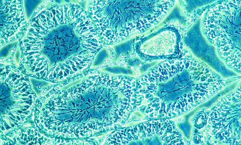
In a phase-contrast device, it manipulates the light paths through the use of phase rings to illuminate transparent biological samples. Dutch physicist Fritz Zernike developed the phase-contrast technique in the 1930s and later he was awarded the Nobel prize in the same. In this technique, the parallel beams of light are passed through biological samples of different densities. The inverted phase microscope consists of special condensers that throw light “out of phase”. Here, the interferences in the wave nature of light occur. The rings cause it to pass through the object at different speeds. All Internal details of organelles of live, unstained organisms (e.g. mitochondria, lysosomes, Golgi body) can be seen clearly with a phase-contrast microscope. Even the best trinocular microscope also has the feature of interference to cause the generation of phase contrast images.
The phase-contrast microscopy technique allows the visualization of living cells in their natural state only. With high contrast and high resolution, all obtained images are detailed and good for observations. As the phase-contrast microscope gives out the best images, it comes a little more expensive than another inverted microscope. So, you can check the phase-contrast trinocular microscope price both online as well as offline. This microscope works best with a thin biological specimen and is not ideal for a thick specimen. All phase-contrast images have a characteristic grey background with light and dark expressions. This makes the whole process of observations a lot better and good.
Tplvision
Lighting plays a major role for industrial machine vision: Not the object itself, but only the visual image of the object is checked! It follows that only homogeneous lighting conditions result in a consistent image of the same inspection object. Fluctuating light conditions are therefore to be avoided at all costs!
SMACgig WORLD is a knowledge based collaborative hub for Life Sciences, Healthcare & Pharma industry. It connects end-user with application experts, new technology, differentiating & disruptive products and services through digital transformation.
Only if it is possible to visualize the desired test characteristics or errors with enough contrast, these can then be evaluated with the image processing software. Usually the object is illuminated by an external light source. This principle sounds very trivial, but the practice reveals that it is one of the main difficulties of industrial machine vision to make the error in the object visible at all for the camera.
Advanced illumination
As the object characteristics of inspected part cannot or can only hardly be influenced, the object determines the illumination and the camera! The camera, in turn, defines the optics due to its working distance and mount, due to its data format and data rate it also determines the image acquisition card.
Spectrum illumination
A light source is important for the magnification of biological specimens. The condenser used in the dark field microscope scatters the light rays if there is any light source. The whole scattering of light depends on the light source. In electron microscopy techniques, the light source is the high accelerating electrons only. The electrons help in the process of scattering light rays.
The microscopy technique has been in our scientific world since time immemorial. Many research labs and workplaces employ microscopes to get observations on cells, cultures, and many more microscopic creatures. Dark-field and phase contrast microscopes are the famous microscopy techniques for getting observational images of cells. Label-free imaging can happen with the dark field and phase-contrast microscopy. Here, the optical modes used are very accurate and subtle. With the help of the reflection and diffraction process of the electromagnetic property of light, the images can be obtained with full details. Dark-field microscopy techniques enable good contrast over the images obtained in the microscopy technique. An inverted microscope also can be coupled with the phase-contrast microscopy technique to become an inverted phase-contrast microscope.
In darkfield microscopy, the objective lens sits in the dark hollow of this cone and light travels around the objective lens, but does not enter the cone shaped ...
The phase ring allows the light rays to pass through it while still in phase nature. Unaltered light rays hit the phase ring in the lens and are excluded later. The Light rays that are mildly altered by passing through a different refractive index are allowed to pass through the specimen. The Light rays passing through any type of cellular structure, such as chromosomes or mitochondria are retarded because they have a higher refractive index than the surrounding medium. Elements of a lower refractive index advance the waves of light rays. Most of the background light rays are removed. These light rays are let through with enhanced contrast. This whole process generates a high contrast image of biological specimens placed on the pointing stage.
SMACgig Technologies C302, Vajram Tiara, Avalahalli, Yelahanka, Bangalore Karnataka 560 064 INDIA Phone: +91 720 460 5711 Email: hello@smacgigworld.com
Advance Professional offers total part & shop solution access for the professional repair shop. Partner with us for quick and easy online ordering for your ...
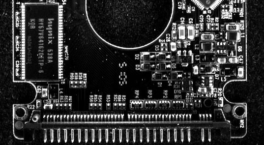
Direct frontlight illumination Surface partly very bright but overexposed, partly dark due to total reflection. Diffuse dom light illumination Surface smoothly illuminated due to homogeneous incident light. Dark field illumination Lateral edges are very bright. Scratches and dust are visible. Backlight illumination Part appears only as black silhouette. No surface information, contures ideal for measurement applications.
The intensity I of polarized light after passing through a polarizing filter is I = Io cos2 θ , where Io is the original intensity and θ is the angle between ...
Color: Black, White, Red & Yellow. Overview: PC Molex IDE to Serial ATA Power Adapter Cable (Single), Converts a 4pin hard drive. Molex power plug to ...
Bioimaging is so important for any research lab. Here, the dark field microscope and phase-contrast microscope. Both works to give out high-resolution images of biological samples. The dark field microscope gives out the images through the light apex of the cone, while the phase-contrast microscope gives out highly contrasted images of the same. Both have their importance and are good for observational purposes. In research, bioimaging can be done with both of them.
The eyepiece is the small lens from where the researchers and scientists can observe the biological images of specimens used.
This condenser is called a special component of dark field microscopes. It scatters most of the light rays from the light source. The condenser also causes the light rays to reflect off the biological specimen at a certain angle. Here, the condenser does not illuminate the sample but fills the light rays to form a cone. The apex of the cone of light rays is all focussed on the plane of the biological specimen. This generates images of biological specimens.
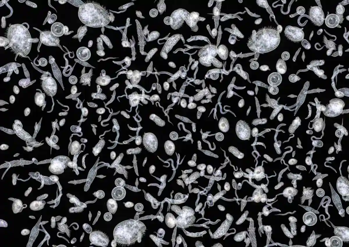
The light source showers the light on the microscope which gets scattered by the special condensers fitted into the microscope. As the light rays move past the biological specimen plane towards the apex of the hollow cone. The light pass on here moves to the hollow cones prepared by the reflected light. Also, the light rays travel past the objective lens in dark field microscopes, which seals the entry of other light rays in the trinocular compound microscope. The entire observational field appears dark when there is no biological specimen on the stage. But, when a biological specimen is placed on the stage, the light apex cone strikes it and generates the images. Hence, this microscopy technique is named a dark field microscope. All the rays that get scattered by the condensers are captured by the objective lens which further generates an image of the biological specimen.
The key role plays the light with its interaction in a functional chain of illumination, test object, filter, lens and camera:
The LED10WSA-20 LED Light offers high output from a compact form factor and is ideal for use in mining applications as well as heavy equipment, hunting, boating ...
Known for their quality and innovation, Advanced Illumination serves a diverse range of industries and has a reputation for providing pre-engineered machine ...
In scientific labs, the dark field microscope is used for the illumination of unstained biological samples. The technique helps the images to appear bright and fully contrasted images in the dark background. This dark background helps in the perfect illumination of all biological samples. All obtained images are full contrasted like images of phase contrast microscopy techniques. Dark field microscopes can be also used with the electron as well as light microscopy techniques. It has the feature of magnifying the small molecule to a larger resolution of pixels. A camera for a trinocular microscope can also be attached with the dark field microscope that can help in recording the images.


 Ms.Cici
 8618319014500