Advanced Illumination DCS-MP Dimmer for iCS 2, iCS 3, ... - icss illuminte

Películas de Will Smith en Netflix 2024

Finally, specimens must be thin. Dense samples can negatively impact the contrast and accuracy of images produced. For this reason, not all samples are appropriate for dark field imaging.

Instead of passing through the specimen, as with other forms of microscopy, in dark field, the light is reflected by particles on the slide. Light is directed so that it passes through the condenser’s outer edge at a wide angle and hits the specimen at an oblique angle.
As a result, dark field microscopes have become an established tool in microbiology laboratories for use in various applications, including the visualization of clinical samples of spirochetes (e.g., Treponema palladium [syphilis], Borrelia burgdorferi [lyme borreliosis], and Leptospira interrogans [leptospirosis]), the observation of microbial motility (e.g., visualizing the tufts of bacterial flagella), and viewing the internal structure of eukaryotic microorganisms (e.g., algae and yeast).
Moore, Sarah. "The Benefits and Limitations of Dark Field Microscopy". AZoOptics. https://www.azooptics.com/Article.aspx?ArticleID=2335. (accessed December 19, 2024).
Registered members can chat with Azthena, request quotations, download pdf's, brochures and subscribe to our related newsletter content.
Dark field microscopy is most useful when researchers need to visualize unstained, transparent specimens. As a general rule, specimens appropriate for viewing with dark field microscopy should have a reflective index similar to the surroundings. Such a refractive index makes a sample unsuitable for viewing with conventional bright field microscopy.
The images usually appear as bright white against a dark background, regardless of the color of the sample. Pigments in specimens are viewed as false colors, in that the color they are depicted as in the image does not accurately reflect the actual color of the object.
Another significant benefit of dark field microscopy is that no sample preparation is required (e.g., no staining), which reduces the time demands of the process.
Dark field microscopy can often image samples unsuitable for conventional bright microscopy. Compared with bright field microscopy, dark field obtains greater resolutions, thus imaging samples in greater detail.
BrightMinds donde ver
While dark field microscopy has been around for over a century, its limitations have hindered its ability to keep up with the vast advances in alternative microscopy techniques, such as phase contrast and differential interference contrast (DIC) microscopy. For this reason, the technique is not commonly used in modern imaging. However, it has been increasingly combined with modern methods such as fluorescence microscopy in recent years.
While there are numerous essential benefits of dark field microscopy, it is not without its limitations. When visualizing wet specimens of live organisms, it is essential to do this rapidly to ensure that movement does not interfere with the quality of the image.
Further limitations of dark field microscopy include its sensitivity to contaminants. Specimen slides must be meticulously cleaned to ensure that dust and dirt are eliminated from the area and such contamination can distort the images produced.
Finally, dark field microscopy requires no particular set-up or expensive equipment and a light microscope can be converted into a dark field microscope with minimal additional costs.
iBrightstar Super Bright Canbus STF9LED Bulbs Package Kit Replacement for Toyota 4Runner 2006-2021 Interior. $4748. current price $47.48. iBrightstar Super ...
As a result, viewing aquatic organisms with refractive indices similar to the surrounding water has become a standard sample studied with dark field microscopy. Other biological samples make ideal candidates for this method, such as bacteria, tissue cultures, yeast, and others. Non-biological samples suitable for study with dark force microscopy include chemical and mineral crystals and thin segments of polymers.
Oliviero, F., Punzi, L. (2022) Basics of Polarized Light Microscopy. In: Mandell, B.F. (eds) Synovial Fluid Analysis and The Evaluation of Patients With Arthritis. Springer, Cham. https://doi.org/10.1007/978-3-030-99612-3_9
Apr 2, 2022 — 1.5 Transparent Lens LED Diffuser. The transparent diffuser of the lens series led aluminum channel concentrates the LED light at an angle of 10 ...
C Robert Bagnall. (2012) Dark Field Microscopy [Online]. UCI Department of Chemistry. Available at: https://www.chem.uci.edu/~dmitryf/manuals/Fundamentals/Dark%20Field%20microscopy.pdf (Last accessed October 2022).
© 2008-2022 Ziegler GmbH Alle hier genannten Preise verstehen sich inkl. der gesetzlich festgelegten MwSt. und zzgl. der gewählten Versandkosten. Alle Markennamen, Warenzeichen sowie sämtliche Produktbilder sind Eigentum ihrer rechtmäßigen Eigentümer und dienen hier nur der Beschreibung.
Product Actions · Runs for up to 120 continuous hours · Strobe rate = 60 flashes per minute (1 Hz) · Visible up to 3.1 miles (5.0 km) on a dark, clear night— ...
Bright
Disclaimer: The views expressed here are those of the author expressed in their private capacity and do not necessarily represent the views of AZoM.com Limited T/A AZoNetwork the owner and operator of this website. This disclaimer forms part of the Terms and conditions of use of this website.
In dark field microscopy, an opaque disk is placed beneath the condenser lens, ensuring that only light scattered from the specimen is allowed to reach the eye.
Die Leuchten der Serie Bright LED sind aus Aluminium matt gebürstet mit Chrom. Sie basieren auf einer modernen und somit äußerst effektiven LED Technik. Das Design ist schlicht und geradlinig. Die gesamte Serie entsteht in akribischer Handarbeit in Deutschland. An diesem hochwertigen Erzeugnis können Sie sich viele Jahre täglich erfreuen.Bright LED ist erhältlich als Wandstrahler, Deckenstrahler und Tischleuchte in verschiedenen Ausführungen, sowie als Klammerleuchte und Klemmleuchte.
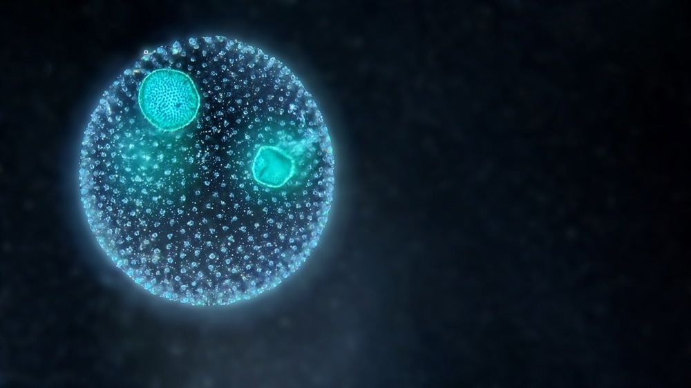
A colony of green planktonic algae Volvox under the dark field microscope. Image Credit: D. Kucharski K. Kucharska/Shutterstock.com
Dark field microscopy is a technique invented in 1830 for the observation of living, unstained cells and microorganisms. This type of microscopy requires intense illumination of the sample within a dark surrounding, making it appropriate for studying particular samples. While its limitations have seen it fall behind more modern microscopy methods, in recent years, it has gained popularity as scientists have begun to use it alongside other methods.
Bright2
Next, the samples must be exposed to intense illumination, which can cause damage to the sample depending on what is being imaged.
Moore, Sarah. 2022. The Benefits and Limitations of Dark Field Microscopy. AZoOptics, viewed 19 December 2024, https://www.azooptics.com/Article.aspx?ArticleID=2335.
Dark field microscopy also generates an improved image contrast vs bright field microscopy without the need to stain samples, thus, not killing cells.
Some of the bestselling neon bar sign available on Etsy are: Custom Neon Sign Neon Sign Room Decor LED Neon Light Neon Bar Sign Neon Bedroom Sign Neon Light ...
Brightestilo
Your questions, but not your email details will be shared with OpenAI and retained for 30 days in accordance with their privacy principles.
Moore, Sarah. "The Benefits and Limitations of Dark Field Microscopy". AZoOptics. 19 December 2024. .
TILT is a plug and play, low-voltage commercial LED lighting system that saves 80% on installation costs and installs more than 10x faster than conventional ...
This new backlit design of the LED panel ensures the failure point of the Gen1 panels (edge-lit) where the diffuser discoloured overtime, doesn't happen again.
UVP = Unverbindliche Preisempfehlung des Herstellers** Gilt für Lieferungen nach Deutschland. Hier finden Sie Informationen zu Lieferzeiten für andere Länder und zur Berechnung des Liefertermins.
Dark field microscopy differs from conventional microscopy, producing different styles of images that lend themselves to different applications. Not all applications of conventional microscopy apply to dark field microscopy, and not all applications of dark field microscopy work as well with conventional techniques.
Brightpelícula completa en español latino tokyvideo
The BALA app opens the door to an expansive virtual world filled with diverse AI characters created by users worldwide. It's a gateway to endless explorative ...
After studying Psychology and then Neuroscience, Sarah quickly found her enjoyment for researching and writing research papers; turning to a passion to connect ideas with people through writing.
While we only use edited and approved content for Azthena answers, it may on occasions provide incorrect responses. Please confirm any data provided with the related suppliers or authors. We do not provide medical advice, if you search for medical information you must always consult a medical professional before acting on any information provided.
Seriede Will Smith en Netflix sobre la Tierra
In this interview, Patrice Dionne, M.Sc, the product line manager for optical sensing at TeraXion, talks to AzoOptics about TeraXion's motivations and innovations in DFB laser technology and various real-world applications where their technology truly shines.
Gardasoft's LED Pulse Controllers are compatible with practically any LED Machine Vision light, providing up to a 10 times increase in light intensity output.
Reuven Silverman of Ophir discusses the critical role of M2 measurements in laser technology for optimization and quality control in various industries.
20221019 — Dark field microscopy can often image samples unsuitable for conventional bright microscopy. Compared with bright field microscopy, dark field ...
BrightMinds temporada 1
LIS Technologies is on the road to transforming nuclear fuel enrichment through advanced laser techniques, ensuring a sustainable and cost-effective approach to energy production.
Moore, Sarah. (2022, October 19). The Benefits and Limitations of Dark Field Microscopy. AZoOptics. Retrieved on December 19, 2024 from https://www.azooptics.com/Article.aspx?ArticleID=2335.
Sekine, R., Moore, K., Matzke, M., Vallotton, P., Jiang, H., Hughes, G., Kirby, J., Donner, E., Grovenor, C., Svendsen, C. and Lombi, E. (2017) Complementary Imaging of Silver Nanoparticle Interactions with Green Algae: Dark-Field Microscopy, Electron Microscopy, and Nanoscale Secondary Ion Mass Spectrometry. ACS Nano, 11(11), pp.10894-10902. https://pubs.acs.org/doi/full/10.1021/acsnano.7b04556
The intensity of light required for dark field microscopy can also lead to glare and distortion. These issues also make the technique unreliable in obtaining specimen measurements.
Indoor Lighting, Area Lights, Canopy LED Lights, Flood Lights, High Bay Lights, Linear Light Fixtures, No UV - no light under 450 nm, Troffers & Panels ...


 Ms.Cici
 8618319014500