
Advanced Illumination - illumination logo png
Oblique lightphotography
Find out about Scientifica's latest product releases, company news, and developments through a range of news articles, customer interviews and product demonstration videos
There are no additional optics in the above sample collection path, making oblique contrast ideal for two-photon and confocal microscopy, as no intensity is lost. It is also used to introduce contrast in patch-clamp experiments.
Obliqueillumination examination
When making adjustments, check that the sector stop is still positioned correctly and that the slider is not too far in or out.
Oblique contrast is a microscopy technique used to increase the contrast of images of thin, transparent, unstained samples, the details of which can be difficult to visualise using brightfield microscopy. Two main advantages of oblique contrast are that it is a cheap and easy way of improving the contrast of your images.
Firstly, pull out the oblique slider and open the oblique iris so the condenser is adjusted for brightfield illumination.

What is obliquelighting in Forensic Science
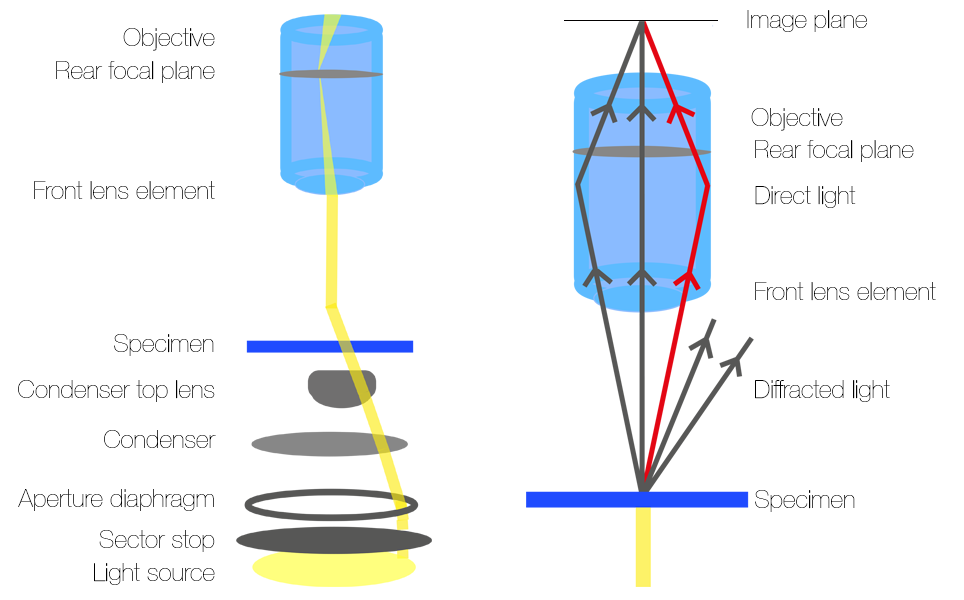
With a standard condenser used in brightfield microscopy, the specimen is axially illuminated; light passes through the centre of the specimen, diffracting at different angles as it hits the specimen before recombining at the back of the image plane. As the light diffracts at different angles and therefore travels a longer distance before reaching the image plane, the phase of the light is affected. However, when light of equal amplitude and wavelength and inverse phase combines it cancels out so there is no overall change in phase. This is called destructive interference. Therefore, there is no overall change in the image.
To make a brightfield microscope capable of oblique contrast, the only additional components needed are an oblique condenser and an infrared or visible light source. The condenser is placed in the condenser housing above the light source. The oblique contrast mechanism (sector stop) of the condenser is located below the condenser lens and aperture diaphragm.
Transmitted lighting
Obliqueillumination microscopy
Then, adjust the direction of the sector stop by turning the diaphragm control knob. Close down or open the stop to adjust the amount of oblique illumination until the desired image is achieved. The amount of oblique illumination needed depends on the specimen and the amount of detail required. The oblique condenser enables the angles of illumination to be altered through 360 degrees without moving the specimen.
Oblique contrast is suitable for imaging thin, transparent or nearly transparent samples such as cell cultures or thin brain slices. It can be used for visualising specimens that can’t be stained, such as living cells, tissues or organisms.
Set up Koehler illumination on the microscope. If using a manual microscope, remove the condenser stop before setting up Koehler illumination and replace it once Koehler illumination has been set up.
Obliquecontrast
Oblique contrast utilises an oblique condenser containing a sector stop with a small off-centre slit which makes the light go through the sample at an angle, so the specimen is illuminated from one direction only, shown in figure 2a. The sector stop is located beneath the lens and aperture diaphragm of the condenser. Light from the source passes through the slit which causes it to pass through the condenser lens at an oblique, off-axis angle. The specimen is then illuminated an angle.
Your microscope is now set up for oblique contrast. Please contact Scientifica if you have any questions or require any further guidance.
Oblique lightexample
As oblique contrast doesn’t use polarised light, it is suitable for imaging vertebrate axons. Vertebrate axons are myelinated by a birefringent material and, unlike polarised light, the unpolarised light doesn’t affect the refractive index of the myelin. As petri dishes are also birefringent, oblique contrast is ideal for imaging samples within these, for example cultures of bacteria.
Similarly to Differential Interference Contrast (DIC), images produced using oblique contrast have shadowed regions, giving a pseudo three-dimensional effect. The light strikes the specimen at an oblique angle, rather than an axial angle, which introduces phase changes to reveal features with a much greater contrast than brightfield illumination.

After passing through the specimen, the light rays enter the microscope objective and converge at the rear focal plane before they are observed through eye pieces or on a camera. As the light in oblique contrast hits the sample at an angle, only the diffracted sidebands of light on one half of the direct beam will be collected by the objective, as shown in figure 2b. This means that there is constructive and destructive interference between the direct light beams and collected sidebands.


 Ms.Cici
 8618319014500